

Article Figures & Data
Figures
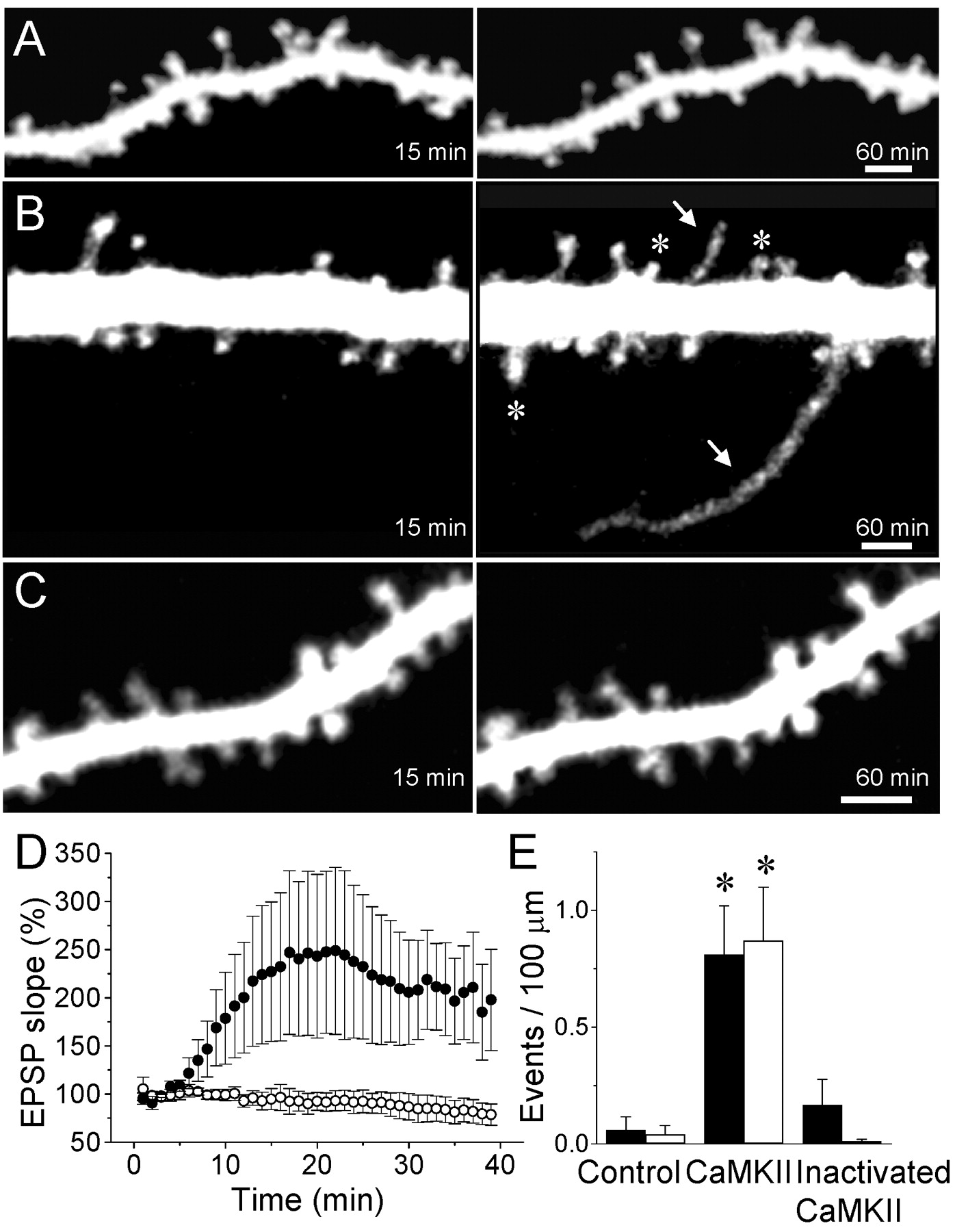
- Figure 1.
Growth of filopodia and formation of new spines induced by intracellular injection of activated CaMKII. A, Confocal microscopic images of dendritic spines taken 15 and 60 min after intracellular injection of the fluorescent dye sulforhodamine in a control experiment. B, Growth of two filopodia (arrows) and the formation of three new spines (asterisks) in a cell injected with activated CaMKII. C, Absence of changes in a cell injected with heat-inactivated CaMKII. Scale bars, 2 μm. D, Changes in EPSC slope measured as a function of time in seven cells injected with activated CaMKII (black circles) and five cells injected with the heat-inactivated enzyme (open circles). Data are means ± SEM. E, Quantitative analysis of the growth of filopodia (black columns) and formation of new spines (open columns) expressed as events per 100 μm of dendritic segment and per 60 min under control condition (n = 16), after the injection of activated (n = 14) or heat-inactivated (n = 9) CaMKII (*p < 0.01; t test).
- Figure 2.
Structural plasticity is induced by the activation of endogenous CaMKII. A, Formation of three new spines (arrows) in a cell injected with the phosphatase inhibitor calyculin A (150 nm). B, Growth of two filopodia in a cell injected with calmodulin (150 nm; bars: 2 μm). C, Growth of filopodia (black columns) and formation of new spines (open columns), expressed as events per 100 μm of dendritic segment and per 60 min, under control conditions (n = 16), after the intracellular injection of activated CaMKII (n = 14), or the phosphatase inhibitor calyculin A (150 nm; n = 13) or calmodulin (150 and 450 nm; n = 15). Data are means ± SEM. *p < 0.01; t test.
- Figure 3.
Blockade of LTP-induced filopodia growth and spine formation by inhibition of CaMKII. A, Formation of two new spines (asterisks) induced by theta burst stimulation. B, Application of high-frequency stimulation in the presence of KN93 (10 μm) did not induce the growth of filopodia or the formations of new spines. Scale bars, 2 μm. C, Changes in EPSC amplitude triggered by theta burst stimulation under control conditions (black circles) or in the presence of KN93 (10 μm, open circles). Data are means ± SEM (n = 5, 6). D, Quantitative assessment of filopodia growth (black columns) and spine formation (open columns) observed under control conditions (n = 16), after the application of theta burst stimulation in the absence of drugs (LTP; n = 10), or after stimulation in the presence of MK801 (40 μm; n = 8) or KN93 (10 μm; n = 10). Data are means ± SEM. *p < 0.05; t test.
- Figure 4.
Blockade of anoxia/hypoglycemia-induced structural plasticity by inhibitors of CaMKII. A, Growth of two filopodia (arrows, top) and formation of two new spines (asterisks, bottom) observed after the application of a brief (5 min) period of anoxia/hypoglycemia. B, Anoxia/hypoglycemia applied in the presence of AIP (200 nm) failed to induce structural plasticity. Scale bars, 2 μm. C, Growth of filopodia (black columns) and formation of new spines (open columns) expressed as events per 100 μm of dendritic segments and per 60 min under control conditions (n = 16), after a short (5 min) period of anoxia/hypoglycemia, (n = 12), or after anoxia/hypoglycemia, but in the presence of KN93 (40 μm; n = 7) or AIP (200 nm; n = 11). Data are means ± SEM. *p < 0.01; t test.


