

Article Figures & Data
Figures
- Figure 1.
An obligatory role of SRF in BDNF stimulation of SRE-mediated transcription in cortical neurons. A, BDNF stimulation of SRE-mediated transcription is blocked by coexpression of a dn SRF. Cortical neurons plated in 24-well plates were transfected using LipofectAMINE 2000 with 0.6 μg of DNA per well of a 5×SRE-driven luc reporter and 0.3 μg of DNA per well of a dn SRF or vector control to measure SRE-mediated transcription. At 60 hr after transfection, cells were placed in serum-free media and treated with 10 ng/ml BDNF or vehicle control (C) where indicated. Reporter gene activity was measured 6 hr later. B, Effects of mutations within the SRE that interfere with the binding of SRF on SRE-mediated transcription. Cortical neurons (3 × 10⁶ cells per 60 mm plate) were transfected using a calcium phosphate coprecipitation method with 1 μg of pAF42.SRE.wt or constructs with mutations in the SRE that disrupt SRF binding (pAF42.SRE.pm1 and pAF42.SRE.mut2). A α-globin gene was cotransfected as a control for transfection efficiency and equal loading. Forty eight to 60 hr after transfection, the cells were treated for 1 hr with vehicle control (-) or BDNF (+). Similar results were obtained from three independent experiments. The transcription from the endogenous fos gene (fosr) serves as a control for BDNF stimulation and RNase protection. C, Expression of a wild-type SRF but not the vector control or the dominant negative SRF, is sufficient to induce SRE-mediated transcription even under trophic withdrawal conditions. Cortical neurons were transfected with a wt or dn SRF or vector control (0.6 μg of DNA per well) as described in A. Data (A, C) are from three independent experiments of quadruplicate determinants. Error bars indicate SEM.
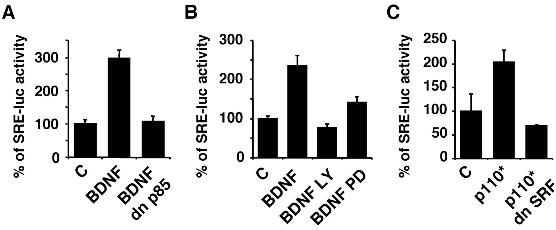
- Figure 2.
BDNF stimulation of SRE/SRF-mediated transcription in cortical neurons requires PI3K activity. Cortical neurons plated in 24-well plates were transfected with a 5×SRE-driven luciferase reporter to measure SRE-mediated transcription as described in Figure 1A. A, BDNF (10 ng/ml) stimulation of SRE-mediated transcription is blocked by coexpression of a dominant negative PI3K (dn p85). B, BDNF stimulation of SRE-mediated transcription is blocked by LY294002 (LY, 30 μm), a PI3K inhibitor, and by PD98059 (PD, 50 μm), an MKK1 inhibitor that inhibits ERK1/2 activation. C, Expression of a constitutive active PI3K (p110*) stimulates SRE-mediated transcription, which is blocked by coexpression of a dominant negative SRF. Data are from three independent experiments of quadruplicate determinants. Error bars indicate SEM.
- Figure 3.
SRF protects cortical neurons from trophic withdrawal. A, Representative photomicrographs of cortical neurons. Primary cortical neurons were transfected with vector control or an expression vector for wt SRF (4 μg). In all cases, a β-galactosidase expression vector was A, Representative photomicrographs of cortical neurons. Primary cortical neurons (-S), serum-containing conditioned medium (+S), or 10 ng/ml BDNF as indicated. Twenty-four hours later, cells were fixed and immunostained with an antibody against β-galactosidase to identify transfected cells (top panels). Hoechst 33342 staining was used to visualize nuclear morphology (bottom panels). Arrows point to the nuclei of transfected cells. The arrow-pointed nuclei in d are fragmented and condensed, characteristic of apoptosis, whereas those in panels b, f, and h are evenly stained, indicating healthy cells. B, Cells expressing SRF maintain viability after serum withdrawal. Cortical neurons were transfected with vector control or wt SRF (4 μg) and subjected to serum deprivation (-S) for 24 hr as in A. The cells were then incubated with 10 nm Mitotracker Red CMXRos to stain mitochondria (middle panels). Thirty minutes later, the cells were fixed and stained with an antibody against β-galactosidase to identify transfected cells (top panels) and with Hoechst dye 33342 to visualize nuclear morphology (bottom panels). Note that the cell transfected with the wt SRF has evenly stained nuclei and exhibits a punctate staining pattern for the mitotracker (arrow), indicating that it is nonapoptotic and viable. In contrast, the control transfected cell has typical apoptotic nuclear morphology and diffuse mitotracker staining, indicating that it is apoptotic and nonviable. C, D, SRF protects cortical neurons from trophic withdrawal in a dose dependent manner. Cortical neurons were transfected at DIV 3 with varying concentrations of a wt SRF DNA (0-4 μg). Cells were also cotransfected with a plasmid DNA encoding β-galactosidase as a marker for transfection. The CGN vector DNA was used as a supplement so that all plates have an equal amount of DNA. Two days after transfection, cells were washed twice with serum-free medium and then placed in serum-free medium (-S) or serum-containing conditioned medium (+S). Apoptosis in the transfected cell population (β-galactosidase-stained neurons; C) and the general cell population (D) was scored 24 hr later. Results are averages of five independent experiments ± SEM; ***p ≤ 0.001(ANOVA). NS, Not statistically significant. E, Western analysis for transfected SRF. Cortical neurons were transfected and treated as in C and D. Cell lysates were prepared 2 d after transfection, and 25 μg of total protein was used for Western blotting against SRF. Western analysis of β-actin was used as a loading control. The transfected SRF is expressed in a dose-dependent manner.
- Figure 4.
SRF is necessary for full neuroprotection provided by BDNF against trophic deprivation. Cortical neurons were transfected with control vector (4 μg) or dn SRF (4 μg) and treated as described in Figure 3. BDNF was added at 10 ng/ml where indicated. Results are averages of three independent experiments ± SEM; ***p ≤ 0.001(ANOVA).
- Figure 5.
Role of PI3K signaling in SRF neuroprotection against trophic deprivation. Cortical neurons were transfected with 4 μg of DNA of a control vector, a dn SRF, or a constitutive active PI3K (p110*) where indicated and treated as described in Figure 3. A, Neuroprotection against trophic deprivation provided by expression of a constitutive active PI3K is reversed by coexpression of a dn SRF, suggesting that SRF contributes to PI3K neuroprotection. B, Neuroprotection afforded by expression of the wild-type SRF is completely reversed by 30 μm LY294002 treatment (LY), suggesting that the activity of the endogenous PI3K is necessary for SRF neuroprotection. C, Anti-phospho-Akt (p-Akt) Western analysis demonstrating complete inhibition of endogenous PI3K activity in the presence of 30 μm LY294002 (LY). Relative levels of p-Akt were normalized to the loading control β-actin. Results are averages of three independent experiments ± SEM; **p ≤ 0.01; ***p ≤ 0.001 (ANOVA).
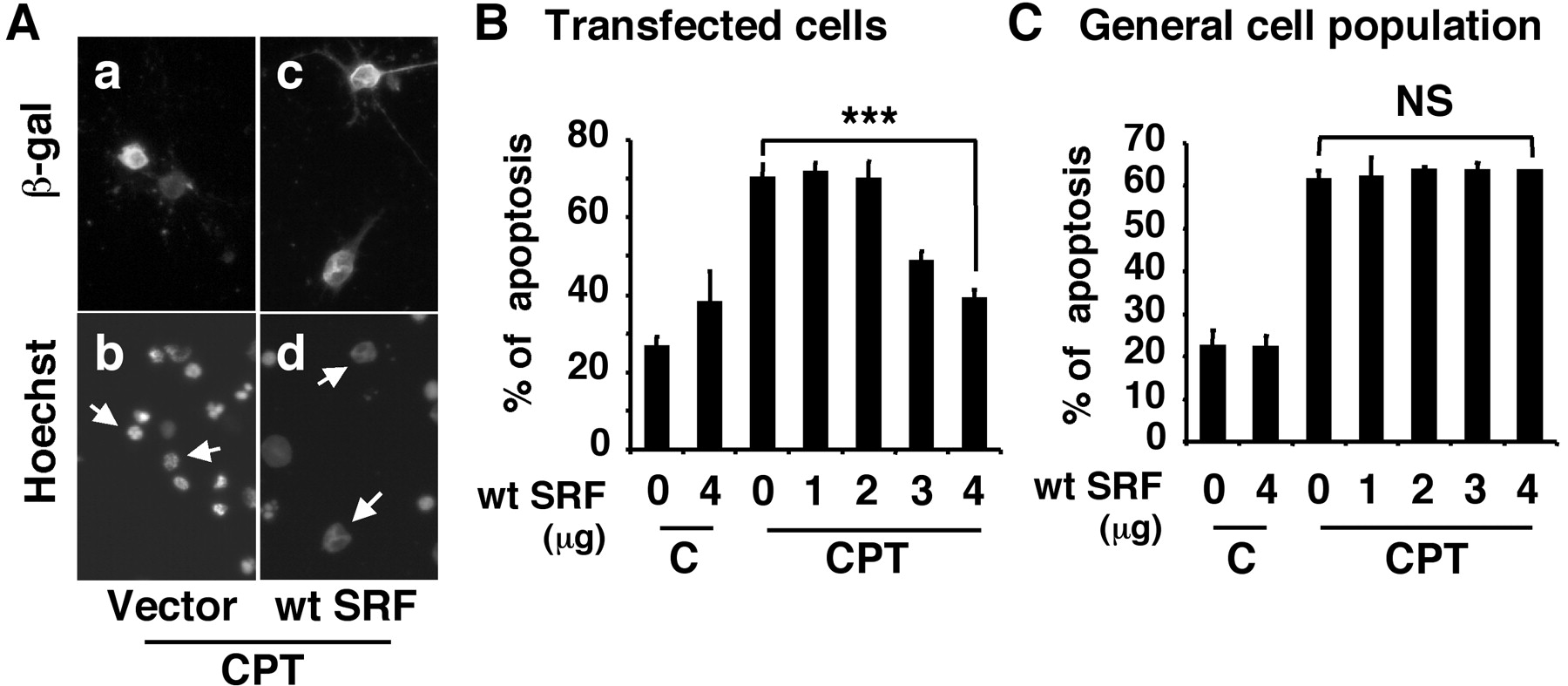
- Figure 6.
Expression of a wild-type SRF is sufficient to protect cortical neurons against camptothecin, a DNA-damaging agent. A, Representative photomicrographs of cortical neurons. Primary cortical neurons were transfected with vector control or an expression vector for wild-type SRF (4 μg). In all cases, a β-galactosidase expression vector was cotransfected as a marker for transfection. Two days after transfection, cells were treated with 5 μm camptothecin (CPT). Twenty-four hours later, cells were fixed and immunostained with an antibody against β-galactosidase to identify transfected cells (a, c). Hoechst 33342 staining was used to visualize nuclear morphology (b, d). Arrows point to the nuclei of transfected cells. The arrow-pointed nuclei in b are apoptotic, whereas those in d are healthy. B, C, Quantitation of SRF neuroprotection against camptothecin. Cortical neurons were transfected with varying concentrations of a wt SRF (0-4 μg). The CGN vector DNA was used as a supplement so that all plates have equal amount of DNA. Two days after transfection, cells were treated with 5 μm camptothecin or vehicle control (C), and apoptosis in transfected cells (B) and in the general cell population (C) was scored 24 hr later. Results are averages of three independent experiments ± SEM; ***p ≤ 0.001(ANOVA). NS, Not statistically significant.
- Figure 7.
BDNF protection against camptothecin is mediated by SRF. A, Expression of a dominant negative SRF reduces BDNF neuroprotection against camptothecin. B, Expression of a dominant negative SRF reduces neuroprotection of a constitutive active MKK1 against camptothecin. Cortical neurons were transfected with control vector (4 μg), a dn SRF (4 μg), or a constitutive active MKK1 (4 μg), a kinase that phosphorylates and activates ERK1/2. Cells were treated with vehicle control (C) or 5 μm camptothecin (CPT) as described in Figure 6. BDNF was added at 10 ng/ml at the time of camptothecin treatment. Results are averages of three independent experiments ± SEM; *p ≤ 0.05; ***p ≤ 0.001(ANOVA).


