

Article Figures & Data
Figures
- Figure 1.
Whole-cell K+ currents recorded from intermediate cells of the stria vascularis of the mouse. A, Representative traces for a family of K+ currents obtained from a holding potential of –80 mV and stepped up to the voltages indicated. Inward sodium and calcium currents, if present, were suppressed by substituting choline and NMG for sodium and calcium ions (see Materials and Methods). B, Similar data obtained from the same cell (A) after application of external solution containing 1 μm E-4031, a delayed rectifier K+ current blocker. C, Difference current traces (A, B) depicting the E-4031-sensitive current traces. For clarity, most of the current traces were not plotted. D, Summary data of the current–voltage relationship of control (•), current after application of E-4031 (▪), and the E-4031-sensitive component (○). The mean steady-state current magnitudes at ∼300–400 ms were measured at the different test potentials. The data represent recordings from seven intermediate cells.
- Figure 2.
RT-PCR for detecting MERG1a expression in the inner ear. RNA from different tissues was used. A band at ∼1.4 kb was obtained for all tested RNA samples. M, DNA molecular weight; StV, stria vascularis; OC, organ of Corti; Br, brain; Ht, heart; NT, no RNA template control.
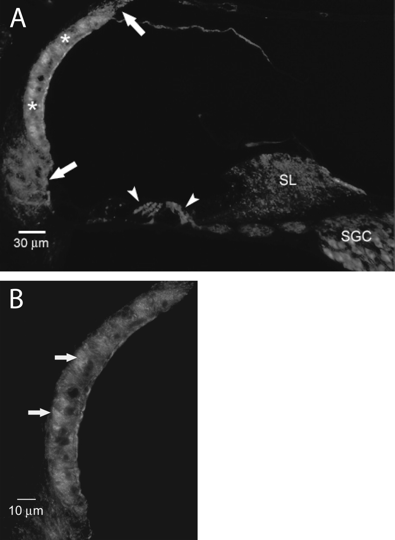
- Figure 3.
Reactivity for ERG in a cross section of a lower apical turn of a mouse cochlea. A, Immunoreactivity for ERG is localized to the middle portion of the stria vascularis (asterisks). Lower levels of reactivity are noted in the spiral ganglion cells and outer and inner hair cells (arrowheads), as well as below and above the stria vascularis in the region of the type II and type V fibrocytes (arrows), respectively. B, Higher magnification of the stria vascularis shows that the immunoreactivity is localized to the region of the interdigitating processes of marginal and intermediate cells, although the apical margin of some marginal cells is also reactive. SL, Spiral ligament; SGC, spiral ganglion cell.
- Figure 4.
Reactivity for ERG in nonstrial cochlear tissue. A, B, Organ of Corti. The specificity of the ERG reactivity in the hair cells is shown in A; an adjacent section stained with hematoxylin is shown in B. Reactivity in the outer hair cells is most pronounced in the perinuclear region (arrow), whereas reactivity is noted throughout the cytoplasm of the inner hair cell (arrow-head). The cups of the Dieter's cells (asterisks) also appear immunofluorescent. ERG is expressed in the eighth nerve fibers under the inner hair cell (double arrows). C, E, Immunofluorescence in the spiral ligament was confined to the type II fibrocytes in the region of the spiral prominence and surrounding the root cells (asterisks) as well in the superficial layer of the type V fibrocytes (arrows) superior to the stria vascularis. D, Rosenthal's canal (double arrows) as well as the spiral ganglion cells of the modiolus are reactive for ERG. E. A cross section of a basal turn lateral wall from a mouse cochlea in which normal serum was substituted for the primary antibody lacks reactivity for ERG. OHC, Outer hair cell; SGC, spiral ganglion cell; SL, spiral limbus; SP, spiral prominence. F, Negative control.
- Figure 5.
Reactivity for ERG in vestibular tissue. A, B, Immunoreactivity for ERG is localized to the hair cells (arrows) and nerve fibers (asterisks) in both the crista ampullaris (A) and the macula (B). C, D, A view of the sensory epithelium at higher magnification indicates ERG reactivity in the basal pole of the hair cells. The staining often shows a punctuate perinuclear pattern. Whether ERG is confined to the hair cell or is also present in the nerve calyx cannot be determined at the light microscopic level. E, F, Adjacent sections at the same magnification stained with hematoxylin.
- Figure 6.
Immunogold localization of MERG. Cochlear sites of MERG expression were examined with immunogold electron microscopy with the postembedding technique. Thin mid-modiolar sections (70 nm) of cochlea used previously for routine ultrastructural examination were etched with sodium metaperiodate before incubation in primary and secondary antibodies conjugated to either 16 nm (A, F, G) or 10 nm (B–E) colloidal gold particles (arrows). A, C, In the stria vascularis, particles were localized in the cytoplasm and processes of the intermediate cells. Occasional particles were noted on the plasmalemma of marginal cells. The number of particles on intermediate cell interdigitations or cytosol around the nucleus exceeds the label on the marginal cell processes by ∼40–45%. Label was seen only in the processes that extend into the interstrial space; no label was seen on the cell body of the marginal cells. Of the marginal or intermediate cells (A, C), 90–95% of the specific label is adjacent to or near the plasmalemma, and, in a few instances, several gold particles are clumped together and it is impossible to determine which cell type they localize. B, In the spiral ligament, labeling was found on the type II fibrocytes in the region in which their amplified processes were interspersed with the upper root cells. D, The area immediately above the nucleus of the outer hair cell showed some gold particles. E, In the inner hair cell, however, gold label was observed at the base of the hair cell and in the cytosol of afferent fibers. A, Afferent nerve; E, efferent nerve. G, Gold particles were also found in nerve fibers in Rosenthal's canal. F, However, no particles were observed in the cytosol or plasmalemma of the basal cells of the stria vascularis. The nuclear location of gold particles may be nonspecific binding associated with the colloidal gold technique (Smith and Jarett, 1993). RC, Root cell; II, type II fibrocytes.
- Figure 7.
Expression of inner ear-specific MERG1a in Xenopus oocytes. Outward K+ currents from Xenopus oocytes were injected with mRNA of StV-specific MERG1a channel. The recordings were obtained from a holding potential of –80 mV to step potentials ranging from –70 to +60 mV (ΔV = 10 mV). A, Uninjected oocytes had no current. B, MERG1a-injected oocytes yielded robust currents. C, Summary data of the current–voltage relationships from 19 oocytes. D, Outward tail currents were elicited from different step potentials to –40 mV. The activation curve was obtained by plotting the normalized tail current (I/Imax) at each test potential and then fitted by the Boltzmann equation {I/Imax = [1 + exp(V1/2 – V)/km]}–1, where V1/2 is the half-activation voltage and km is the maximum slope. The estimated V1/2 and km from the activation curve were –47.4 ± 1.4 and 10.6 ± 1.3 mV (n = 10), respectively.
- Figure 8.
Pharmacology of inner ear-specific MERG1a. Outward K+ current from Xenopus oocytes injected with mRNA of StV-specific MERG1a channel was sensitive to rBeKm-1 and E-4031. A, B, Examples of current traces recorded from a holding potential of –80 mV to a step potential of 30 mV for control and after application of rBeKm-1 and E-4031, respectively. The concentrations of the drugs are indicated. C, D, Dose–response curves. The IC50 values for rBeKm-1 and E-4031 were ∼16 and 165 nm, respectively.
- Figure 9.
Effects of [K+]e on the magnitude of MERG1a currents. A, Exemplary MERG1a current traces elicited from a holding potential of –80 mV to step potentials ranging from –80 to +40 mV with ΔV = 10 mV. The current was recorded in an external solution with no added K+ (∼0mm). B, C, Similar current traces elicited after perfusing external solutions containing 5 and 10 mm K+, respectively. Unlike most K+ currents, the current magnitudes increase with increasing [K+]e. Group summary data of the MERG1a current magnitude versus voltage at different [K+]e (0–50 mm; n = 7) are shown. The currents were substantially enhanced as [K+]e was increased.
- Figure 10.
Single-channel currents of MERG1a expressed in CHO cells. A, Representative and consecutive single-channel traces recorded in a cell-attached patch with a pipette ([K+], 140 mm). The bath solution contained 140 mm K+, and the resting potential of CHO cells was ∼0 mV. The holding potential was –50 mV, and the step potentials are indicated. The I–V relationships are shown in B. The inset in B is an example of an amplitude histogram used to generate I–V relationships at 50 mV. The single-channel conductance was 14.3 ± 2.8 pS (n = 6). C, Closed; O, open.


