

Article Figures & Data
Figures
- Figure 1.
Upregulation of NR2C mRNA by BDNF stimulation. Representative data of NR2C and GAPDH mRNA blots and ethidium bromide-stained 18s rRNA and quantitative data of NR2C mRNA blotting analysis are indicated. A, Cells were treated with or without 50 ng/ml BDNF in low (5 mm) and high (25 mm) KCl for 96 h. Levels of NR2C mRNA were quantitated by RNA blotting of total RNAs (15 μg per lane; n = 4). **p < 0.01. B, Time course of NR2C mRNA upregulation by the addition of BDNF (100 ng/ml) in granule cells cultured in low KCl (n = 4). *p < 0.05, **p < 0.01, BDNF stimulated versus unstimulated. C, Dose-response relationship of BDNF-induced NR2C mRNA upregulation. Cells were cultured in low KCl and treated with the indicated concentrations of BDNF for 96 h (n = 4). *p < 0.05, **p < 0.01, BDNF stimulated versus unstimulated. D, PCR-based nuclear run-on assays. Cells were cultured in low KCl and treated with or without 50 ng/ml BDNF for 96 h. Nuclei were isolated from BDNF-treated and untreated cells and incubated for 50 min in the presence and absence of four nucleotide triphosphates (NTPs). RNA was extracted, and levels of run-on NR2C mRNA were quantitated by PCR analysis (n = 3). *p < 0.05. Error bars indicate SEM.
- Figure 2.
BDNF-induced NR2C upregulation by TrkB activation. Cells were cultured in low KCl and treated with or without BDNF (100 ng/ml in A and 50 ng/ml in B-D and F) for 96 h. A, Levels of indicated mRNAs were quantitated by RNA blotting (n = 4). B, Cell lysates (40 μg) were immunoblotted with anti-panNR2 antibody or anti-NR1 antibody. Molecular sizes (kilodaltons) of protein makers are indicated on the left. C, P2 membrane fractions were isolated, solubilized, and immunoprecipitated (IP) with anti-NR1 antibody, followed by immnoblotting with anti-panNR2 antibody. D, Cell-surface proteins were biotinylated with Sulfo-NHS-SS-Biotin. Cell lysates were solubilized, precipitated with NeutrAvidin beads, and immunoblotted with anti-panNR2 antibody. E, Cells were cultured in low KCl and treated with BDNF, NGF, NT-3, or NT-4 (50 ng/ml each) for 96 h. Levels of NR2C mRNA were quantitated (n = 4). F, Granule cells were prepared from TrkB-/- knock-out mice(KO) and their wild-type (WT) littermates. NR2C and TrkB mRNAs were analyzed by RNA blotting. Ethidium bromide-stained 18s rRNA is also indicated. *p < 0.05; **p < 0.01. Error bars indicate SEM.
- Figure 3.
BDNF-induced upregulation of NR2C mRNA via the ERK1/2 signaling cascade. A, Cells were cultured in low and high KCl and treated with 50 ng/ml BDNF for the indicated times. Cell lysates (40 μg) were immunoblotted with antibody against either phosphorylated TrkB at Tyr 515 (p-TrkB) or TrkB. stim., Stimulation. B, Cells were cultured in low KCl. These cells were pre incubated for 30 min with 50 nm K252a, 20 μm U0126, 5 μm SB203580 (SB), 10 μm LY294002 (LY), or 2 μm U73122 and further incubated with the addition of 50 ng/ml BDNF for 48 h. Inhibitors were supplied at 24 h after BDNF stimulation. Representative blots of NR2C mRNA and ethidium bromide-stained 18s rRNA and quantitative data of NR2C mRNA blotting analysis are indicated (n = 3-5). C, Cells were transfected with GFP, GFP-dnMEK1/2, or GFP-dnMEK5. Transfected cells were cultured in low KCl and treated with or without 50 ng/ml BDNF for 48 h. GFP-positive cells were collected by FACS, and RNA was extracted from GFP-positive cells. Levels of NR2C mRNA were quantitated by PCR analysis (n = 3). **p < 0.01. Error bars indicate SEM.
- Figure 4.
BDNF-induced NR2C mRNA upregulation by L-VSCC inhibitor or calcineurin inhibitors under the depolarizing condition. Representative blots of NR2C mRNA and quantitative data of RNA blotting are indicated. A, Cells were cultured in high KCl and treated with or without 50 ng/ml BDNF for 4 8 h in the presence or absence of 1 μm nifedipine (Nif) (n = 4). B, Cells were cultured in high KCl. These cells were preincubated for 30 min with 10 μm KN62 (KN), 1 μm FK506 (FK), 1 μm bisindolylmaleimide I (Bis I), 500 nm cyclosporin A (CsA), or 1 μm rapamycin (Ra) and further incubated with or without 50 ng/ml BDNF for 96 h (n = 4). **p < 0.01. Error bars indicate SEM.
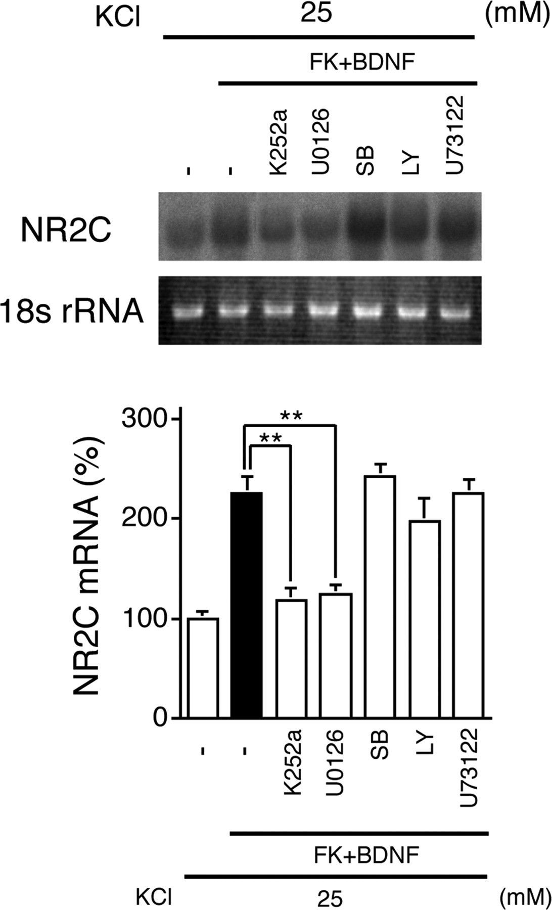
- Figure 5.
TrkB-ERK signaling cascade for BDNF-induced upregulation of NR2C mRNA under the depolarizing condition. Cells were cultured in high KCl. These cells were preincubated for 30 min with 50 nm K252a, 20 μm U0126, 5 μm SB203580 (SB), 10 μm LY294002 (LY), or 2 μm U73122 and further incubated with the addition of 50 ng/ml BDNF plus 1 μm FK506 (FK) for 48 h. Inhibitors were supplied at 24 h after treatment with BDNF plus FK506. Representative blots of NR2C mRNA and ethidium bromide-stained 18s rRNA and quantitative data of NR2C mRNA blotting analysis are indicated (n = 3-4). **p < 0.01. Error bars indicate SEM.
- Figure 6.
Involvement of de novo synthesized BDNF in NR2C mRNA upregulation under the depolarizing condition. Representative blots of NR2C or BDNF mRNA and quantitative data of RNA blotting analysis are indicated. A, Cells were cultured in high KCl and treated with 50 ng/ml BDNF, 1 μm FK506, and 50 ng/ml BDNF plus 1 μm FK506 for 96 h (n = 3). B, Cells were cultured in high and low KCl and treated with or without BDNF (50 ng/ml) and/or the indicated inhibitors [1 μm FK506 and 10 μm KN62 (KN)]. Levels of NR2C and BDNF mRNAs were analyzed by RNA blotting. The CaMK inhibitor KN62 blocked both BDNF and NR2C mRNA upregulation (n = 3). The addition of exogenous BDNF recovered NR2C mRNA upregulation in cells treated with KN62 and FK506 (n = 3). C, Cells were cultured in high KCl. These cells were preincubated for 30 min with or without 40 μg/ml anti-BDNF antibody (Ab) and further incubated for 48 h in the presence or absence of 1 μm FK506 (n = 3-4). D, Cells were prepared from TrkB-/- knock-out mice (KO) and their wild-type littermates (WT). These cells were cultured in high KCl and treated with or without 1 μm FK506 for 48 h. Upregulation of NR2C mRNA by FK506 treatment was significantly suppressed in cells prepared from TrkB-/- knock-out mice (n = 3). *p < 0.05; **p < 0.01. Error bars indicate SEM. FK, FK506.
- Figure 7.
Decrease in NR2C mRNA and protein levels in the TrkB-/- cerebellum and increase in NR2C mRNA levels in FK506-treated cerebellar slice culture. A, In situ hybridization analysis of NR2C mRNA was performed with a digoxigenin-labeled antisense probe for NR2C mRNA in parasagittal sections of littermates of TrkB-/- and TrkB+/+ mice at P14. Hybridization signals (blue) of NR2C mRNA were greatly reduced in granule cells of TrkB-/- mice compared with those of TrkB+/+ mice. Note also that granule cells comparably migrated into the IGL between the two genotypes. B, Total RNA was isolated from cerebella of littermates of TrkB-/-, TrkB+/-, and TrkB+/+ mice at P8, P11, and P14 and subjected to PCR analysis (n = 3). *p < 0.05; **p < 0.01. C, NR2B mRNA levels in the cerebellum of the three genotypes at P14 were quantitated by PCR analysis as described in B. D, Cerebellar extracts of the three genotypes at P14 were subjected to immunoblot analysis with anti-panNR2 antibody. E, Slice cultures were conducted in high KCl and treated with or without 1 μm FK506 (FK) for 96 h. RNA was extracted, and levels of NR2C mRNA were quantitated by PCR analysis (n = 4). **p < 0.01. Error bars indicate SEM.


Additional Files
Supplemental data
Files in this Data Supplement:
- supplemental material - Supplemental material. A Model for sSignaling mMechanisms of BDNF-iInduced NR2C uUpregulation in gGranule cCells. For detailed explanation, see the text. Activation of signaling cascades and their blockade by selective inhibitors are indicated by arrows and blocked bars, respectively. The target of calcineurin (CaN) dephosphorylation remains elusive and is presumed at downstream of the ERK1/2 signaling.


