

Article Figures & Data
Figures
- Figure 1.
Diagram of primary tissue dissected for in vitro assay of stem-cell activity. A2-mm-thick coronal slice was cut between -1.22 and -2.70 mm, relative to bregma, as determined from an adult mouse brain atlas (Paxinos and Franklin, 2001). The slice was placed caudal side up and the tissue dissected using two pairs of curved forceps. First, the midbrain was removed, and then the hippocampus, as a whole, was separated gently from the corpus callosum. Finally, the pLV, including the corpus callosum, was pinched away from the cortex. Hippo, Hippocampus; LV, lateral ventricle. This image is from Paxinos and Franklin (2001) with permission.
- Figure 2.
Proliferative cell frequency when primary tissue was cultured with both EGF and FGF-2. A, Primary hippocampal neurosphere limiting dilution assay (n = 9; mean ± SEM). The intercept of log(37% negative wells) gave a neurosphere-forming frequency of one neurosphere for every 9229 cells plated. Inset, Representative photograph of a primary hippocampal neurosphere. Scale bar, 100 μm. B, Primary pLV neurosphere limiting dilution assay (n = 6; mean ± SEM). Neurosphere-forming frequency was calculated as one neurosphere for every 766 cells plated. Inset, Representative photograph of a primary pLV neurosphere. Scale bar, 100 μm.
- Figure 3.
Potential for long-term proliferation in vitro in the presence of both EGF and FGF-2. A, Colony diameters (in millimeters) when primary hippocampal and pLV cells were cultured in the neural colony-forming cell assay (mean ± SEM). B, Expansion of cell numbers when primary hippocampal and pLV cells were cultured in the neurosphere assay through multiple passages (mean ± SEM). Hippo., Hippocampus.
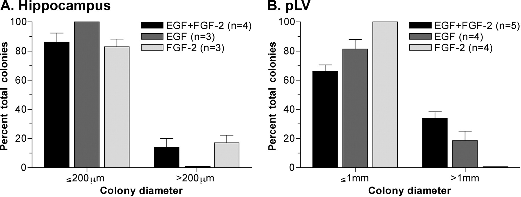
- Figure 4.
Differential effects of EGF and FGF-2 on proliferation. A, Colony diameters (in micrometers) produced by primary hippocampal cells when cultured in the presence of EGF plus FGF-2 or each growth factor separately in the neural colony-forming cell assay (mean ± SEM). B, Colony diameters (in millimeters) when primary pLV cells were cultured in the presence of EGF plus FGF-2 or each growth factor alone in the neural colony-forming cell assay (mean ± SEM).
- Figure 5.
Differentiation of primary neurospheres. A-C, Representative differentiated neurospheres immunolabeled for GFAP (green) and βIII tubulin (red) and counterstained with DAPI (blue). Primary pLV neurosphere (A), primary hippocampal neurosphere grown in the presence of EGF and FGF-2 (B), and a primary hippocampal neurosphere cultured in the presence of EGF, FGF-2, and BDNF (100 ng/ml) (C) are shown. A, B, Insets, Immunolabeling for the oligodendrocyte marker O4 (green) in representative differentiated pLV (A) and hippocampal (B) neurospheres. D, Percentage of differentiated neurospheres containing cells immunopositive for either GFAP alone (astrocytes) or GFAP plus βIII tubulin (neurons) after growth in the presence of EGF and FGF-2 or with BDNF (100 ng/ml) added to hippocampal cultures at different time points during neurosphere proliferation (n = 3; mean ± SEM). E, Percentage of differentiated neurospheres containing astrocytes alone or astrocytes plus neurons when primary hippocampal cells were cultured in increasing concentrations of BDNF (n = 3; mean ± SEM). Hippo., Hippocampus.
- Figure 6.
Effect of BDNF on hippocampal neurosphere frequency and proliferative potential. A, Primary hippocampal neurosphere frequency when cells were cultured in the presence of EGF and FGF-2 plus BDNF at increasing concentrations (n = 4; mean ± SEM). B, Colony diameters (in micrometers) when primary hippocampal cells were cultured in the neural colony-forming cell assay in the presence of EGF and FGF-2 or EGF and FGF-2 plus BDNF (100 ng/ml). Values are mean ± SEM.


