

Article Figures & Data
Figures
- Figure 1.
Recording with rhodamine-phalloidin-filled electrodes does not disrupt basic physiology or LTP. A, EPSCs elicited by stimulation of the Schaffer-commissural projections to the inner portion of stratum radiatum. Recordings were collected during the 20 min baseline period and for 60 min after TBS. The traces at the top show representative EPSCs collected from a control cell (no theta bursts; left) and from a cell in which LTP was induced by a single train of 10 theta bursts (right). Traces collected 60 min apart are superimposed in both cases. The graph in A summarizes results for a group of neurons that received control stimulation alone (open circles) or theta bursts at the 20 min time point (closed circles). Error bars represent SEM. B, Phalloidin-labeled cells from two control cases: laminar placements are indicated by arrows. s. pyram, Stratum pyramidale; s. rad, stratum radiatum; s. molec, stratum lacunosum-moleculare. Photomicrographs are at 10× objective magnification.
- Figure 2.
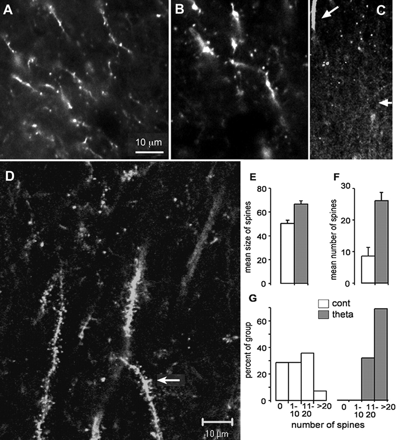
Theta bursts increase labeling of dendritic spines. A, B, Two cases, photographed at 40× objective magnification, in which densely labeled spine heads are located on stained segments of secondary dendritic branches. Note in B that the punctate spine heads are absent from most of the field. C, Lower-power photomicrograph (10× objective magnification) illustrates the location of labeled elements within s. radiatum. The primary branch of the apical dendrite can be seen emerging from s. pyramidale at the top of the figure (top arrow). A small cluster of labeled spines is located to the right of the branch; the distribution of these profiles is restricted to the proximal portion of s. radiatum (the mid proximodistal level of the layer is marked by small arrow at right). D, The relationship of labeled spine heads to secondary dendritic branches is evident in confocal images: the arrow indicates labeled dendrites decorated with numerous densely labeled spines (40× objective magnification). E, Size of labeled spines in a 5300 μm2 sampling zone from control (cont; n = 14) and potentiated (theta; n = 19) slices (mean ± SEM); the group difference is highly significant (p < 0.005). F, Number of labeled spines in the same areas from the two groups (p < 0.0001). G, Distribution of spine numbers across slices in the control and experimental groups. As shown, <10% of the control slices had 10 or more labeled spines in the proximal half of s. radiatum, whereas >60% of the theta burst cases fell into this category.
- Figure 3.
Theta burst stimulation causes actin polymerization in dendritic segments. A, A primary (apical) dendritic branch from a cell with potentiated synapses. Note the numerous fine oblique dendritic branches. SP, Stratum pyramidale; SRp, proximal stratum radiatum; SRd, distal stratum radiatum; SM, stratum lacunosum-moleculare. B, A cluster of labeled segments from a theta burst slice in which multiple cells had been clamped. Note that the labeled segments are restricted to the inner half of the s. radiatum, the area innervated by the fibers that had received theta bursts. C, Mean number of labeled dendritic segments in control (cont) and theta-stimulated (theta) slices; the difference between groups is highly significant (p = 0.001). Error bars represent SEM. D, Distribution of numbers of stained segments across control and theta burst slices (bars represent numbers of segments per slice sample field, in nonoverlapping categories). Slightly <40% of the potentiated slices did not have more labeled branches than found in controls; this value is higher than that for labeled spines (Fig. 2G). E, F, Labeled basal dendrites in two cells expressing LTP; s. pyramidale is at the bottom of each micrograph. Photographs were taken at 10× objective magnification. Scale bar: (in A) A, B, E, F, 20 μm.
- Figure 4.
Blocking LTP induction does not prevent the labeling of cell bodies and primary dendritic branches. The rhodamine-phalloidin-labeled cell to the left (A) is from a slice that had been infused with the NMDA receptor antagonist APV from 15 min before the delivery of theta bursts to its Schaffer-commissural afferents, whereas the labeled cell on the right (B) was clamped at -80 mV during the theta burst train. There was no evidence of increased spine staining in these cases, although, as shown, labeling of the cell bodies and primary dendritic branches was robust. so, Stratum oriens; sr, stratum radiatum. Magnification, 10×.
- Figure 5.
Effects of theta burst stimulation on labeling after topical application of rhodamine-phalloidin. A, Field EPSPs recorded at a single site in s. radiatum of CA1b and elicited by stimulating electrodes in CA1a (S1) or CA1c (S2). Theta bursts were delivered to S1 and then 2 min later to S2. The induction of short-term potentiation in S1 had no evident effect on the response in S2 recorded 2 min later. Calibration bars: 0.5 mV, 5 ms. B, D, Labeling in the standard-sized sampling zone placed over areas of afferent stimulation in the proximals. radiatum of two control slices. C, E, Labeling in fields comparable with those shown in B and D in two slices that had received theta burst stimulation. A fine dendritic segment is labeled in C; similar processes are very faint or not labeled in E. F, Number of labeled spines and dendritic branches in the sampling zone in slices given control (C; n = 4) or theta burst (T; n = 5) stimulation. Note that the large increase (∼14-fold; p < 0.002) in phalloidin-positive spines was elicited by the latter treatment without any evident increase in labeled dendritic segments. Magnification, 20×. Scale bar: B-E, 50 μm. Error bars represent SEM.
- Figure 6.
Theta stimulation-induced increases in rhodamine-phalloidin labeling persist for at least 30 min and are blocked by the NMDA receptor antagonist APV in slices from young adult rats. Photomicrographs of labeling within the sampling zone in stratum radiatum after topical application of rhodamine-phalloidin are shown; stimulating and recording electrodes were located as described in Figure 5. Top panels (from left to right) show the sampling zone in a control slice, in three slices (a, b, c) that received TBS, and in a fourth slice in which TBS was delivered in the presence of 100 μm APV. The slices were fixed 30 min after theta burst or (for control) low-frequency stimulation. As shown, relative to control, there was a large increase in punctate labeling with TBS stimulation but not with TBS plus APV. The graph at the bottom describes the effects of theta burst stimulation on the group mean ± SEM of the initial slopes of fEPSPs recorded from the proximal s. radiatum and elicited by stimulation of the Schaffer-commissural projections. Traces are representative fEPSP recordings from ACSF- and APV-infused slices that were collected during the period of baseline recordings (a) and 30 min after theta burst stimulation (b). Both the graph and traces demonstrate that APV completely blocked the induction of LTP.


