

Article Figures & Data
Figures
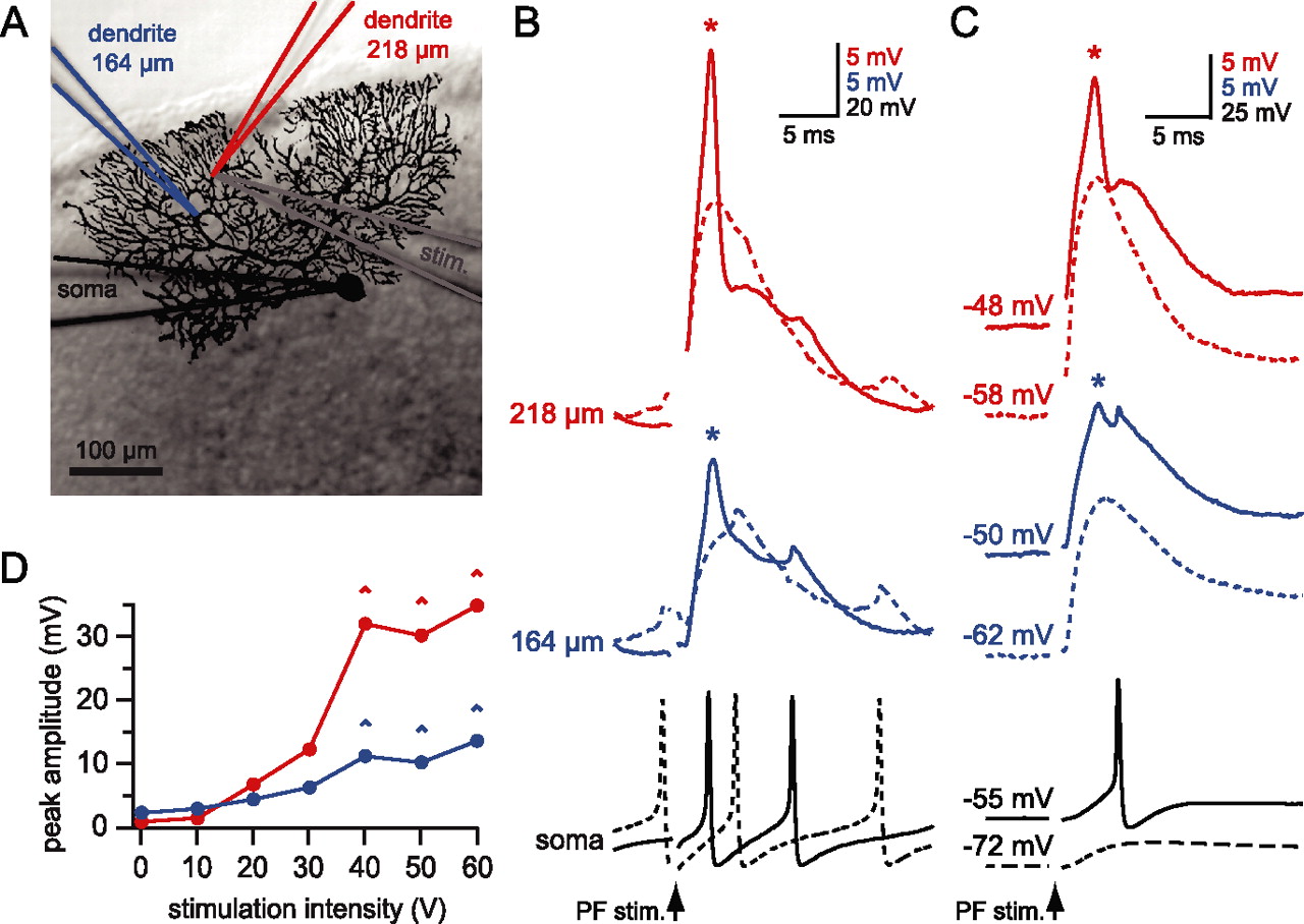
- Figure 1.
Local calcium spikes in Purkinje cell dendrites triggered by PF stimulation. A, Simultaneous triple whole-cell dendritic and somatic recording from a Purkinje cell. An image of the biocytin-filled cell is superimposed on the DIC image of the slice taken at the end of the recording. The recording electrodes and the corresponding traces are color-coded: red for the distal dendrite (218 μm from the soma), blue for the more proximal dendrite (164 μm), and black for the soma. The stimulating electrode (stim; gray) was buried in the slice directly underneath the most distal dendritic recording electrode. B, Traces from the recording shown in A; raising the PF stimulation (arrow) intensity from 30 V (dashed lines) to 40 V (solid lines) resulted in the initiation of a dendritic calcium spike (asterisk) near the distal dendritic recording site. Note the small amplitude of backpropagating APs at the dendritic recording sites. C, Suprathreshold stimulation evoked calcium spikes at rest (spontaneous firing with no holding current, solid lines) that are blocked when the cell is hyperpolarized (dashed lines). D, Graph showing the nonlinear relationship between stimulation intensity and peak depolarization evoked by the PF stimulation. The depolarization was measured at both dendritic locations (red, distal; blue, proximal) as the difference between a 5 ms baseline before the stimulation and the peak voltage reached in a 10 ms window after the stimulation. Note the step-like increase in peak depolarization at the distal recording site associated with initiation of the dendritic calcium spike (^) and the strong attenuation of the calcium spike at the more proximal recording site (54 μm away from the distal dendritic recording).
- Figure 2.
The amount of DSE depends on the number of dendritic calcium spikes. A, Dendritic recording 134 μm from the soma during DSE induction. Different stimulation intensities were used (black, 30 V; blue, 40 V; red, 50 V) during a train of 10 PF stimuli (arrowheads) at 100 Hz. Note the calcium spikes (asterisks) evoked at the higher stimulus intensities. B, Time course of changes in normalized dendritic PF EPSP amplitude (black, n = 4 trials; blue, n = 5 trials; red, n = 3 trials). All data are from the same cell as in A; error bars indicate SEM. C, Relationship between the number of dendritic calcium spikes during the train and the amount of DSE averaged across 12 cells. Open circles show the block of DSE by the CB1 antagonist AM251 (1 μm; n = 4 cells). The control data points were fit with a sigmoidal curve with baseline constrained to 1.
- Figure 3.
DSE and dendritic calcium spikes have a similar voltage threshold. A, Dendritic recording 108 μm from the soma. The same stimulation intensity was used to evoke 10 PF EPSPs at 100 Hz (arrowheads) when the dendrite was held at different membrane potentials (black, −90 mV; blue, −70 mV; red, no holding current, approximately −50 mV). Note that calcium spikes (asterisks) were observed only at the most depolarized potentials. B, Time course of changes in normalized dendritic PF EPSP amplitude (black, n = 3 trials; blue, n = 3 trials; red, n = 3 trials). All data are from the same cell as in A; error bars indicate SEM. C, Relationship between the number of calcium spikes during the train and the amount of DSE averaged across seven cells. The data points were fit with a sigmoidal curve with the baseline constrained to 1.
- Figure 4.
Dendritic spikes evoked by current injection or CF synaptic input fail to trigger DSE. A, Dendritic recording 112 μm from the soma. Dendritic calcium spikes (asterisks) were evoked by injecting a train of EPSC waveforms (shown below the dendritic recording; see Materials and Methods). B, Dendritic calcium spikes (asterisks) evoked in the same cell by stimulating PFs adjacent to the recording electrode. C, Comparison of the effect of calcium spikes evoked by current injection (red) or PF stimulation (black) on PF EPSP amplitudes averaged across six cells. D, Dendritic recording 114 μm from the soma. Stimulating CF input evoked a complex spike, which was associated with three dendritic calcium spikes (asterisks). E, Dendritic recording from the same cell showing a PF train that evoked dendritic calcium spikes (asterisks). F, Time course of PF EPSP amplitude before and after the two different induction protocols (blue, CF stimulation; black, PF stimulation) averaged across six cells. C, F, Error bars indicate SEM.
- Figure 5.
Presynaptic activity during calcium spikes evoked by current injection does not rescue DSE. A, Dendritically recorded calcium spikes (150 μm from the soma) during synaptic stimulation (black) and combined current injection and synaptic stimulation (red). The current injection was adjusted to evoke several calcium spikes. Shown in the bottom panel is the time course of normalized synaptic strength of four cells. Weak synaptic stimulation during current injection-evoked calcium spikes failed to depress the stimulated synapses. B, When the current injection (same as in A) was paired with stronger synaptic stimulation, which alone evoked calcium spikes and some degree of DSE, the amount of depression was increased (bottom panel, pooled data from 4 cells; asterisks denote p < 0.05 between the two conditions). C, When we used synaptic stimuli evoking six or more calcium spikes, pairing with current injection (same as in A, B) failed to increase the degree of DSE (pooled data from 4 cells). EPSP amplitudes were measured at −70 mV. Error bars indicate SEM.
- Figure 6.
Calcium spikes evoked by dendritic current injection are segregated spatially from synaptically evoked calcium spikes. A, Purkinje cell filled with Alexa 594 and fluo-5F (the image shows the Alexa fluorescence). The positions of the PF stimulation electrode (blue) and the dendritic recording electrode (red; 90 μm from the soma) are indicated. B, The area of interest (shown by the white rectangle in A) was imaged for calcium signals during synaptic stimulation, triggering calcium spikes. The image is a maximum intensity projection of a stack of 41 consecutive images acquired at 40 Hz. C, Maximum intensity projection of a stack recorded during calcium spikes evoked by current injection. Two ROIs outlining the branchlets showing calcium signals during synaptic stimulation (ROISYN) or current injection (ROIINJ) were selected. The time course of the fluorescent signal in the two ROIs is plotted below the images. The same number of calcium spikes was evoked by the synaptic stimulation and the current injection. Scale bars: B, C, 10 μm. D, Pooled data from eight cells showing the peak fluorescence change during synaptic stimulation-evoked calcium spikes at the two ROIs selected in the same way as in B, C. E, Pooled data from the same eight cells showing fluorescence changes during current injection-evoked calcium spikes.
- Figure 7.
BK channels control dendritic calcium spike propagation. A, Dendritic recording from a Purkinje cell filled with Alexa 594 and fluo-5F, recorded 140 μm from the soma. The image was taken with Alexa fluorescence. The stimulating electrode is drawn in blue. Scale bar, 50 μm. Inset, High-magnification image of the area of interest (dotted box) with five ROIs indicated. B, Maximum intensity projections of image stacks acquired during synaptically or current injection-evoked calcium spikes in control ACSF and 100 nm penitrem A. Scale bar, 20 μm. C, Calcium transients recorded from the five respective ROIs in the inset of A during synaptically evoked calcium spikes before (black) and after the BK channels (red) were blocked. D, The similarly selected five ROIs were averaged over three cells and normalized to the synaptic maximum in control ACSF. E, Calcium transients recorded from the five respective ROIs in the inset of A during current injection-evoked calcium spikes before (black) and after (red) the BK channels (were blocked. F, The similarly selected five ROIs were averaged over several cells and normalized to the synaptic maximum in control ACSF. Blocking the BK channels clearly improved the spatial spread of calcium spikes. Asterisks (in D, F) denote statistical significance (p < 0.05); error bars indicate SEM. G, The change in the fluorescence signal after penitrem A application was quantified in every ROI under control conditions (open circles) and BK channel block (filled circles). H, Dendritic calcium spikes (aligned at threshold and averaged) evoked with current injection before (black) and after penitrem A application (red). The bottom panel shows the second derivative of the calcium spikes, with a dotted vertical gray line indicating the threshold (see Materials and Methods).
- Figure 8.
Blocking BK channels enables calcium spikes evoked by current injection or CF activation to trigger DSE. A, Dendritic calcium spikes evoked by dendritic current injection in control ACSF (black trace) and 100 nm penitrem A (red trace), recorded 130 μm from the soma. The amplitude of the injected dendritic current waveform (blue traces) was adjusted to evoke similar numbers of calcium spikes under the two conditions. B, Time course of PF synaptic strength before and after the calcium spikes evoked by the current injection shown in A. The same number of calcium spikes, which were unable to trigger DSE in control ACSF, produced strong short-term depression of PF EPSPs when the BK channels were blocked. C, The amount of DSE was plotted versus the number of calcium spikes evoked by current injection. An increasing amount of DSE was triggered with an increasing number of calcium spikes but only when BK channels were blocked by 100 nm penitrem A. Pooled data are from six cells. D, The amount of DSE plotted against the number of synaptically evoked calcium spikes. Block of BK channels did not change the efficacy of synaptically evoked calcium spikes to evoke DSE. Pooled data are from four cells. E, Time course of PF synaptic strength shows prominent DSE after 20 CF stimuli when the BK channels were blocked, but not in control conditions. Single trials are from the same cell. F, The amount of DSE was plotted versus the number of CF stimuli delivered during the induction. Pooled data are from five cells. C F, Error bars indicate SEM, and asterisks denote p < 0.05.


Additional Files
Supplemental data
Files in this Data Supplement:
- supplemental material - Figure 1
- supplemental material - Figure 2


