

Article Figures & Data
Figures
- Figure 1.
HIV-infected PBMC transmigration across an in vitro BBB model in response to CCL2 is greatly increased compared with that of uninfected cells. Uninfected (C) or HIV-infected human PBMCs were added to the top chamber of the BBB model, consisting of cocultured human ECs and astrocytes in the absence (filled bars) or presence (open bars) of CCL2 (100 ng/ml) in the bottom chamber. PBMCs were infected with the R5 viral strains ADA, 92BR021, and JR-CSF, the X4 viral strains 92UG029 and 92UG021, and the R5X4 strain 92RW009. After 24 h, transmigrated cells in the bottom chamber were immunostained with PE-conjugated anti-CD14 (a monocyte marker) and FITC-conjugated anti-CD45 (a pan leukocyte marker) antibodies and analyzed by flow cytometry to quantify the number of monocytes (CD14+) (A) and lymphocytes (CD14−) (B) in the transmigrated population of cells. The data are expressed as mean ± SD of 21 experiments. *p < 0.005 of CCL2 compared with no CCL2 in each group, #p < 0.005 of HIV-infected cells with CCL2 compared with uninfected (C) cells with CCL2; &p < 0.005 of cells infected with JR-CSF, 92UG029, 92UG021, and 92RW009 with CCL2 compared with ADA-infected cells with CCL2.
- Figure 3.
Unlike CCL2, CXCL10, CCL3, and CCL5 did not induce high levels of HIV-infected PBMC transmigration or increase BBB permeability. After the addition of HIV-infected PBMC to cocultures, monocyte (A) and lymphocyte (B) transmigration (24 h) in the presence of CCL2, CXCL10, CCL3, or CCL5 (100 ng/ml) was quantified by flow cytometry. The permeability of the BBB model (C) was analyzed after HIV-infected PBMC transmigration for 24 h in the presence of CCL2, CXCL10, CCL3, or CCL5 (100 ng/ml). The data represent the mean ± SD. *p < 0.005 of CXCL10, CCL3, or CCL5 compared with CCL2 (n = 5).
- Figure 4.
Immunofluorescence and Western blot analysis of BBB cocultures after HIV-infected PBMC transmigration in response to CCL2 indicates that TJP expression in BBB cells is reduced. After transmigration (24 h) of uninfected (Control) or HIV-infected PBMC in response to CCL2, cocultures were immunostained with occludin, claudin-1, and ZO-1 antibodies and examined by confocal microscopy. After transmigration of uninfected cells, surface scanning of our coculture model showed a characteristic distribution of occludin (A) and claudin-1 (D) in cell-to-cell apposition membranes, whereas ZO-1 immunoreactivity (G) exhibited a diffuse pattern of expression. This is attributable to the reactivity of astrocyte foot processes, positive for TJP, that protrude into the EC layers. After HIVADA-infected (similar results were obtained for HIVJR-CSF) or HIV92UG021-infected PBMC transmigration, the expression of occludin (B, C), claudin-1 (E, F), and ZO-1 (H, I) was greatly reduced. The inset in H represents the staining for VWF (red) and GFAP (green) to demonstrate astrocyte end foot processes on the endothelial monolayer. Scale bar, 50 μm. These results are representative of 12 separate experiments. Protein lysates of BBB cells were prepared from cocultures by scraping off the cells from 16 inserts after transmigration for 24 h of uninfected (Con) and HIV-infected PBMCs in the absence (−) or presence of CCL2 (M). Lysates were analyzed by Western blotting for occludin, claudin-1, and ZO-1. Occludin, claudin-1, and ZO-1 expression in cocultures was reduced only after HIVADA-, HIVJR-CSF-, and HIV92UG021-infected PBMC transmigration in the presence of CCL2 (J). EC protein lysate (S) was used as a positive control to identify the electrophoretic mobility of TJP reactive bands. Blots were stripped and incubated with a histone-1 antibody to document equivalent protein loading. These results are representative of five separate experiments.
- Figure 6.
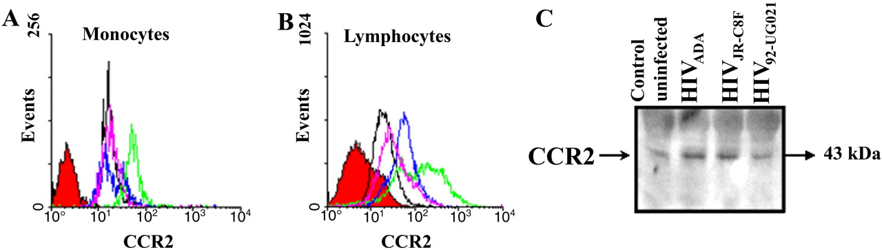
Infection of PBMC with different HIV strains increases CCR2 expression. FACScan analysis of nonpermeabilized monocytes (A) and lymphocytes (B) immunostained with a CCR2 antibody indicate that infection of PBMC with ADA (green line), JR-CSF (blue line), and 92UG021 (pink line) viral strains increases the surface expression of CCR2 on lymphocytes compared with uninfected cells (black line), whereas ADA and JR-CSF viral strains increased CCR2 on monocytes. Cells were also incubated with an isotype-matched mouse myeloma antibody as a negative control for nonspecific staining (red-filled curve). These results of surface FACS analyses were corroborated by quantification of total CCR2 protein by Western blot analysis for CCR2 in uninfected (Control) and HIVADA-, HIVJR-CSF-, and HIV92UG021-infected PBMCs. These results are representative of five separate experiments.


Tables
- Table 1.
BBB permeability after HIV-tat and/or gp120 treatment or treatment with supernatants from HIV-infected PBMCsa
Conditions Direct effect of viral proteins Without CCL2 With CCL2 Control 1.3 ± 0.37 1.9 ± 0.47 HIV-tat 4.9 ± 0.095b 4.4 ± 0.19b gp120 (JRFL or HxB) 4.7 ± 0.45b 4.5 ± 0.89b Tat plus gp120 5.2 ± 0.67b 5.3 ± 0.8b Supernates from HIV-infected leukocytes 4.8 ± 1.3b 5.1 ± 1.9b


