

Article Figures & Data
Figures
- Figure 1.
RPA analysis of estrogen effects. A–C, Representative RPA assay of total RNA (5 μg/lane) from vehicle- and E2-treated (25 μg; 24 h) animals illustrating the levels of Cav 3.1 α1 mRNA detected in the POA, MBH, and pituitary (Pit) from individual animals. A, Sense RNA (125–4000 fg) was used to construct a standard curve. B, Linear regression analysis of the Cav3.1 α1 RNA sense standard curve using a PhosphorImager revealed r = 0.983. D, Distribution and quantitative analysis of Cav3.1 α1 mRNA in POA, MBH, and Pit tissues obtained from oil-treated (n = 3) and E2-treated (n = 3) guinea pigs 24 h after the injection. Each α1 band was quantified from the sense RNA standard curve and the values normalized to the corresponding cyclophilin band. The asterisks denote a significant change (paired t test; **p < 0.01) in the level of Cav3.1 α1 mRNA caused by E2 relative to that observed in oil-treated controls. UP, Undigested probe; DP, digested probe.
- Figure 2.
Distribution of Cav3.1 α1 subunit mRNA by in situ hybridization. Dark-field photomicrographs of coronal sections through the POA from rostral to caudal (a–d) and through the rostral and more caudal MBH (e, f) that illustrate the distribution of autoradiographic grains indicative of Cav3.1 α1 mRNA in an E2-treated animal. AC, Anterior commissure; DMH, dorsomedial nucleus; OC, optic chiasm; PVHap, paraventricular nucleus of the hypothalamus anterior parvocellular part; PVT, paraventricular nucleus of the thalamus; 3V, third ventricle. Scale bars: (in a) a, b, d–f, 200 μm; c, 100 μm.
- Figure 3.
Film images Cav3.1 α1 mRNA distribution in rostral and more caudal brain regions. Bright-field view of film autoradiograms illustrating the distribution of Cav3.1 α1 mRNA in coronal sections from the POA (A) and the MBH (B) area in an E2-treated animal. The darker the images, the denser the mRNA expression. AM, Amygdala; CTX, cortex; Fx, fornix; S, septum; Thal, thalamus. See Figure 2 for additional abbreviations.
- Figure 4.
Emulsion autoradiograms of Cav3.1 α1 mRNA expression in the AVPV, MPN, and BST in ovariectomized oil- or E2-treated guinea pigs. Dark-field photomicrographs of matched sections from oil-treated (a, c, e) and E2-treated (b, d, f) guinea pigs illustrating autoradiographic grains indicative of Cav3.1 α1 mRNA expression in the AVPV, MPN, and BST. Scale bar, 200 μm (for all photomicrographs). BSTpr, Principal nucleus of the BST; BSTdl, dorsolateral nucleus of the BST.
- Figure 5.
Emulsion autoradiograms of Cav3.1 α1 mRNA expression in the arcuate nucleus in E2-treated guinea pigs. Dark-field photomicrographs of matched sections from oil-treated (a, c) and E2-treated (b, d) guinea pigs illustrating low-power (a, b) and high-power (c, d) views of Cav3.1 α1 mRNA expression in the arcuate region of the hypothalamus. Scale bars: a, b, 200 μm; c, d, 100 μm.
- Figure 6.
Film images of Cav3.1 α1 mRNA expression in ovariectomized oil- or E2-treated guinea pigs. Bright-field view of film autoradiograms illustrating the distribution of Cav3.1 α1 mRNA in coronal sections from rostral (a, b) to caudal (g, h) in oil-treated (a, c, e, g) and E2-treated (b, d, f, h) animals. The darker the images, the denser the mRNA expression. For abbreviations, see Figures 3 and 4.
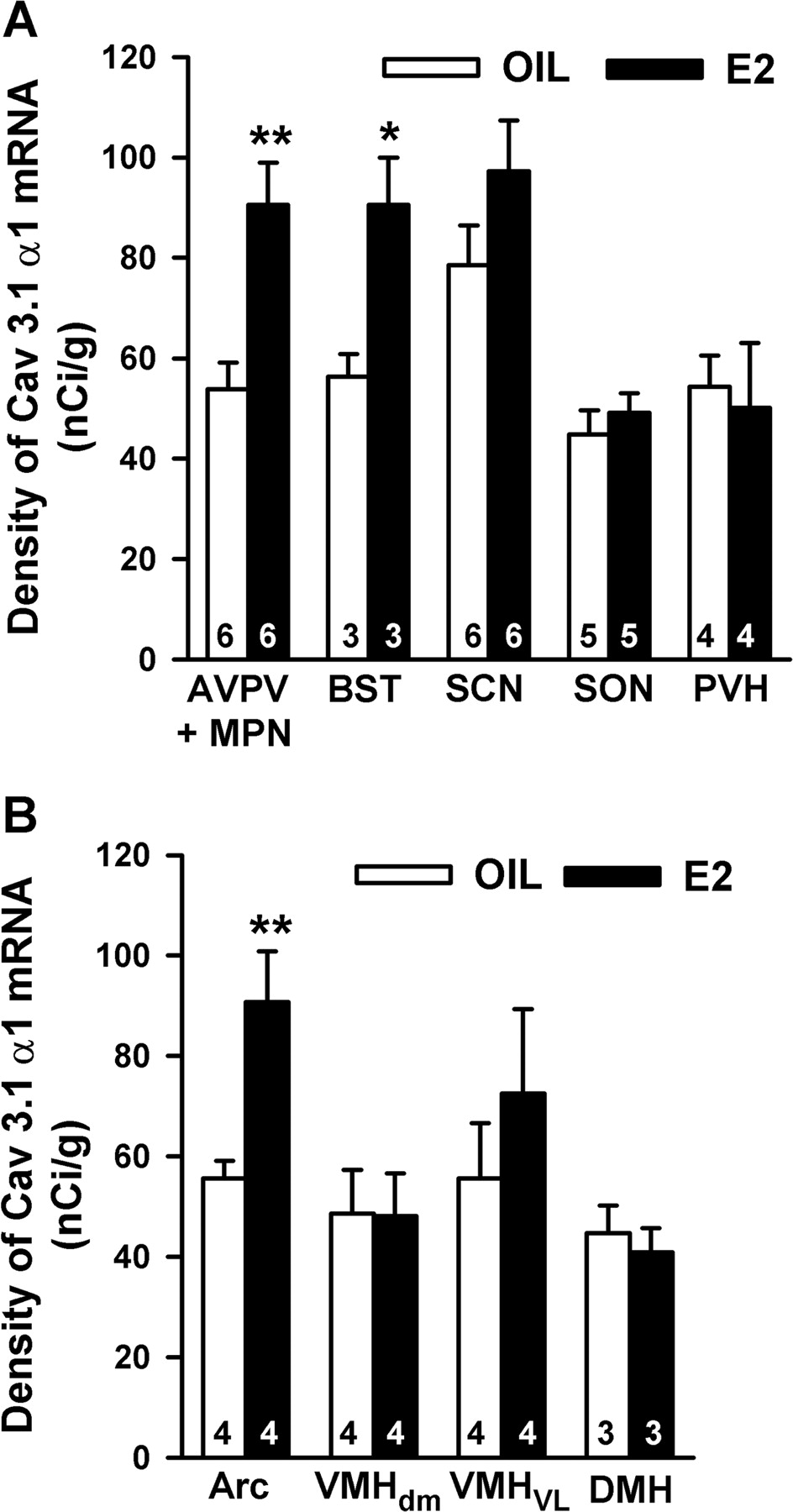
- Figure 7.
Group data of E2-induced increase in Cav3.1 α1 mRNA expression in the female guinea pig. Distribution and quantitative analysis of Cav3.1 α1 mRNA in tissue sections through the POA (A) and BH (B) regions obtained from oil- and E2-treated animals (n = 3–6). Values represent the mean density ± SEM of Cav3.1 α1 mRNA levels (nanocuries per gram of tissue) in film images from the different brain regions. (*p < 0.05, **p < 0.01; two-tailed Student's t test). For abbreviations, see Figures 3 and 4. Error bars represent the mean ± SEM.
- Figure 8.
Lack of an effect of estrogen on voltage dependence of T-type channel activation and inactivation. a, Procedures of activation of the transient Ca2+ current (T-type current) in the arcuate neurons. T-type current was isolated using the extracellular medium that suppressed Na+ and K+ currents and electrodes containing Cs+ solution. Depolarizing steps, 300 ms in duration, were applied from holding potentials of −120 mV (top traces) or −60 mV (bottom traces). Test potentials varied from −85 to −45 mV in 5 mV increments. On the right, traces obtained at the two different holding potentials were subtracted to isolate T-type currents. b, Voltage dependence of the inactivation and activation of the Ca2+ T-type current in arcuate neurons from oil- and E2-treated females. Activation of T-type calcium current was analyzed from neurons recorded as indicated in a. Steady-state inactivation of T-type calcium current was analyzed from neurons recorded as indicated in Figure 9a. Peak amplitude of current is plotted in relation to the maximum current obtained. The mean curve was drawn using the Boltzmann equation as described in Materials and Methods. The mean V1/2 values for T-type channel activation were not significantly different for control cells (V1/2 = −59.1 ± 1.0 mV; slope factor, 2.1 ± 0.4; n = 7) and estrogen-treated cells (V1/2 = −59.6 ± 1.1 mV; slope factor, 2.4 ± 0.7; n = 6). Similarly, V1/2 values for channel steady-state inactivation were similar for both groups (V1/2 = −85.8 ± 0.8 mV, slope factor, 4.8 ± 0.3, n = 12 for control vs V1/2 = −87.5 ± 0.9 mV, slope factor 5.1 ± 0.5, n = 6 for E2-treated group).
- Figure 9.
Estrogen increases the whole-cell T-type calcium current in arcuate neurons. a, The peak current was obtained by applying conditioning pulses, 1 s in duration, ranging from −120 to −65 mV in 5 mV increments and then a test pulse to −60 mV (inset), and the T-type calcium current in the arcuate neuron was completely blocked by 100 μm nickel chloride. b, The current amplitudes were normalized to the cell capacitance in all cases to calculate current density. Bar graphs summarize the density of T-type calcium current in arcuate neurons from oil-treated and E2-treated animals. The mean density was significantly greater in E2-treated (0.97 ± 0.17 pA/pF; n = 25) than in oil-treated (0.48 ± 0.10 pA/pF; n = 23) ovariectomized females. Error bars represent the mean ± SEM tested per group. *p < 0.05 versus vehicle. c, Double labeling of arcuate dopamine and POMC neuron that expressed a T-type current. Top, Biocytin-streptavidin-Cy2 labeling of arcuate neurons after whole-cell recording. Labeled fibers are extending toward medial (third ventricle, 3V), lateral, and ventral regions in both cells. Bottom, Immunocytochemical staining of TH and β-endorphin in the respective cells (arrows). Scale bar, 50 μm (in all panels). d, Expression of Cav3.1 α1 mRNA in arcuate neurons. Representative gel illustrating that single arcuate neurons expressed Cav3.1 α1 mRNA including TH, POMC, and unidentified neurons. One cell contained no RT as a negative control (-RT). In addition, the following controls were included: aCSF from the dispersed cellular milieu and a water blank, both of which were negative after RT-PCR (data not shown). The expected size of the PCR products is as follows (in bp): Cav3.1, 228; TH, 223; POMC, 206.
- Figure 10.
E2 increases rebound burst firing in arcuate neurons. Representative traces recorded in whole-cell voltage and current clamp from arcuate neurons in oil-treated (a, b) and E2-treated (c, d) animals. a, In voltage-clamp mode in an arcuate neuron from oil-treated animal, depolarizing voltage commands stepped from −120 mV elicited a peak amplitude T-type current of 20 pA. b, In the current-clamp mode, the cell exhibited an LTS, which triggered a single Na+ action potential after a hyperpolarizing current pulse to −100 mV. c, Similar protocols in current-clamp and voltage-clamp modes were applied to another arcuate neuron from an E2-treated animal. In voltage-clamp mode, the voltage command evoked a peak amplitude T-type current of 44 pA. d, The rebound from a hyperpolarization current pulse evoked a burst of Na+ action potentials on the crest of a LTS in this arcuate neuron. NiCl2 (100 μm) abolished the LTS and accompanying Na+ spikes (bottom trace). e, Left, Box plot of the peak amplitudes of T-type current used for Na+ spike analysis in cells from oil-treated (n = 9) and E2-treated (n = 7) groups. [The median is the center line in the box. The edges of the box represent the 75th and the 25th percentiles of the distribution. The vertical lines extending from the box represent the upper (90th percentile) and lower (10th percentile) limits of the distribution.] Right, Summary histograms of cells from oil-treated (n = 9) and E2-treated (n = 7) groups showing a significant increase in the number of rebound Na+ spikes within the time course of the LTS (500 ms). Error bars represent the mean ± SEM tested per group. ***p < 0.005 versus vehicle.


