

Article Figures & Data
Figures
- Figure 1.
Expression levels of Kv4.2 and Kv4.3 in mouse primary visual and primary somatosensory cortex are layer specific. A, B, Immunofluorescence images of Kv4.2 and Kv4.3 expression in coronal sections through mouse primary visual cortex. Staining is densest in layers 1 and 4 and the layer 5/6 border. C, D, Kv4.2 and Kv4.3 immunofluorescence in tangential sections through mouse primary somatosensory cortex. Labeling is most intense in layer 4, in the center of barrels. Expression in septa between barrels is sparse. Scale bars: A, B, 0.2 mm; C, D, 1 mm.
- Figure 2.
Kv4.2 clusters are expressed in pyramidal cell somata, dendrites, and spines. A, Confocal microscopic images of punctate Kv4.2 immunolabeling (red) in layers 4 and 5 of mouse primary visual cortex. The distribution of Kv4.2 clusters is non-uniform, showing a higher density over the neuropil than the pyramidal cell soma in the same focal plane (arrows; A, B). B, C, Kv4.2 clusters (red, yellow) are associated with YFP-labeled (green) pyramidal cell somata and with basal and apical dendrites. D–F, High-magnification images of the boxed apical dendrite of a YFP-labeled layer 5 pyramidal neuron (B, C), showing close association of Kv4.2-labeled clusters (red and yellow puncta marked with arrows) with dendritic shaft and spines. The numbers indicate layers. Scale bars: A–C, 10 μm; D–F, 2 μm.
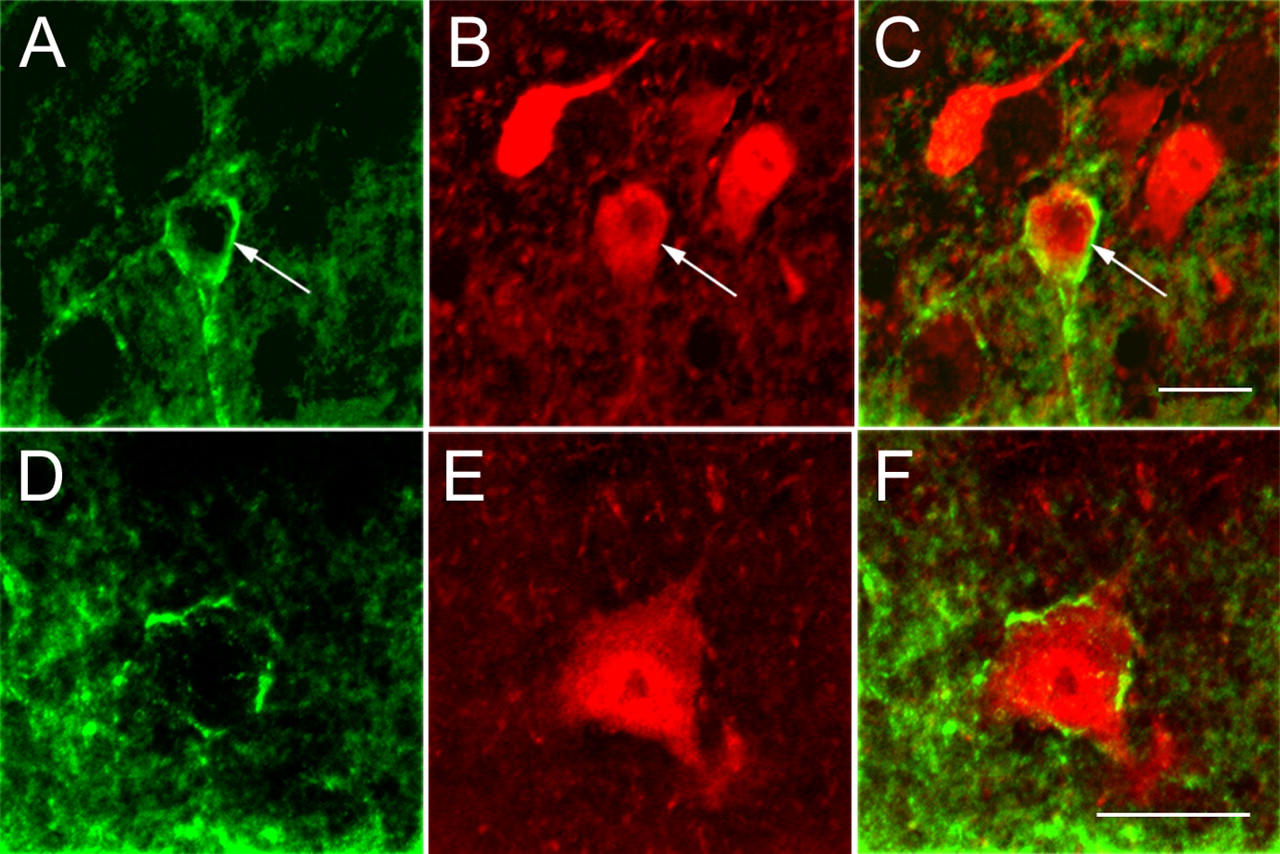
- Figure 3.
Kv4.2 expression in GABAergic neurons of mouse primary visual cortex. Confocal microscopic images of immunolabeled GABAergic neurons (red) in layer 2/3 (A–C) and layer 5 (D–F). Kv4.2 (green) is coexpressed in somata and dendrites of a subset of GABAergic neurons. Arrows in A–C mark Kv4.2-expressing GABAergic neuron. Scale bars, 10 μm.
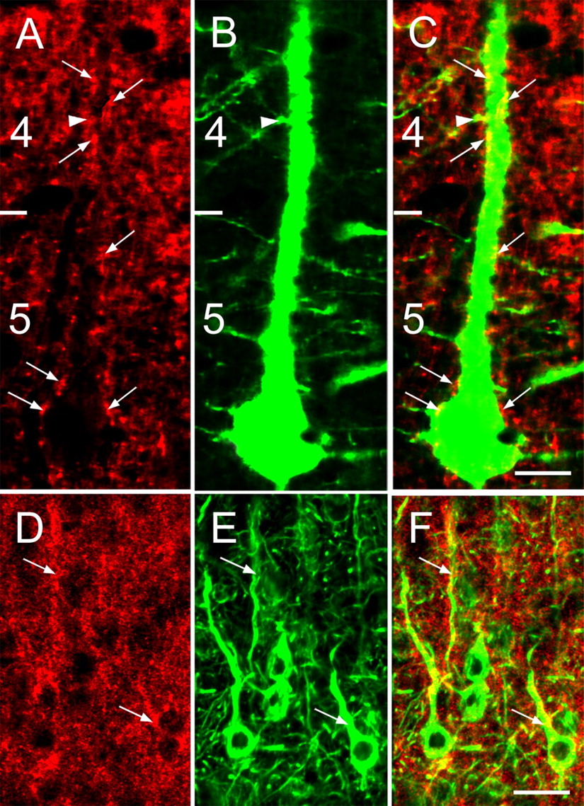
- Figure 4.
Confocal microscopic images of Kv4.3 immunolabeling in pyramidal neurons of mouse primary visual cortex. A–C, YFP-labeled pyramidal neuron with a soma in layer 5 and sparsely spiny apical dendrite in layers 4 and 2/3 that are decorated with Kv4.3 clusters (red and yellow puncta marked with arrows). The arrowhead indicates a spine, the neck of which contains a Kv4.3-positive cluster. D–F, Kv4.3 expression (red; D) in layer 2/3 pyramidal neurons that are stained with an antibody against neurofilament (green; E). Kv4.3 clusters (arrows) that are associated with somata and dendrites appear yellow (F). The numbers indicate layers. Scale bars: A–C, 10 μm; D–F, 20 μm.
- Figure 5.
Confocal microscopic images of Kv4.3 immunolabeling in GABAergic neurons of mouse primary visual cortex. A–C, Coexpression (arrows) of Kv4.3 (green; A) and GABA (red; B) in a subset of layer 2/3 nonpyramidal cells (C) in which Kv4.3 labeling extends into distal branches of the dendritic tree. D–F, Coexpression (arrows) of Kv4.3 (green; D) and GABA (red; E) in a subset of layer 5 nonpyramidal neurons (F). Scale bars, 10 μm.
- Figure 6.
Electron micrographs showing that Kv4.2 is differentially expressed at putative inhibitory and excitatory synapses in mouse primary visual cortex. A, Extrasynaptic localization of Kv4.2 immunoperoxidase labeling (arrows) at an asymmetric synapse onto a thin dendrite (D). The dark immunoperoxidase reaction product is easily distinguished from the much lighter postsynaptic density. B, Kv4.2 immunoperoxidase at the base and neck of spine. Kv4.2 is absent from the asymmetric synapse. C, Extrasynaptic localization of Kv4.2 at an asymmetric synapse onto spine. D, Extrasynaptic localization of Kv4.2 (arrows) at an asymmetric synapse onto GABA immunogold (black dots)-labeled dendrite. E, Synaptic localization of Kv4.2 at a symmetric synapse (AT2) onto a pyramidal cell soma (P). Notice that Kv4.2 is contained in the postsynaptic membrane associated with the synaptic cleft, the margins of which are indicated by arrowheads. Scale bars, 0.5 μm. AT1, Asymmetric synapse; Sp, spine.
- Figure 7.
Electron micrographs showing that Kv4.3 is differentially expressed at putative inhibitory and excitatory synapses in mouse primary visual cortex. A, B, Kv4.3 immunoperoxidase labeling (dark reaction product marked by arrows) in thin dendrites is excluded from asymmetric synapses (AT1). The thick dendrite (D) does not express Kv4.3. C, D, Kv4.3 immunoperoxidase labeling (arrows) in postsynaptic membranes directly across Gray type 2 axon terminals (AT2) that contain flattened vesicles and form symmetric, putative inhibitory synapses with spine (Sp) and perikaryon (P) of a nonpyramidal cell [identified by infoldings of nuclear (Nu) membrane]. Scale bars, 0.5 μm.
- Figure 8.
Electron micrographs showing differential localization of Kv4.2 and Kv4.3 at GABAergic and non-GABAergic synapses in mouse primary visual cortex. A, Kv4.2 immunoperoxidase (arrows and arrowheads) expression in a GABA immunogold (black dots)-labeled section. Notice that Kv4.2 (arrows) is expressed in the spine (Sp) membrane but is excluded from the asymmetric synapse formed by a non-GABAergic Gray type 1 axon terminal (AT1). In contrast, Kv4.2 is expressed at a symmetric synapse formed by GABA immunogold-labeled Gray type 2 axon terminal. B, C, Postsynaptic localization of Kv4.3 immunoperoxidase labeling (arrows) at symmetric synapses formed by GABAergic (black dots) Gray type 2 axon terminals onto dendrite (D) and perikaryon (P). Scale bars, 0.5 μm.
- Figure 9.
Schematic representation of Kv4.2 (red dots) and Kv4.3 (black dots) subunit distribution in neocortical pyramidal cells (top) and GABAergic interneurons (bottom). A close-up at the bottom right applies to both Kv4.2 and Kv4.3 and shows that Kv4 channels are excluded from excitatory synapses (green) but are present at GABAergic inhibitory synapses (purple). PC, Pyramidal cell.


Tables
- Table 1.
Percentage of Kv4.2- and Kv4.3-immunolabeled electron microscopically identified neuronal profiles in mouse visual cortex
Dendrites Antibody Total number of stained profiles Somata Thina Thickb Spines Kv4.2 129 3% (4 of 129) 77% (99 of 129) 8% (10 of 129) 12% (16 of 129) Kv4.3 116 5% (6 of 116) 72% (83 of 116) 12% (14 of 116) 11% (13 of 116)
Supplemental Data
Files in this Data Supplement:
- supplemental material - Figure legends
- supplemental material - Figure 1
- supplemental material - Figure 2


