

Article Figures & Data
Figures
- Figure 1.
Muscarinic effects in the absence of background activity. A, Firing of a layer 5 pyramidal neuron in forelimb motor cortex before (Control) and after (Oxo-M) application of Oxo-M in response to a DC step. In this case, muscarinic activation reversed spike frequency adaptation, causing the instantaneous frequency to increase rather than decrease over the course of the spike train. (In all figures, when data from control and Oxo-M conditions are plotted together, the former are in black, and the latter are in gray.) B, Muscarinic receptor activation increased the average number of spikes produced by a 1 s DC step consistently over a wide range of amplitudes (top panel). On average, it also reduced, but did not reverse, spike frequency adaptation. Plotted in the bottom panel are the instantaneous spike frequencies produced by a 200 pA current step as a function of spike interval number, normalized to the frequency for the first interval (n = 20 for both graphs). C, Muscarinic receptor activation replaced the burst afterhyperpolarization with a burst afterdepolarization. The size of the afterdepolarization varied with the intensity of the burst and could give rise to prolonged spike firing. Shown is the response of one neuron in the presence of Oxo-M to current injections of three different amplitudes (bottom traces). D, In some neurons, the afterdepolarization produced persistent graded activity. The top trace (Vm) shows spike firing induced by the current injections of the middle trace (I). Note that the instantaneous firing rate (FR) shown in the bottom trace could be ramped up or down in a controlled, step-like manner. Calibration: 25 mV, 100 pA; 6 Hz vertical; 10 s horizontal.
- Figure 2.
Voltage- and calcium-dependent currents responsible for observed effects. The voltage- and calcium-dependent currents responsible for the effects of muscarinic receptor activation were isolated and measured using standard voltage-clamp techniques. The traces at the left illustrate the voltage-clamp commands used to elicit different currents. Example currents of each type are shown at the right and include currents recorded before (black) and after (gray) application of Oxo-M. A, The potassium leak current was reduced by Oxo-M. B, The potassium M current was also reduced. C, A calcium-activated potassium current (KCa) was blocked and replaced by a calcium-activated nonspecific cation current (NCC). D, There was no effect on the hyperpolarization-activated current (H). E, There was no evidence for a voltage-dependent, nonspecific cation current (V).
- Figure 3.
Dynamic clamp simulations. A, The noisy background input that motor cortical neurons receive in vivo was mimicked in vitro by injecting excitatory (gray) and inhibitory (black) conductance trains. These were generated independently as stochastic processes. Depicted at the left are typical examples. Fluctuations were normally distributed (middle) and were correlated at short times (right). B, Trains of synaptic inputs were generated by combining unitary AMPA and NMDA conductance transients. As shown, AMPA and NMDA transients were each modeled as a difference of exponentials and were activated simultaneously (left). The AMPA transients had much faster kinetics and were larger by a fixed amplitude ratio (10:1). The magnesium block of NMDA receptors was mimicked by multiplying NMDA conductances by a sigmoidal function (right). C, Slow ADP and AHP currents were introduced by simulating a spike-dependent conductance. Every time a neuron fired an action potential (black dots), this conductance was incremented. Between spikes, it decayed exponentially back to zero with a slow time constant (500 ms). The reversal potential was set to +50 mV (ADP) or −85 mV (AHP), depending on which current was being simulated.
- Figure 4.
Subthreshold behavior in the presence of background activity. A, Membrane potential recordings from one neuron before (Control; black trace) and after (Oxo-M; gray trace) application of Oxo-M. B, Examples of input resistance and membrane potential measurements, with and without background activity and/or muscarinic receptor activation. In the absence of background activity (top panels), the input resistance was large, and the membrane potential fluctuated only in a very narrow band. Application of Oxo-M produced marked changes in both (Control, black traces; Oxo-M, gray traces). Addition of background activity (bottom panels) reduced the input resistance, depolarized the membrane potential, and introduced wide fluctuations in its value. In this case, muscarinic effects were still stereotyped but much smaller. Input resistance was estimated from the voltage deflection produced by a hyperpolarizing current step (top, 20 pA; bottom, 150 pA). C, The effect of muscarinic receptor activation on membrane potential was small but reliable. Left, Values for individual cells before and after Oxo-M (gray traces) for one choice of excitatory/inhibitory parameters (those shown in Fig. 3A). The black trace is the average over all cells (n = 8). Right, Average change in membrane potential resulting from Oxo-M application for four different choices of background parameters. Excitatory and inhibitory mean conductances were independently varied between low (L) (gexc = 6 nS; ginh = 24 nS) and high (H) (gexc = 8 nS; ginh = 32 nS) values (n = 7). D, The autocorrelation of the membrane potential and its cross-correlation with the excitatory and inhibitory background conductances were similar before (black) and after (gray) muscarinic receptor activation. Correlations for a representative cell are shown.
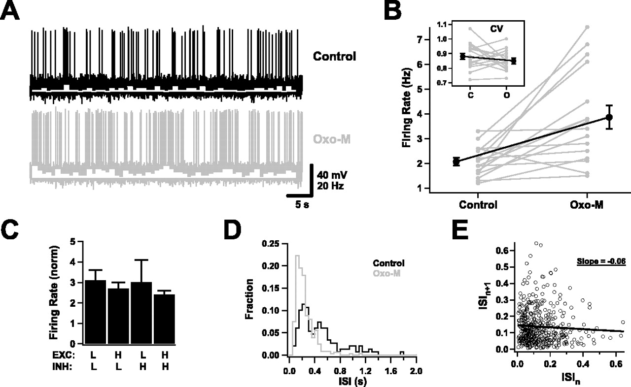
- Figure 5.
Suprathreshold behavior in the presence of background activity. A, Spiking activity of a neuron in response to background inputs, before and after application of Oxo-M. Spike rate histograms are superimposed in white (bin size, 1 s). B, Average firing rates were reliably increased by muscarinic receptor activation. Gray traces are data for individual cells; the black trace is the average of all cells. The inset shows the coefficient of variation of ISIs (n = 16). C, The increase in firing rates was not sensitive to the exact choice of background parameters. The average firing rates in Oxo-M normalized to the control rates as the mean excitatory and inhibitory conductances were varied as in Figure 4C (n = 7). D, ISI histogram for a representative cell before and after Oxo-M. E, An example of an ISI return map, measured in the control condition. The circles are individual interspike intervals; the line is a linear fit, which has a negative slope.
- Figure 6.
Dissecting the effects of conductance, depolarization, and fluctuations. Changes in membrane potential (ΔVm, in mV) and input resistance (ΔRN, as a percentage of control values) produced by Oxo-M application were measured as conductance, depolarization, and the size of membrane potential fluctuations were independently varied. A, A DC conductance reversing at the original resting potential was added (n = 7). B, A DC was used to fix the membrane potential at −70, −60, or −50 mV (n = 6). C, A fluctuating current was added (n = 5). The largest SD produced membrane potential fluctuations of ∼5 mV.
- Figure 7.
Increased sensitivity to correlated inputs with muscarinic activation. A, Sensitivity to correlated inputs was probed by combining compound AMPA-NMDA transients in Poisson trains and adding these to the fluctuating background. An example of an excitatory conductance constructed in this way is shown in the bottom trace (the inhibitory background conductance is not shown); the simulated train is present during the time marked by the thick bar. Also shown are the responses of a neuron to this input before (top trace) and after (middle trace) activation of muscarinic receptors. B, Muscarinic receptor activation increased neuronal sensitivity to synaptic trains. Each train was constructed by simulating 20 presynaptic inputs, each with a maximal AMPA value of 1.5 nS, firing independently at a Poisson rate between 0 and 50 Hz. The response of each neuron to a train of a given frequency was measured over 10–50 trials, and a mean firing rate was calculated. Input trains lasted 300 ms. Shown are the firing rates, averaged over all cells, in response to trains of different frequencies. The rates at a stimulus frequency of 0 Hz are nonzero because of firing in response to background inputs (n = 9). C, Individual spike times were generally not precise because of the fluctuating background. The raster plot shows individual spikes elicited in one neuron by injecting the same train 100 times; it begins at time t = 150 ms and lasts 300 ms. The background was allowed to fluctuate randomly between trials. At the bottom is a spike histogram constructed using 1 ms bins. Only occasionally, as at t = 211 ms in this example, were spikes notably precise. D, Muscarinic inhibition of synaptic inputs might increase sensitivity to correlated inputs. Spike histograms constructed in response to 300 ms input trains (100 trials, 20 ms time bins) in the presence of high or low background activity.
- Figure 8.
More sustained firing with muscarinic activation. A, Spike histograms for a neuron in the control condition and after application of Oxo-M (100 trials, 100 ms time bins). A 1 s input train was applied at t = 500 ms. Note the marked drop in numbers of spikes later in the train in the control case and contrast it with the stability of the muscarinic case. Note also that the spontaneous spike rate is depressed immediately after the input terminates in the control case. B, An example of the response of a neuron to two short inputs separated by 300 ms (10 trials are overlaid) in control conditions.
- Figure 9.
Varying membrane conductances with the dynamic clamp. A, A slow ADP conductance was introduced through the dynamic clamp and mimicked many aspects of the muscarinically activated slow ADP. The response of a neuron to a DC step (in the absence of background activity) before (Control) and after (ADP) introduction of the simulated conductance is shown. Note the reduced spike frequency adaptation and the appearance of an afterdepolarization. B, Mimicking the suppression of the potassium M conductance of Oxo-M, by subtracting a simulated M-type conductance through the dynamic clamp, had only small effects. Response to a DC step before and after subtraction of an M conductance (gmax = 1 nS). Scale bars are the same as in A. C, Change in membrane potential produced by systematically varying leak and AHP/ADP conductances through the dynamic clamp, with background activity set to produce 1–3 Hz spiking. Negative numbers indicate the specified conductance was being subtracted. Conductances are given in nanosiemen (n = 7). D, Effect on average firing rates of systematically varying leak and AHP/ADP conductances. Rates are shown relative to the rate when no conductance was added or subtracted (n = 7). E, Replacing the endogenous AHP conductance (black) by a strong, simulated ADP conductance (gray) introduced temporal correlations into firing trains produced by background activity. This neuron displayed an increased tendency to fire in sustained bouts as a result. Calibration: 25 mV vertical, 1 s horizontal. F, The correlations introduced by the simulated ADP conductance were reflected by changes in the coefficient of variation of interspike intervals (n = 7).


