

Article Figures & Data
Figures
- Figure 1.
Persistence of electrical excitability in nonafferented utricle hair cells in culture. A, Diagram showing the percentage of utricle hair cells expressing INa as a function of rat age. Data in open bars were taken from Chabbert et al. (2003). Data in filled bars were recorded in hair cells from utricle acutely isolated at P0, P1, and P10 and from cultured utricle (P0+10DIV) (**p < 0.01). B, Comparison of INa density (mean ± SD) at indicated stages (*p < 0.05, **p < 0.01). Mean membrane capacitances (Cm) did not differ significantly between stages: 4.3 ± 1.4 pF at P0, 3.7 ± 0.9 pF at P10, and 4.0 ± 1.1 pF at P0+10DIV. In A and B, the total number of cells studied at each stage of development is shown at the top of each bar. C, Plot of relative activation (•; between −70 and +20 mV from a Vhold of −110 mV) and inactivation (■; test potentials, 5 ms at −30 mV; conditioning prepulses, 10 ms between −120 and −30 mV from a Vhold of −110 mV) of INa at indicated stages, expressed as G/Gmax and I/Imax as a function of test potentials and conditioning prepulses, respectively. D, Representative voltage responses (top traces) in the current-clamp mode of the whole-cell patch-clamp technique, following a given protocol and at indicated stages of development. Underlying whole-cell currents recorded in voltage-clamp mode are given below. Capacitive transients have been truncated for clarity. Depolarizing current injections (100 pA steps) were applied to cells held under a hyperpolarizing holding current. Traces shown in the figure are single traces.
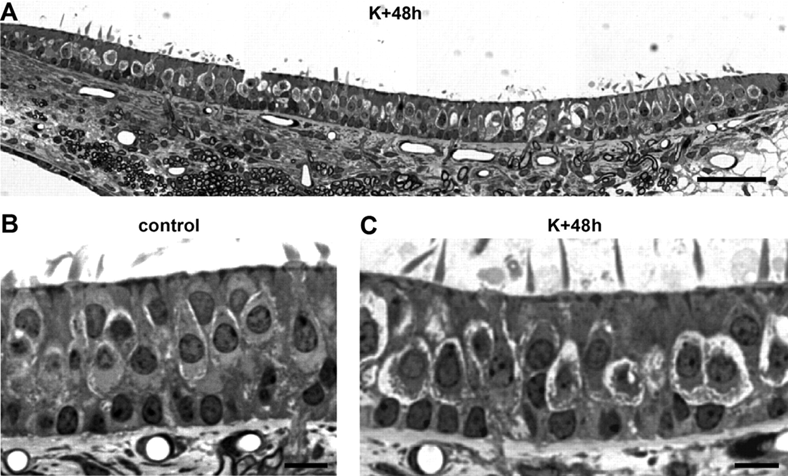
- Figure 2.
Morphological evaluation of excitotoxic damages. A, Longitudinal sections through the entire utricle revealing numerous swollen nerve terminals below hair cells at K+48h. B, C, High-magnification observations in control and K+48h conditions. Scale bars: A, 30 μm; B, C, 10 μm.
- Figure 3.
Morphological consequences of transient nerve terminal impairment. Electron microscopy observations of vestibular nerve terminals at type I (hcI; top) and type II (hcII; bottom) hair cells in the peristriolar region of utricles from adult rats in given conditions are shown. A, B, Illustration of intact nerve calyx, afferent, and efferent bouton terminals in control utricles. C, D, Forty-eight hours after kainate application, both calyx and bouton terminals displayed characteristic swellings. The calyx membrane often came off from the hair cell membrane (arrows). Bouton swellings resulted in indentations of type II hair cell basal membrane. E–H, No damage persisted in the sensory epithelium 1 week after kainate application (E, F) or if DNQX was applied with kainate (G, H). nc, Nerve calyx; a, afferent; e, efferent; sc, supporting cells. Scale bars, 1 μm.
- Figure 4.
Synapse ultrastructure after kainate treatment. A–D, Synaptic ribbons in type I hair cells in given conditions. A, B, Synaptic ribbon (arrow) in a type I hair cell surrounded by a swollen nerve calyx at K+48h. Synaptic vesicles (arrowhead) are close to the hair cell basal membrane facing the postsynaptic density (asterisk). C, Two synaptic ribbons in a single type I hair cell at K+48h. D, Ribbon structure in the control condition. E–H, Synaptic ribbons in type II hair cells. E, G, Postsynaptic densities in swollen afferent boutons contacting type II hair cells at K+48h. The ribbon is closed to basal membrane and surrounded by synaptic vesicles. H, Ribbon and postsynaptic density in a control type II hair cell. I–K, No damage of efferent terminals was observed at K+48h. hcI, type I hair cell; hcII, type II hair cell; nc, nerve calyx; a, afferent; e, efferent.
- Figure 5.
Behavioral consequences of transient nerve terminal impairment. Rating scores for vestibular dysfunction from a behavioral test battery are shown. Rats were assessed at 2 d (K+48h) or 7 d (K+1 week) after kainate application or 2 d after kainite plus DNQX (K+DNQX+48h) application. The kainate-induced vestibular dysfunctions were transient and prevented by DNQX. Data from a positive control group of animals treated with a dose of 3,3′-iminodipropionitrile (IDPN) that induces a complete loss of hair cells are shown. The total number of cells studied at each stage of development is shown above each bar.
- Figure 6.
Transient recovery of excitability in adult utricle hair cells after nerve terminal impairment. A, Diagram showing the percentage of utricle hair cells expressing INa in given conditions. The total number of recorded cells is shown at the top of each bar. **p < 0.01. B, Representative voltage responses (top traces) for the given protocol, with the underlying whole-cell currents (bottom traces). Capacitive transients have been truncated for clarity. All traces are single traces. A high-density INa supporting a sodium-based AP was observed only 48 h after kainate application (K+48h). Voltage and current traces shown under neuroprotection (K+DNQX+48h) and recovery (K+1 week) conditions are those of the only cells expressing INa in these two sets of conditions (mean Cm of 3.3 and 3.9 pF, respectively).
- Figure 7.
Transient recovery of Nav1.2 expression in adult utricle hair cells after nerve terminal impairment. Immunolocalization of neurofilament-N52 (NF; red) and Nav1.2α (green) subunits in adult rat utricles in given conditions is shown. A, Nav1.2 labeling is not detected in utricle hair cells or in supporting cells in control. B, Forty-eight hours after kainate application, a strong Nav1.2 immunoreactivity was observed at the basal hair cell membrane (arrows). C, D, Nav1.2 immunoreactivity does not persist 1 week after kainate application (C) and is absent in K+DNQX+48h experimental conditions (D). Scale bar (in D), 10 μm.


Tables
Conditions V1/2 act (mV) V1/2 inact (mV) P0 −40.1 ± 2.3 (10) −77.6 ± 2.9 (6) P0+10DIV −43.1 ± 4.0 (6) −74.1 ± 4.8 (4) K+48h −43.4 ± 6.6 (10) −72.6 ± 4.6 (3) -
Mean potentials at which half of the sodium channels of the cell are activated (V1/2 act) or inactivated (V1/2 inact) in given experimental conditions are shown. No significant difference in V1/2 act and V1/2 inact was observed between the studied conditions. Data are given as mean ± SD, and the total number of cells studied is given in parentheses.
-
Conditions Calyces (μ m) Boutons (μ m) Control 0.61 ± 0.03 (49) 0.84 ± 0.03 (36) K+48h 2.09 ± 0.14 (39) 2.32 ± 0.26 (25) K+1week 0.65 ± 0.02 (12) 0.75 ± 0.03 (32) K+DNQX+48h 0.54 ± 0.02 (23) 0.86 ± 0.10 (11) -
Sizes of calyx and bouton terminals measured in given experimental conditions are shown. Nerve terminal size is significantly larger in the K+48h condition relative to other given conditions (p < 0.001). Values are given as mean ± SD in each condition, and the total number of cells studied is given in parentheses.
-


