

Article Figures & Data
Figures
- Figure 1.
Minocycline inhibits proNGF expression after SCI. A , B , NGF mRNA ( A ) and protein expression ( B ) were increased after SCI. B , C , Anti-mature-NGF antibody detected both mature (14 kDa) and proNGF (26 kDa) ( B ), and anti-proNGF antibody detected the high-molecular-weight band of 26 kDa ( C ). Note that the extent of induction of proNGF was greater than that observed with mature NGF ( B ). D , Minocycline treatment decreased the level of proNGF expression compared with that observed in vehicle-treated control at 5 d after injury. E , Quantitative analysis of Western blots shows that minocycline significantly inhibited proNGF expression when compared with that in vehicle control at 5 d after injury. Values are mean ± SD of three separate experiments. *p < 0.001. F–I , Immunocytochemical analysis shows that microglia in the white (WM) ( F ) and gray (GM) ( G ) matters, and neuron ( H ) and astrocyte ( I ) located near the lesion site expressed proNGF at 5 d after injury. J , K , Sham-operated ( J ) and negative control ( K ) were negative for proNGF. Double labeling using specific cell markers revealed that proNGF-positive cells shown in F–I were indeed microglia ( F , G ), neuron ( H ), and astrocyte ( I ), respectively (data not shown). Scale bars, 10 μm.
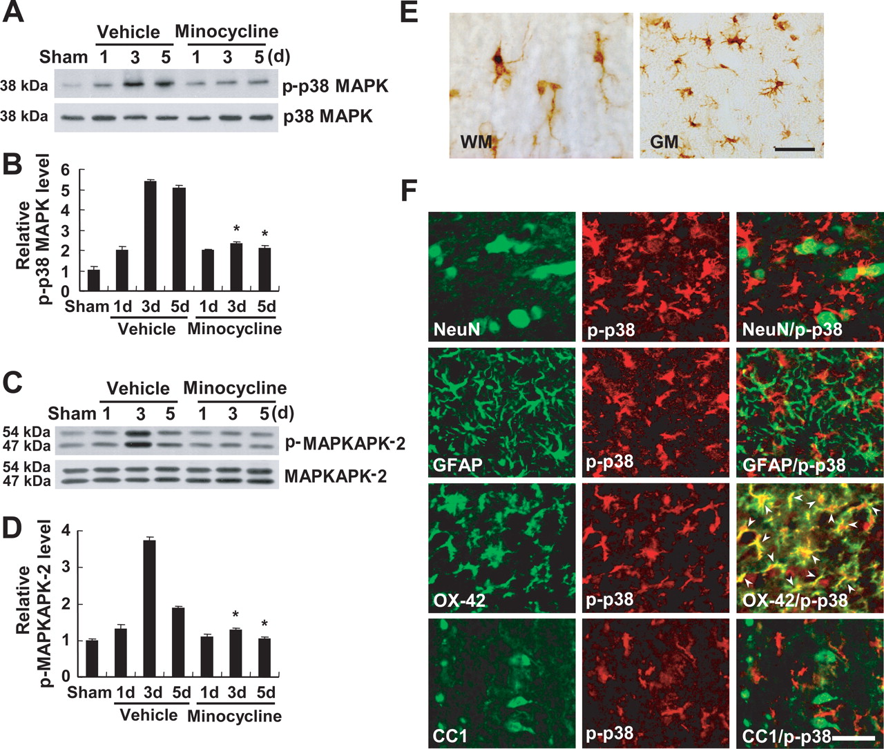
- Figure 2.
Minocycline inhibits p38MAPK and MAPKAPK-2 activation after SCI. A , C , SCI increased the levels of p-p38MAPK ( A ) and p-MAPKAPK-2 ( C ). B , D , Quantitative analysis of Western blots shows that minocycline treatment significantly inhibited both p-p38MAPK and p-MAPKAPK-2 when compared with those observed in vehicle control at 3 and 5 d after injury. Values are mean ± SD of three separate experiments. *p < 0.001. E , Immunocytochemical analysis shows that cells resembling microglia in the white (WM) and gray (GM) matters expressed p-p38MAPK at 5 d after injury. Double-labeled immunocytochemical analysis shows that only OX-42-positive microglia expressed p-p38MAPK (arrowheads), whereas neurons (NeuN), astrocytes (GFAP), and oligodendrocytes (CC1) were negative at 5 d after injury ( F ). Scale bars, 20 μm.
- Figure 3.
Minocycline inhibits p38MAPK-dependent proNGF expression in microglia, BV2 cells. A , Cells were plated onto six-well plates and treated with LPS. Note that BV2 cell treated with LPS for 4 h exhibited cellular processes, a characteristic of activated microglial cells. B , The level of p-p38MAPK was increased and peaked at 30 min after LPS treatment. C , Minocycline (1 and 5 nm) treatment decreased the level of p-p38MAPK at 30 min after LPS treatment when compared with that in cultures treated with LPS only. D , Quantitative analysis of Western blots shows that minocycline significantly inhibited p-p38MAPK expression at 30 min after LPS treatment when compared with that in cultures treated with LPS only. Values are mean ± SD of three separate experiments. *p < 0.001. E , The level of p-MAPKAPK-2 was increased and peaked at 30 min after LPS treatment. F , Minocycline (1 and 5 nm) or SB203580 treatment (1 and 5 μm), an inhibitor of p38MAPK, decreased the level of p-MAPKAPK-2 at 30 min after LPS treatment when compared with that in cultures treated with LPS only. G , Quantitative analysis of Western blots shows that minocycline and SB203580 significantly inhibited p-MAPKAPK-2 expression at 30 min after LPS treatment when compared with that in cultures treated with LPS only. Values are mean ± SD of three separate experiments. *p < 0.001. H , I , NGF mRNA ( H ) and proNGF protein (26 kDa) ( I ) expression were increased and peaked at 2 and 4 h, respectively, after LPS treatment. J , Minocycline (1 and 5 nm) and SB203580 (1 and 5 μm) treatment decreased the level of proNGF expression at 4 h after LPS treatment compared with that in cultures treated with LPS only. K , Quantitative analysis of Western blots shows that minocycline and SB203580 significantly inhibited proNGF expression at 4 h after LPS treatment when compared with that in cultures treated with LPS only. Values are mean ± SD of three separate experiments. *p < 0.001.
- Figure 4.
SB203580 inhibits proNGF expression after SCI. SB203580 (1 and 5 μg) was injected directly into the spinal cord at the lesion epicenter after injury. Spinal cord tissues were harvested at 5 d after injury. A , SB203580 treatment decreased the level of proNGF expression when compared with that in vehicle-treated control after injury. B , Quantitative analysis of Western blots shows that SB203580 significantly inhibited proNGF expression compared with that in vehicle-treated control after injury. Values are mean ± SD of three separate experiments. *p < 0.05; **p < 0.001. C , Double-labeled immunocytochemical analysis showed that p-p38MAPK-positive microglia expressed proNGF (arrows) at 5 d after injury. Scale bar, 20 μm.
- Figure 5.
A , Microglia-derived proNGF induces apoptosis of oligodendrocytes in culture. Immunocytochemical analysis shows that MBP-positive oligodendrocytes expressed p75NTR (arrows). Scale bar, 20 μm. Differentiated oligodendrocytes were treated with LPS-treated BV2 cell culture medium. After 24 h, cells were processed for TUNEL and MBP staining. B , Representative photographs show that LPS-treated BV2 cell culture medium or recombinant NGF (as a positive control) induced the apoptotic cell death of oligodendrocytes as revealed by the presence of both TUNEL- and MBP-positive cells (arrows). Note that control shows no TUNEL/MBP-positive cells. Scale bar, 20 μm. C , Quantitative analyses of TUNEL-positive oligodendrocytes show that LPS-induced oligodendrocyte cell death was significantly inhibited by minocycline or SB203580 treatment. Also, oligodendrocyte cell death was significantly attenuated when LPS-treated BV2 cell culture medium subjected to immunoprecipitation using a neutralizing anti-NGF polyclonal antibody or when oligodendrocytes were treated with anti- p75NTR antibody before treatment with LPS-treated BV2 cell culture medium ( C ). Values are mean ± SD of three separate experiments. *p < 0.001 compared with LPS.
- Figure 6.
Minocycline inhibits p75NTR expression after SCI. A , B , The levels of p75NTR mRNA ( A ) and protein ( B ) were increased and peaked at 5 d after SCI. C , E , Minocycline treatment decreased the levels of p75NTR mRNA ( C ) and protein ( E ) expression compared with those observed in vehicle control at 5 d after injury. D , F , Quantitative analyses of Western blots show that minocycline significantly inhibited p75NTR mRNA ( D ) and protein ( F ) expression when compared with those observed in vehicle control at 5 d after injury. Values are mean ± SD of three separate experiments. *p < 0.001. G , Immunocytochemical analysis showed that CC1-positive oligodendrocytes expressed p75NTR at 5 d after injury (arrows). Scale bar, 20 μm.
- Figure 7.
Minocycline inhibits RhoA activation after SCI. GTP-bound RhoA was isolated by pull-down assay and detected by Western blot using anti-Rho antibody as described in Materials and Methods. A , RhoA was activated and peaked at 5 d after injury. B , Minocycline decreased the level of RhoA activation compared with that in the vehicle control at 3 and 5 d after injury. Total Rho level determined from total tissue lysates was not changed after injury ( A , B ). C , Quantitative analysis of Western blots shows that minocycline significantly inhibited RhoA activation when compared with that in the vehicle control at 3 and 5 d after injury. Values are mean ± SD of three separate experiments. *p < 0.05; **p < 0.001.
- Figure 8.
Minocycline and SB203580 inhibits apoptosis of oligodendrocytes after SCI. Rats receiving the 25 mm insult were treated twice per day with minocycline, beginning 2 h after injury. SB203580 (5 μg) was injected directly into the spinal cord at the lesion epicenter at 2 h after injury. After 5 d injury, spinal cord sections were processed for double labeling using CC1 antibody, a marker for oligodendrocytes, and anti-cleaved capase-3 antibody. A , B , Immunocytochemical analysis shows that a number of cleaved (activated) caspase-3-positive oligodendrocytes (red, arrows) was observed in the injured spinal cord ( B ), whereas no cleaved caspase-3-positive cell was observed in the sham-operated cord ( A ) (longitudinal sections). C–E , Minocycline ( D ) or SB203580 ( E ) treatment decreased the number of cleaved caspase-3-positive oligodendrocytes (arrows) compared with that observed in the vehicle control ( C ) (transverse sections). Scale bar, 20 μm. F , Quantitative analysis of caspase-3-positive oligodendrocytes shows that minocycline or SB203580 significantly inhibited the death of oligodendrocytes when compared with that in the vehicle control after injury. Cleaved caspase-3-positive oligodendrocytes were counted as described in Materials and Methods. *p < 0.001.
- Figure 9.
Minocycline reduces myelin and axonal loss after SCI. Spinal cords at 38 d after injury were processed for Luxol fast blue and neurofilament staining. Transverse cryosections were selected 2000 μm rostral to the lesion site. A , B , Luxol fast blue staining shows that myelin loss in lateral funiculus was extensive in the vehicle control ( B ) when compared with that in sham control ( A ) after injury. C , Minocycline treatment decreased the extent of myelin loss after injury. Scale bar, 30 μm. D , E , Neurofilament staining shows that fewer axons were observed in the vehicle control ( E ) when compared with those in sham control ( D ) after injury. F , Minocycline treatment decreased the extent of axonal loss after injury. Scale bar, 30 μm. G , Quantitative analysis of neurofilament-stained axons within the vestibulospinal tract showed that the number of axons in minocycline-treated spinal cord was significantly higher than that in the vehicle control. Neurofilament-positive axons were counted as described in Materials and Methods. *p < 0.05.
- Figure 10.
Minocycline improves functional recovery after SCI. After SCI, minocycline or MP was administered either immediately or 2 h after injury, and recovery was assessed by BBB, inclined plane test, footprint analysis, and grid walk test (n = 20). A , B , Minocycline treatment (given immediately or after a 2 h delay) significantly improved locomotor function as assessed by BBB score ( A ) and inclined plane test ( B ) when compared with that of vehicle control. Note that MP treatment had no significant effect on recovery. *p < 0.05; **p < 0.01. C , The minocycline-treated group (given immediately or after a 2 h delay) shows a significantly lower error percentage when compared with that of vehicle-treated group, whereas MP-treated group shows no significant improvement in the grid walk test. The grid walk test was performed at 5 weeks after injury. *p < 0.05. D , Representative footprints obtained from each group at 5 weeks after SCI show that minocycline treatment (given after a 2 h delay) improved foot coordination, whereas vehicle- and MP-treated animals showed inconsistent coordination and toe drags.


