

Article Figures & Data
Figures
- Figure 1.
MSK1 expression and brain morphology. A, MSK1 is highly expressed in the brain and hippocampus in wild-type mice, whereas MSK1 expression is absent in MSK1 knock-out mice. MSK2 expression is equivalent in wild-type and MSK1 knock-out mice. TBP was used as a loading control. B, Nissl stains of sagittal brain sections, showing no difference in brain morphology between wild-type and MSK1 knock-out mice. C, Close-up view of the hippocampus.
- Figure 2.
MSK1 knock-out mice have a contextual fear conditioning deficit. A, Quantification of freezing behavior in wild-type (n = 19) and MSK1 knock-out mice (n = 13) during training, and contextual and cued tests 24 h post-training. MSK1 knock-out mice displayed significantly less freezing than wild-type mice during the contextual test. B, Quantification of freezing behavior in wild-type (n = 10) and MSK1 knock-out mice (n = 9) during contextual and cued tests 1 h post-training. C, Quantification of freezing behavior in wild-type (n = 12) and MSK1 knock-out mice (n = 11) during contextual and cued tests 7 d post-training. Error bars indicate SEM. An asterisk denotes a significant difference (p < 0.05) as determined by Tukey's multiple-comparison test.
- Figure 3.
Analysis of select baseline control behaviors in wild-type and MSK1 knock-out mice. A, Plot of latency times spent on rotarod device for wild-type (n = 12) and MSK1 knock-out mice (n = 8) over eight trials. B, Measurement of shock thresholds to flinching, jumping, and vocalization behaviors for wild-type (n = 13) and MSK1 knock-out mice (n = 11). C, Comparison of percent inhibition of acoustic startle response for wild-type (n = 18) and MSK1 knock-out mice (n = 12) with different prepulses. Error bars indicate SEM.
- Figure 4.
MSK1 knock-out mice have impaired passive avoidance and spatial learning. A, Latency times for wild-type (n = 23) and MSK1 knock-out mice (n = 13) to step into the dark compartment of the training chamber in the passive avoidance task. MSK1 knock-out mice have significantly shorter latency time on day 2 after training. B, Plot of latency times to find the escape platform for wild-type (n = 14) and MSK1 knock-out mice (n = 18) over eight trial blocks in the Morris water maze. C, Time spent in each quadrant of the Morris water maze pool for wild-type (n = 14) and MSK1 knock-out mice (n = 18) during the probe trial. MSK1 knock-out mice spent significantly less time in the target quadrant than wild-type mice, lacking a spatially selective search strategy. D, Number of platform crossings in each quadrant for wild-type (n = 14) and MSK1 knock-out mice (n = 18) during the probe trial. MSK1 knock-out mice had significantly fewer platform crossings in the target quadrant than wild-type mice. Error bars indicate SEM. Asterisks denote a significant difference (p < 0.05) as determined by Tukey's multiple-comparison test.
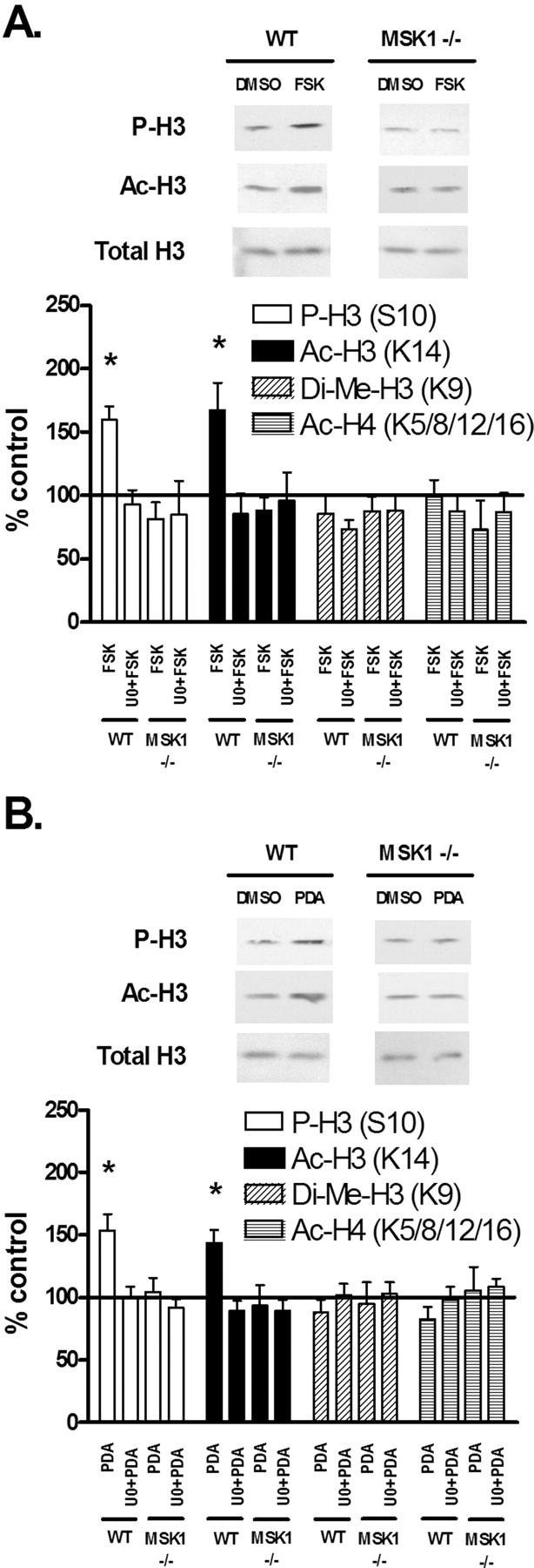
- Figure 5.
Regulation of histone modifications after in vitro stimulation of hippocampal slices. A, Quantification of immunoblot densities for phospho-histone H3 (P-H3), acetyl-histone H3 (Ac-H3), dimethyl-histone H3 (Di-Me-H3), and acetyl-histone H4 (Ac-H4). Treatment of wild-type hippocampal slices (n = 7) with FSK (50 μm) plus Ro20–1724 (100 μm) for 1 h significantly increased H3 phosphorylation and acetylation, whereas preincubation with U0126 (20 μm) for 10 min followed by FSK blocked these changes. Treatment of MSK1 knock-out hippocampal slices (n = 7) with FSK plus Ro20–1724 for 1 h did not result in any significant changes in histone modifications, and preincubation with U0126 for 10 min followed by FSK had no significant effect. Total histone H3 was unchanged. Representative immunoblots for P-H3, Ac-H3, and total H3 are shown for FSK treatments. B, Quantification of immunoblot densities for phospho-histone H3, acetyl-histone H3, dimethyl-histone H3, and acetyl-histone H4. Treatment of wild-type hippocampal slices (n = 8) with PDA (3 μm) for 1 h significantly increased H3 phosphorylation and acetylation, whereas preincubation with U0126 (20 μm) for 10 min followed by PDA blocked these changes. Treatment of MSK1 knock-out hippocampal slices (n = 8) with PDA for 1 h did not result in any significant changes in histone modifications, and preincubation with U0126 for 10 min followed by PDA had no significant effect. Total histone H3 was unchanged. Representative immunoblots for P-H3, Ac-H3, and total H3 are shown for PDA treatments. Drug-treated and vehicle-treated slices for each condition came from one individual animal. All drug-treated slices were compared with vehicle-treated (DMSO) controls and normalized to total protein loaded. Error bars indicate SEM. Asterisks denote significant differences (p < 0.05) as determined by Tukey's multiple-comparison test.
- Figure 6.
Regulation of histone modifications and ERK phosphorylation after fear conditioning training. A, Quantification of immunoblot densities for phospho-histone H3 (P-H3), acetyl-histone H3 (Ac-H3), dimethyl-histone H3 (Di-Me-H3), and acetyl-histone H4 (Ac-H4). Fear conditioning (2 pairs of tone and shock) in wild-type mice (n = 11) resulted in a significant increase in H3 phosphorylation and acetylation 1 h after training, whereas training in MSK1 knock-out mice (n = 11) did not result in significant changes in histone modifications. Total histone H3 was unchanged. Representative immunoblots for P-H3, Ac-H3, and total H3 are shown for fear-conditioned animals. B, Quantification of immunoblot densities for phospho-ERK (P-ERK) and total ERK. Fear conditioning (2 pairs of tone and shock) resulted in a significant increase in ERK phosphorylation 1 h after training for both wild-type mice (n = 11) and MSK1 knock-out mice (n = 11). Total ERK was unchanged. Representative immunoblots for P-ERK and total ERK are shown for fear-conditioned animals. Each sample came from one individual animal. All samples were compared with naive controls and normalized to total protein loaded. Error bars indicate SEM. Asterisks denote significant differences (p < 0.05) as determined by Tukey's multiple-comparison test.
- Figure 7.
NaB enhances fear memory in wild-type mice, but not in MSK1 knock-out mice. A, Representative immunoblots for Ac-H3 and total H3 are shown 1 h after NaB treatment compared with saline. Histone H3 acetylation increased in wild-type and MSK1 knock-out animals with NaB compared with saline-injected controls. Each sample came from one individual animal. B, Quantification of freezing behavior in wild-type (n = 12) and MSK1 knock-out mice (n = 18) during training. NaB had no significant effect on freezing in either group compared with saline. C, Quantification of freezing behavior in wild-type (n = 12) and MSK1 knock-out mice (n = 18) during the contextual test 24 h post-training. With saline injection, MSK1 knock-out mice displayed significantly less freezing than wild-type mice. However, with NaB injection, freezing in wild-type animals was enhanced, whereas freezing in MSK1 knock-out animals was unchanged. D, Quantification of freezing behavior in wild-type (n = 12) and MSK1 knock-out mice (n = 18) during the cued test 24 h post-training. E, Quantification of freezing behavior in wild-type (n = 8) and MSK1 knock-out mice (n = 11) during the contextual test 7 d post-training. F, Quantification of freezing behavior in wild-type (n = 8) and MSK1 knock-out mice (n = 11) during the cued test 7 d post-training. Error bars indicate SEM. Asterisks denote significant differences (p < 0.05) as determined by Tukey's multiple-comparison test.
- Figure 8.
Regulation of CREB phosphorylation after fear conditioning training and HDAC inhibition. A, Quantification of immunoblot densities for phospho-CREB (P-CREB) and total CREB. Fear conditioning (2 pairs of tone and shock) in wild-type mice (n = 6) resulted in a significant increase in CREB phosphorylation 1 h after training, whereas training in MSK1 knock-out mice (n = 6) did not result in significant changes in CREB phosphorylation. Total CREB was unchanged. Representative immunoblots for P-CREB and total CREB are shown for fear-conditioned animals. B, Representative immunoblots for phospho-CREB and total CREB are shown 1 h after NaB treatment compared with saline. There was no change in CREB phosphorylation in either wild-type or MSK1 knock-out animals with NaB compared with saline-injected controls. C, Quantification of immunoblot densities for phospho-CREB and total CREB. In wild-type mice (n = 6), fear conditioning resulted in increased CREB phosphorylation in both saline- and NaB-treated animals. In MSK1 knock-out mice (n = 6), training did not result in significant changes in CREB phosphorylation in both saline- and NaB-treated animals. Thus, the deficit in CREB phosphorylation persists in MSK1 knock-out mice even after administration of NaB. Representative immunoblots for P-CREB and total CREB are shown for each treatment. Each sample came from one individual animal. All samples were compared with naive controls and normalized to total protein loaded. Error bars indicate SEM. Asterisks denote significant differences (p < 0.05) as determined by Tukey's multiple-comparison test.
- Figure 9.
Model of MSK1 in chromatin regulation in memory formation. The activity of cell surface receptors is coupled with the activation of PKC, PKA, MEK, ERK, and finally MSK1. MSK1 regulates a diverse set of targets in the nucleus, including histones (phosphorylation and acetylation), transcription factors (e.g., CREB), and HATs, leading to upregulation of gene transcription. GLU, Glutamate; DA, dopamine; 5HT, 5-hydroxy-tryptophan; NMDA-R, NMDA receptor; AMPA-R, AMPA receptor; NT-R, neurotransmitter receptor; AdC, adenylyl cyclase.


