

Article Figures & Data
Figures
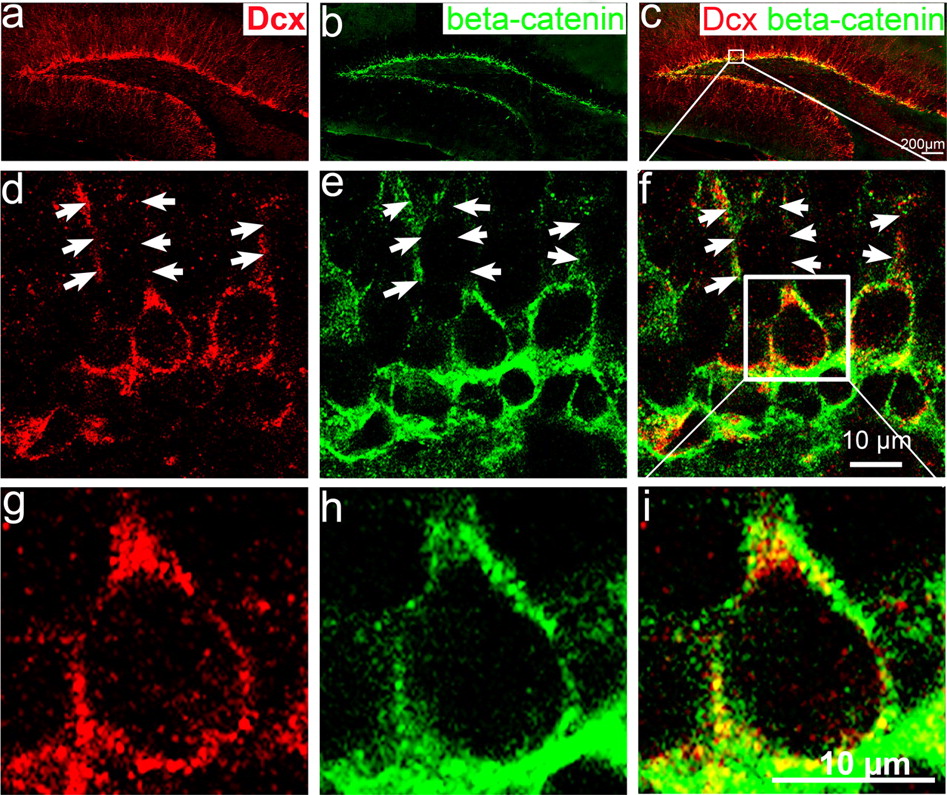
- Figure 1.
β-Catenin is expressed in postnatal dentate gyrus and colocalizes with Dcx-positive newborn neurons at the inner granular cell layer. Immunohistochemistry of hippocampal sections from wild-type mouse brain at P25 labeled with antibodies to the newborn neuronal marker Dcx (red) and to β-catenin (green). a, Dcx positive-cells located in the inner cell layer of dentate gyrus. b, β-Catenin is highly expressed in the SGZ and inner granular cell layer. c, Merged image of a and b showing that β-catenin is expressed in Dcx-positive newborn neurons adjacent to the SGZ. d, e, Confocal images of single focal section further shows that Dcx (d) and β-catenin (e) are highly expressed in the cytoplasm and in the processes of newborn neurons (arrows). f, Merged image of e and d showing that β-catenin colocalizes with the Dcx-positive newborn neurons in the dentate gyrus. g–i, Enlarged view of single newborn neuron within the white box in f showed that Dcx-positive newborn neurons (g) expresses β-catenin (h) in the cytoplasm (i), but not detectable in the nucleus.
- Figure 2.
Expression of Cre recombinase in the hippocampal dentate gyrus of POMC-Cre transgenic mice. a, Cre (red) is expressed in a few cells in the inner granule cell layer in 5-d-old (P5) transgenic mice. b–d, The number of Cre-expressing cells increases in P15 mice (b), then decreases at P25 (c) and remains low in P35 mice (d). Nuclei are stained with Hoechst 33258 in blue to show the dentate gyrus. Scale bar, 100 μm.
- Figure 3.
Cell-type specificity of Cre expression in POMC-Cre transgenic mice. a1–a4, Immunohistochemistry of hippocampal sections from POMC-Cre mice at P15, labeled using antibodies for Cre (red) and a neuron specific marker, NeuN (green). NeuN-positive granular neurons (a1; green) and Cre-positive newborn neurons (a2; red) are located in the postnatal dentate gyrus. In a3, the merged image of a1 and a2 shows that Cre-positive cells are located in the inner granular layer and are adjacent to NeuN-positive mature granular neurons. a4 is an amplified view of a3 of Cre (red) and NeuN (green) double staining to show that Cre is not expressed in NeuN positive cells. b1–b4, Immunohistochemistry of hippocampal sections using antibodies for Dcx (green), a marker of migrating newborn neurons, and Cre (red). The Dcx-positive neurons (b1; green) are located in the same layer as Cre positive cells (b2; red). The merged image (b3) and the enlarged view (b4) of these pictures show that Cre is highly expressed in Dcx-positive newborn neurons in the dentate gyrus. c1–c4, Immunofluorescence labeling for Nestin (green), a marker of neural progenitor cells, and Cre (red). The Nestin-positive cell bodies (green; b1, b3, and b4) are located below the Cre-positive cell layer (red; c2, c3, and c4). The enlarged view (c4) of the merged image (c3) shows that the Nestin is highly expressed in the processes of neural progenitors, but is very weakly expressed in the cell bodies. We do not see any colocalization of Nestin signals and Cre signals. These results suggest that Cre is specifically expressed in the Dcx positive-newborn neurons in the postnatal dentate gyrus. Scale bars: a3, b3, c3, 100 μm; a4, b4, c4, 20 μm.
- Figure 4.
Quantification of newborn neurons expressing Cre recombinase. a–d, Immunostaining to show newborn neurons (Dcx-expressing cells; red) and Cre-expressing cells (green) in the dentate gyrus at p5 (a), p15 (b), p25 (c), and p35 (d). Scale bars: 100 μm. a1–d1, Enlarged views of a–d, respectively within the white boxes. Scale bars, 20 μm. e, The quantification of Cre-positive cells that were colocalized with Dcx at different ages (P5, 10.77 ± 0.37%; P15, 43.13 ± 3.93%; P25, 41.97 ± 1.66%; P35, 44.43 ± 2.23%; n > 5000 cells, from six sections).
- Figure 5.
Cre recombines is expressed in BrdU-labeled newborn cells in the postnatal dentate gyrus (3 weeks old). a, Immunostaining to show BrdU-labeled newborn cells (red) and Cre-expressing cells (green) in the postnatal hippocampal dentate gyrus. b–d, High magnification of confocal image showing colocalization of Cre and BrdU within the white box in a from x, y, and z axes. Scale bars, 10 μm. GCL, Granule cell layer.
- Figure 6.
EGFP reporter (green) is expressed only in granule neurons not in proliferating neural progenitor cells (BrdU-positive cells; red). Immunohistochemistry of POMC-Cre;eGFP double transgenic mice (P35), in which eGFP was expressed after Cre-mediated deletion of a loxP-flanked stop codon. A, Immunofluorescence staining for eGFP (green) to show that the Cre recombinase can functionally delete the targeted gene fragment and induce the expression of eGFP in vivo. eGFP is highly expressed in granular neurons, including in dendrites and axons in the dentate gyrus. b, An enlarged view of a shows eGFP expression in the granular neurons of dentate gyrus. c, Double immunofluorescence staining for eGFP (green) and BrdU (red), a marker of proliferating cells, shows that BrdU-positive cells are located below the eGFP-positive cell layer. There are no eGFP and BrdU double-labeled cells in the dentate gyrus. d, An enlarged image of c. Scale bars: a, 200 μm; b, c, 100 μm; d, 20 μm. e, Quantification of neurons in DG expressing eGFP in the POMC-Cre;eGFP (Z/EG) double transgenic mice at various ages.
- Figure 7.
Dendrite malformation in mice lacking β-catenin in newborn granule neurons in the DG. a, b, Cresyl-violet staining for DG morphology. We did not find any obvious global malformation in DG between control and KO mice. c–f, Immunofluorescence labeling for Dcx (green) highly labeled the cell bodies and dendrites of newborn neurons. c, d, Dcx staining of control mice shows that most of the Dcx-positive newborn neurons develop long primary dendrites with extensive branching at the age of P35. e, f, The newborn neurons in the dentate gyrus of KO mice have no dendrites or have dendrites that lack branching. g, Diagram illustrating dendrite morphology in the early stages of neurogenesis in postnatal hippocampus. At stage A of neurogenesis, the axes of newborn neuron bodies are parallel to the granule cell layer but are becoming perpendicular to the granule cell layer. This is also the stage at which newborn neurons initiate dendrite outgrowth. At stage B of neurogenesis, newborn neurons extend dendrites, which approach the molecular layer and begin branching, thus developing a more complex dendrite morphology. h, Comparing the number of newborn neurons in stage A and late stage B, as determined by the length or morphology of dendrites, between the control mice and KO mice. Approximately 500 newborn neurons were counted and categorized as stage A or stage B. Scale bars: b, e, 100 μm; f, 20 μm.
- Figure 8.
Dendritic morphologies of newly generated neurons labeled by retrovirus-mediated eGFP expression. eGFP-expressing retroviruses were injected into the hippocampus to label newly generated neurons in control and β-catenin KO mice at 5 weeks old. a, Examples of eGFP-labeled newborn neurons in control mice 10 d after retrovirus injection. b, Drawings of representative neurons in control mice. c, Examples of eGFP-labeled newborn neurons in β-catenin KO mice 10 d after retrovirus injection. d, Drawings of representative neurons in β-catenin KO mice. Scale bars, 20 μm.


Additional Files
Supplemental Data
Files in this Data Supplement:
- supplemental material - Figure legends
- supplemental material - Figure 1
- supplemental material - Figure 2
- supplemental material - Figure 3
- supplemental material - Figure 4


