

Article Figures & Data
Figures
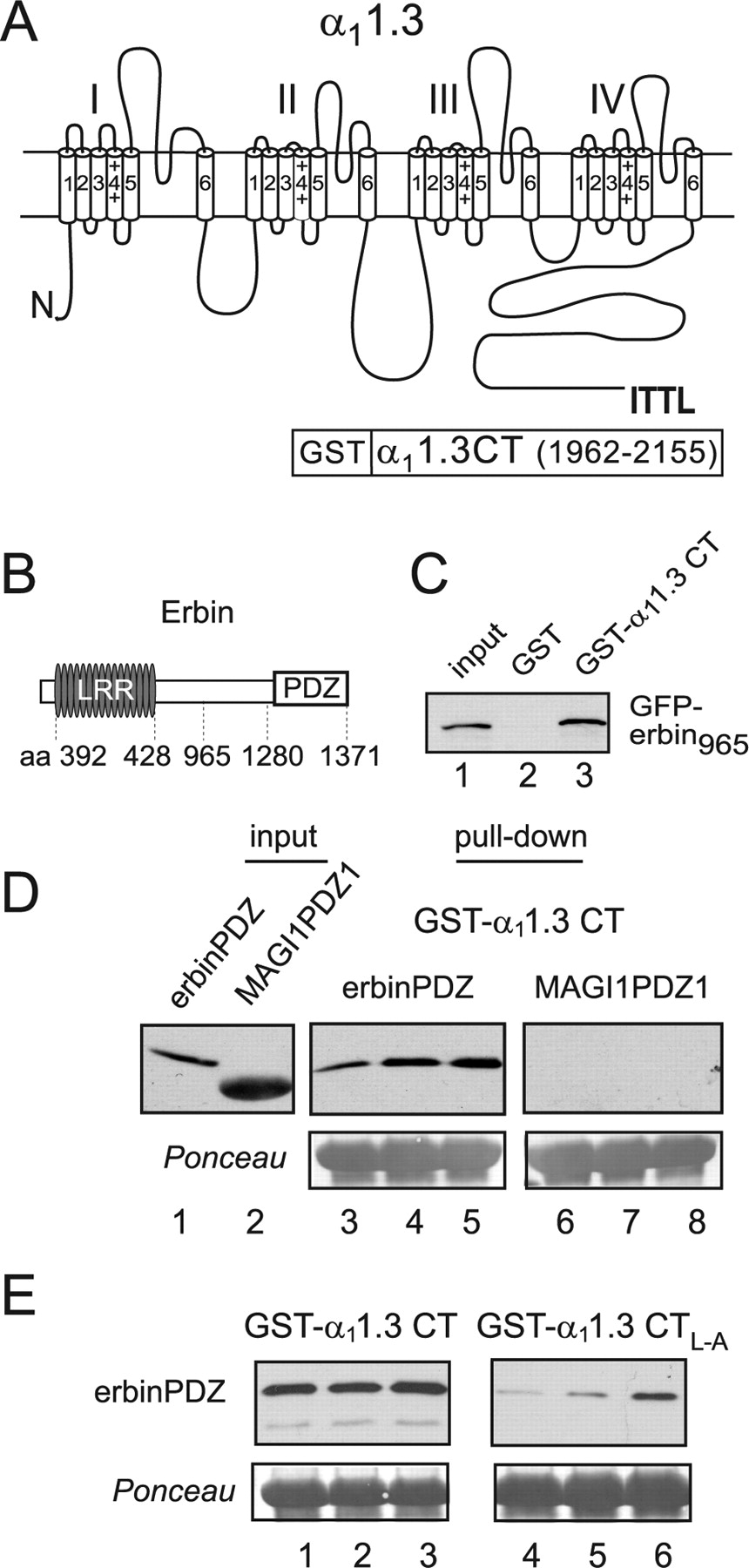
- Figure 1.
The PDZ domain of erbin binds to the C-terminal domain of α11.3. A, Schematic of rat α11.3 showing class I PDZ-binding consensus sequence (ITTL) in the cytoplasmic C-terminal domain and the region used for GST-α11.3 CT in pull-down assays (amino acids 1962–2155). B, Schematic of mouse erbin indicating position of LRR (amino acids 392–428) and the PDZ domain (amino acids 1280–1371). C, Binding of erbin to α11.3 CT. GST-α11.3 CT (lane 3) or GST alone (lane 2) was immobilized on glutathione-agarose beads and incubated with lysates from cells transfected with GFP-erbin965 (amino acids 965–1371). Bound GFP-erbin965 was detected by Western blot with anti-GFP antibodies. Lane 1 shows ∼15% of GFP-erbin965 input used in the pull-down assay. D, Binding of erbinPDZ but not MAGI1PDZ1 to α11.3 CT. GST-α11.3 CT was incubated with purified his-S-erbinPDZ (lanes 3–5) or his-S-MAGI1PDZ1 (lanes 6–8). Input his-S-tagged protein was the following (in μg): 1 (lanes 3, 6), 2 (lanes 4, 7), and 4 (lanes 5, 8). Input lanes 1 and 2 show his-S-tagged proteins used in the assay (1 μg). E, Impaired binding of erbinPDZ to α11.3 CTL–A. His-S-erbinPDZ protein (lanes 1, 4, 1.5 μg; lanes 2, 5, 3 μg; lanes 3, 6, 6 μg) was incubated with GST-α11.3 CT (lanes 1–3) or GST-α11.3 CTL–A (lanes 4–6). In D and E, bound proteins were detected by Western blot (top) with anti-S-protein antibodies, and Ponceau staining (bottom) shows equal levels of GST-α11.3 CT or GST-α11.3 CTL–A in each group.
- Figure 2.
Coimmunoprecipitation of erbin with Cav1.3 from transfected cells and brain. A, Schematic of α11.3 and α11.2 showing C-terminal PDZ-binding motifs. B, Coimmunoprecipitation of erbin and Cav1.3 from transfected cells. HEK293T cells were cotransfected with myc-erbin and Cav1.3 (FLAG-α11.3, β1b, and α2δ; lanes 1, 4, 6) or Cav1.2 (FLAG-α11.2, β1b, and α2δ; lanes 2, 3, 5) and subjected to lysis and immunoprecipitation using rabbit antibodies against α11.3 (lanes 1, 2) or α11.2 (lanes 3, 4). Immunoprecipitated proteins (I.P.; lanes 1–4) were detected by Western blotting (WB) with antibodies recognizing FLAG (top) or myc (bottom) epitopes. Lanes 5 and 6 represent 5% of the cell lysate input used for coimmunoprecipitation. C, Coimmunoprecipitation of erbin and Cav1.3 from the brain. Rat brain lysates were incubated with goat α11.3 antibodies or control goat IgG for immunoprecipitation, and α11.3 and erbin were detected by Western blotting with rabbit α11.3 and erbin antibodies, respectively. D, Lack of α11.3 immunoprecipitation in Cav1.3−/− mouse brain. α11.3 immunoprecipitation protocol used in C was applied to brain lysates from Cav1.3+/+ or Cav1.3−/− mice. An ∼200 kDa protein corresponding to α11.3 was detected in the brain lysate, and samples were immunoprecipitated with α11.3 antibodies from Cav1.3+/+ but not Cav1.3−/− mice. Lysate lanes represent 3% of the input used for coimmunoprecipitation.
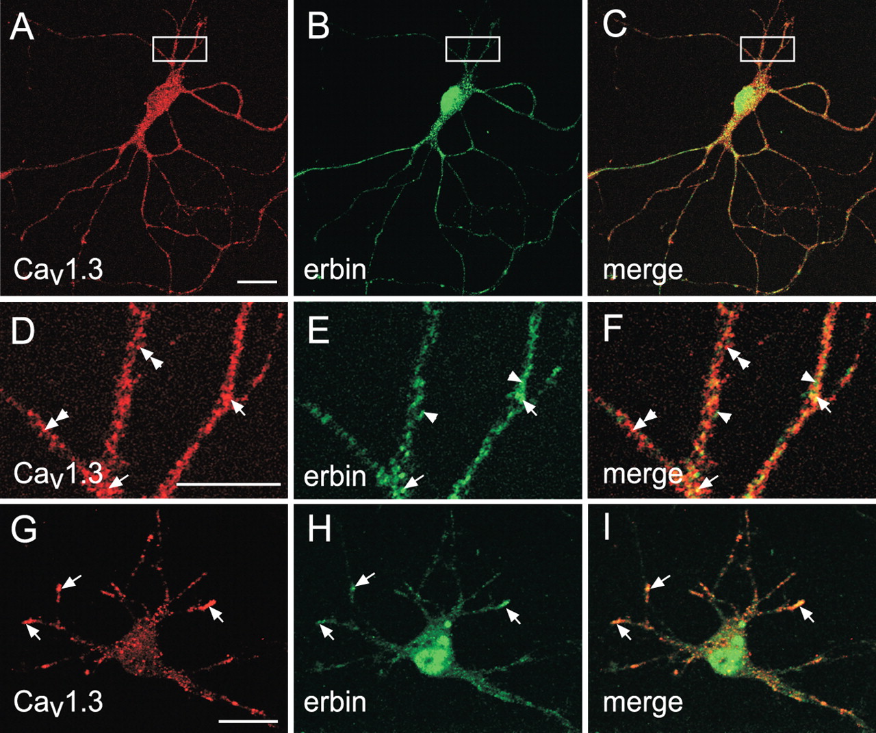
- Figure 3.
Cav1.3 and erbin colocalize in somatodendritic domains of cortical neurons in culture. A–I, Confocal images of primary cortical neuron cultures (A–F, 14 d in culture; G–I, 8 d in culture) sequentially double labeled with antibodies against α11.3 (to detect Cav1.3) and erbin are shown. Immunofluorescence was viewed under optics for rhodamine for Cav1.3 (A, D, G) or fluorescein for erbin (B, E, H). Regions of colocalization appear yellow in the merged images (C, F, I). Extensive colocalization of erbin and Cav1.3 was detected in cell bodies (A–C, G–I) and dendrites (D–F; higher-magnification view of boxed region in panels A–C). In D–I, elements immunoreactive for Cav1.3 alone (double arrowheads), erbin alone (arrowheads), or Cav1.3 plus erbin (arrows) are indicated. Scale bars: A (for A–C), G (for G–I), 20 μm; (in D) D–F, 10 μm.
- Figure 4.
Erbin does not affect voltage-dependent activation or mamplitude of Cav1.3 currents. A, B, Normalized tail current (Norm. Itail) activation curves for HEK293T cells transfected with Cav1.3 alone (n=9) (A) or cotransfected with Cav1.3 plus erbin (n=6) (B). Test pulses (10 ms) were applied from a holding voltage of −90 mV to various voltages, and peak tail currents were measured after repolarization to −70 mV for 2 ms. Tail currents were normalized to the largest in the series and plotted against test voltage. Representative current traces are shown at top. C, D, Current–voltage relationships for Cav1.3 alone (C) and Cav1.3 plus erbin (D). Data were from same voltage protocol as in A and B, except that peak current amplitudes during the test pulse were plotted against test voltage. Error bars represent SEM.
- Figure 5.
Erbin augments VDF of Cav1.3 Ba2+ currents. A, Representative Ba2+ current traces and double-pulse voltage protocol for measuring VDF in cells transfected with Cav1.3 alone or cotransfected with erbin. Currents were evoked by 10 ms test pulses from −90 to −20 mV before (P1) and after (P2) a conditioning 20 ms prepulse (Pre) to +60 mV. B, No effect of erbin on prepulse voltage dependence of facilitation. Percentage of facilitation was calculated as [(IP2/IP1) − 1] × 100 for cells for different prepulse voltages, normalized to that for a +60 mV prepulse, and plotted as percentage of maximum (Max.) facilitation against prepulse voltage for cells transfected with Cav1.3 alone (n=11) or cotransfected with erbin (n=10). C, Increased net facilitation attributable to erbin. Fratio represents the ratio of the P2 and P1 test currents and is plotted against prepulse voltage for Cav1.3 alone (n = 12) or Cav1.3 plus erbin (n = 10). *p < 0.05. Error bars represent SEM.
- Figure 6.
Erbin increases VDF of Cav1.3 Ca2+ currents. A, Representative Ca2+ current traces and double-pulse voltage protocol for measuring VDF in cells transfected with Cav1.3 alone (n=6) or cotransfected with erbin (n=7). Currents were evoked by 5 ms test pulses from −90 to −10 mV before (P1) and after (P2) a conditioning 10 ms prepulse (Pre) to varying voltages. B, Increased net facilitation for prepulse voltages greater than +40 mV in cells cotransfected with erbin. Fratio was determined and plotted against prepulse voltage as in Figure 5C. The dotted line indicates Fratio=1. Points falling below the line result from Ca2+-dependent inactivation with prepulse voltages evoking significant Ca2+ entry. *p < 0.05 by two-factor ANOVA and Tukey's post-test. Error bars represent SEM.
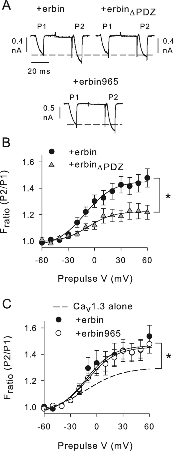
- Figure 7.
Increased VDF depends on PDZ domain of erbin. A, Representative current traces obtained with the same voltage protocol as in Figure 5A for cells cotransfected with Cav1.3 plus erbin, erbinΔPDZ, or erbin965 (amino acids 965–1371). B, Deletion of erbinPDZ prevents effects on VDF. Fratio is plotted against prepulse V for cells cotransfected with Cav1.3 plus erbin (n=10) or erbinΔPDZ (n=8). *p < 0.05. C, Deletion of LRR of erbin does not prevent effects of erbin on VDF. Fratio was plotted against prepulse voltage for cells cotransfected with Cav1.3 plus erbin (n=10) or erbin965 (n=5). The dashed line represents data replotted from Figure 5C for cells transfected with Cav1.3 alone. *p < 0.05 for Cav1.3 plus erbin965 compared with Cav1.3 alone. Error bars represent SEM.
- Figure 8.
Erbin does not cause increased VDF of Cav1.3b channels. A, Schematic showing differences in C-terminal domain of α11.3 and α11.3b. Parentheses indicate unique sequence including the PDZ-binding site (ITTL) in α11.3 but not α11.3b. Splicing of this region in α11.3b results in a truncated C-terminal domain with a distinct C-terminal sequence (ERML). B, ErbinPDZ interacts weakly with α11.3b CT. GST-α11.3 CT or GST-α11.3b CT was used in pull-down assay with his-S-erbinPDZ (1.5 or 3.0 μg). Top, Western blot detection of erbinPDZ. Bottom, Ponceau staining showing equal levels of immobilized GST-α11.3 CT (left) and GST-α11.3b CT (right) used in assay. C, No effect of erbin on VDF of Cav1.3b. Fratio was plotted against prepulse voltage for cells transfected with Cav1.3b alone (n = 14) or cotransfected with erbin (n=8). The dashed line represents data replotted from Figure 5C for cells transfected with Cav1.3 alone. Representative current traces obtained with a +60 mV prepulse are shown above. Error bars represent SEM.
- Figure 9.
Autoinhibition of VDF by the CT of α11.3. A–C, Cotransfection of α11.3 CT fragment (CT500; amino acids 1655–2155) suppresses VDF. Effect of CT500 on Fratio is plotted against prepulse V in cells with Cav1.3b (n=6) (A), Cav1.3 (n=4; B), or Cav1.3 plus erbin (n=10; C). Representative current traces obtained with a +60 mV prepulse are shown above. Vertical scale bars, 0.8 nA; horizontal scale bars, 40 ms. *p < 0.01. D, Voltage-dependent inhibition of VDF by CT500. Percentage inhibition was measured as [1 − (Fratio+CT500/Fratio)] × 100, where Fratio is for Cav1.3b, Cav1.3, or Cav1.3 plus erbin, and Fratio+CT500 represents the mean Fratio for these groups cotransfected with CT500. Stronger prepulse voltage dependence of CT500 inhibition is observed for Cav1.3b and Cav1.3 plus erbin compared with Cav1.3. *p < 0.001. Error bars represent SEM.
- Figure 10.
Effect of erbin but not α11.3 CT depends on auxiliary Ca2+ channel β subunit. A, Erbin does not increase VDF of Cav1.3 channels containing the β4 subunit (Cav1.3-β4). Fratio was determined from double-pulse protocol as described in Figure 5, except that P1 and P2 currents were evoked by 7 ms test pulses from −90 to −20 mV in cells transfected with Cav1.3-β4 (n=7) or cotransfected with erbin (n=8). B, CT500 inhibits VDF of β4-containing Cav1.3b channels (Cav1.3b-β4). Fratio was determined and plotted as in A for cells transfected with Cav1.3b-β4 (n=6) or cotransfected with CT500 (n=7). *p < 0.001. For A and B, representative current traces obtained with +80 mV prepulse are shown above. Error bars represent SEM.


Additional Files
Supplemental Data
Files in this Data Supplement:
- supplemental material - Figure 1
- supplemental material - Figure 2


