

Article Figures & Data
Figures
- Figure 1.
Ectopic expression of NgR1 blocks FGF2-elicited differentiation of PC12 cells and axonal branching in primary cortical neurons. A , FGF2-elicited differentiation is suppressed in PC12 cells. PC12 cells were transfected with NgR1 or eGFP plasmid DNA and then cultured in the presence of FGF2. Double immunofluorescence with anti-NgR1 and TuJ1 or anti-GFP and TuJ1 antibodies identified NgR1+ and GFP+ cells. Quantification of PC12 cell differentiation in the presence of FGF2 revealed significantly fewer processes in NgR1+ cells compared with GFP+ cells, whereas in the absence of FGF2 no cells with processes longer than two cell bodies in diameter were found. B , FGF2-mediated axon branching of rat E18 cortical neurons is suppressed in neurons transfected with NgR1. Neurons transfected with NgR1 or eGFP were cultured in the presence (+FGF2) or absence (−FGF2) of FGF2 and immunostained as described above. C , Quantification of neurons with two or more axonal branches observed in GFP+ (white) and NgR1+ (gray) neurons. D , Frequency histogram of axonal branches. In the presence of FGF2, GFP+ but not NgR1+ neurons show significantly enhanced axonal branching. Data plotted in C and D were from the same experiments. Error bars indicate SEM. *p < 0.05. Scale bars: A , 100 μm; B , 50 μm.
- Figure 2.
NgR1 supports binding of select members of the FGF family. A , NgR1 expressed on the surface of COS-7 cells supports binding of select members of the FGF family. None of the FGF family members tested binds to NgR2. Binding of MAG-Fc to NgR1 and NgR2 is shown as a positive control. B , C , Scatchard plot analysis of AP-FGF1 ( B ) and AP-FGF2 ( C ) binding to NgR1 expressed in COS-7 cells. The insets show saturation binding curves. D , Pull-down experiments using NgR1-Fc and AP-fusion proteins revealed a direct interaction of NgR1 with FGF2 and Nogo-66, but not NiG, or VEGF. Excess IgG competes with NgR1-Fc for binding to protein A/G beads and blocks the pull down of AP-FGF2. E , Cross-linking of 125I-FGF2 to NgR1-Fc and FGFR1-Fc, in the presence or absence of excess “cold” FGF2 or insulin. Troy-Fc and ephrinB3-Fc were used as negative controls. Complexes of 125I-FGF2:NgR1-Fc (∼220 kDa) and 125I-FGF2:FGFR1-Fc (∼250 kDa) were resolved by 7% SDS-PAGE, and the dried gel was exposed to x-ray film. The arrowhead denotes radiolabeled complexes. Scale bar, 30 μm.
- Figure 3.
Structural basis of the NgR1–FGF2 association. A , Chimeric Nogo receptors were expressed on the surface of COS-7 cells and assayed for binding of AP-FGF2, AP-Nogo-66-myc, or AP-OMgp. NgR1 sequences are labeled in red and non-NgR1 sequences are labeled in gray. LRR, Leucine-rich repeats 1–8; NT, LRRNT cap domain; CT, LRRCT cap domain. Full-length NgR1 (construct I), but not NgR3 (construct II), supports binding of FGF2, Nogo-66, and OMgp. The NgR1 LRR cluster (composed of domains LRRNT-LRR-LRRCT) fused to the stalk region of NgR3 is sufficient to confer high-affinity ligand binding (see construct III). Construct IV, composed of the NgR3 LRR cluster and the NgR1 stalk region, does not support binding of FGF2, Nogo-66, or OMgp. Furthermore, the NgR1 LRRNT cap domain and the first three LRRs (construct V) and the NgR1 LRRCT distal portion and stalk region (construct VI) are dispensable for AP-FGF2, AP-Nogo-66-myc, or AP-OMgp binding. The NgR1 LRRCT distal region and stalk are not sufficient to support ligand binding (construct VII). Cell surface expression of chimeric Nogo receptor constructs was confirmed by ICC under nonpermeabilizing conditions. Scale bars: A , D , 30 μm. B , Quantification of ligand binding to chimeric Nogo receptors, normalized to wild-type NgR1 binding (=100%). Of note, the molecular basis for AP-FGF2-myc, AP-Nogo-66, and AP-OMgp is very similar. C , An NgR1-Fc pull-down (PD) assay was used for affinity precipitation of AP-Nogo-66-myc in the presence of increasing concentrations of AP-FGF2. AP-FGF2 competes with AP-Nogo-66-myc for NgR1 binding. D , Binding of AP-FGF2 to full-length NgR1 and NgR1Δstalk (Δstalk) transiently expressed on COS-7 cells. Binding of AP-FGF2 to wild-type NgR1 was normalized to 100%, and no significant change in AP-FGF2 binding was observed after deletion of residues T373–G448 of the NgR1 stalk (110 ± 9%). E , Experiments with PC12 cells stably expressing either NgR1 or NgR1Δstalk revealed that the NgR1 stalk region T373–G448 is important for the inhibition of FGF2-elicited PC12 cell differentiation. *p < 0.05, NgR1 versus vector. Error bars indicate SEM.
- Figure 4.
NgR1 mutant mice show altered distribution of CA1 dendritic spine morphologies in the adult hippocampus. A , Nissl staining of coronal sections of adult wild-type (NgR1 +/+) and mutant (NgR1 −/−) hippocampus. B , Time course of rat hippocampal NgR1 protein expression from E18 to P70 assessed by immunoblotting and normalized to actin. C , Golgi-Cox staining of dendrites of adult NgR1 +/+ and NgR1 −/− CA1 pyramidal neurons revealed no obvious morphological differences between the two genotypes. D , Representative images of dendritic spines of adult NgR1 +/+ and NgR1 −/− CA1 pyramidal neurons along apical dendrites. E , Morphological categories to which individual spines were assigned (Harris et al., 1992). F , Quantification of spine morphologies: assigning individual spines into different classes (stubby, mushroom, and thin) revealed a significantly altered spine distribution profile in NgR1 −/− (n = 12 mice) compared with NgR1 +/+ controls (n = 8 mice). **p < 0.001; *p < 0.05. G , Ultrastructural image of synapses in area CA1 of adult NgR1 +/+ (1730 synapses/n = 4 animals) and NgR1-null mutants (1864 synapses/n = 4 animals). Calculation of synapse density per square micrometer revealed no significant difference (p = 0.939) between NgR1 +/+ (0.68 ± 0.05) and NgR1 −/− (0.69 ± 0.06) mice. Scale bars: A , 200 μm; C , 25 μm; D , 5 μm; G , 0.2 μm. Error bars indicate SEM.
- Figure 5.
FGFR1 shows postsynaptic colocalization with NgR1. Hippocampal homogenate (homog) was used to isolate synaptosomes from 2-week-old and adult rats. Synaptosomes (synapto) were further separated into extrasynaptic junction (extra SJ), synaptic junction (SJ), presynaptic (pre), and postsynaptic (post) fractions and analyzed by immunoblotting. Fractions were probed with antibodies specific for NgR1, Lingo-1, Nogo-A, FGFR1, FRS2α, and the synaptic markers, Synaptophysin, Synthaxin 1A, and PSD-95. At both 2 weeks and in adulthood, NgR1 is enriched at synaptic junctions. In adult hippocampus, NgR1 is preferentially localized to postsynaptic sites and colocalizes with FGFR1 and FRS2α. Of note, Lingo-1 is almost exclusively found presynaptically. Nogo-A is found at synapses but is not enriched compared with crude hippocampal homogenate.
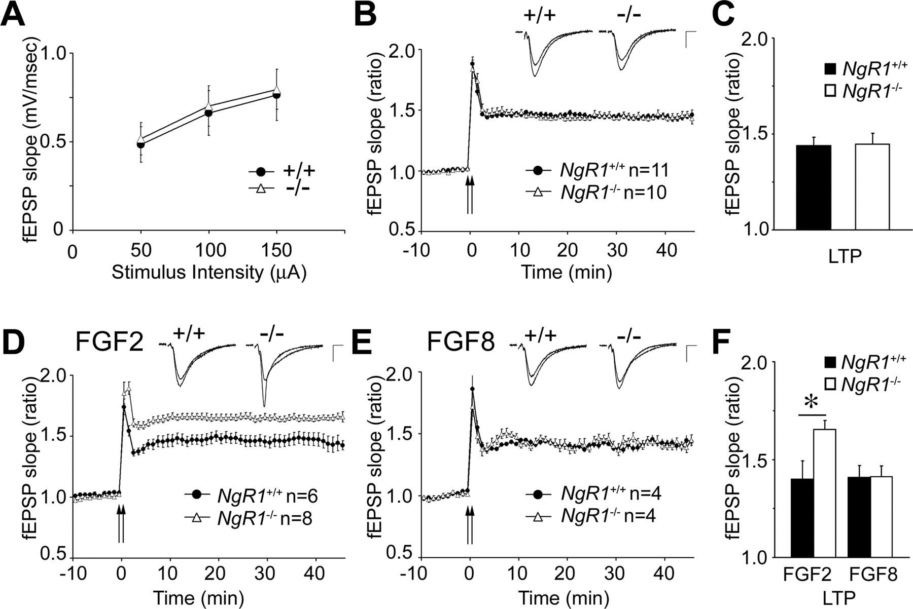
- Figure 6.
FGF2 enhances hippocampal LTP in NgR1 mutants. Recording of fEPSPs at Schaffer collateral–CA1 synapses in acute hippocampal slices from NgR1 wild-type (+/+) and mutant (−/−) mice. A , Input–output curves for basal synaptic transmission revealed no differences between NgR1 +/+ and NgR1 −/− slices. B , Summary of LTP experiments in NgR1 +/+ and NgR1 −/− slices. fEPSPs were recorded at CA1 synapses and slopes were plotted against time before and after tetanic stimulation (2 trains of stimuli at 100 Hz for 1 s, separated by a 10 s interval; 144 ± 4.5 vs 145 ± 5.8%). C , Quantification of LTP at 40–45 min in NgR1 +/+ and NgR1 −/− slices revealed no difference in fEPSP slope ratio (p = 0.978). D , In the presence of FGF2 locally applied via the recording electrode, NgR1 −/− slices show significantly enhanced LTP compared with NgR1 +/+ (165 ± 4.6 vs 140 ± 9.4%; p < 0.05). E , Local application of FGF8, an FGF family member that does not bind to NgR1, does not result in enhanced LTP in NgR1 −/− slices (141 ± 5.7 vs 141 ± 5.9%). F , Quantification of LTP at 40–45 min in the presence of FGF2 and FGF8 in NgR1 +/+ and NgR1 −/− slices. Representative traces before and after LTP are shown as insets. Calibration: 0.5 mV, 5 ms. All values are mean ± SEM. *p < 0.05.
- Figure 7.
Loss of NgR1 attenuates hippocampal LTD. A , Summary of LTD experiments in NgR1 +/+ and NgR1 −/− hippocampal slices after LFS (900 pulses; 1 Hz). Evoked fEPSP slope ratios are shown as a function of time. Representative traces before and after LTD are shown as insets. Calibration: 0.5 mV, 5 ms. B , Quantification of LTD at 55–60 min after LFS in NgR1 +/+ and NgR1 −/− reveals significantly reduced depression in NgR1 −/− slices compared with NgR1 +/+ slices (96.9 ± 4.6 vs 84.9 ± 2.6%; *p < 0.05). Error bars indicate SEM. C , D , PPF, the increase in the second fEPSP slope over the first, was calculated in NgR1 +/+ ( C ) and NgR1 −/− ( D ) slices in the presence (+) or absence (−) of locally applied FGF2. Mean values were plotted against different interpulse intervals (25–500 ms).
- Figure 8.
FGFR kinase activity is necessary for FGF2-enhanced LTP in NgR1 mutants. A–C , LTP is not significantly altered in NgR1 +/+ ( A ) and NgR1 −/− ( B ) slices in the presence of SU5402. Induction of LTP in the presence of the FGFR kinase inhibitor, SU5402, locally applied via the recording electrode (1 mm; concentration in recording electrode), in NgR1 +/+ (−SU5402, n = 11 slices/9 animals; +SU5402, n = 6 slices/4 animals) and NgR1 −/− slices (−SU5402, n = 8 slices/8 animals; +SU5402, n = 6 slices/3 animals). The small insets show traces. Calibration: 0.5 mV, 5 ms. C , Quantification of LTP at 45 min shown in A and B . D , The FGF2-elicited enhancement of LTP in NgR1 −/− hippocampal slices (165.4 ± 4.6%) is not observed in the presence of the FGFR kinase inhibitor SU5402 (137.0 ± 3.7%). fEPSPs were recorded as described for Figure 6. Representative traces before and after LTP are shown as insets. Calibration: 0.5 mV, 5 ms. E , Quantification of LTP at 40–45 min in NgR1 −/− slices after local application of FGF2 in the presence of SU5402 revealed a significant reduction of the fEPSP slope ratio compared with application of FGF2 alone (*p < 0.05). Error bars indicate SEM. F , Dose-dependent inhibition of FGF2- but not EGF-elicited ERK1/2 activation in PC12 cells treated with SU5402. Cell lysates were analyzed by anti-phospho-ERK1/2 blotting and normalized to actin.


Additional Files
Supplemental Data
Files in this Data Supplement:
- supplemental material - Supplemental Legend
- supplemental material - Supplemental Figure 1
- supplemental material - Supplemental Figure 2
- supplemental material - Supplemental Figure 3


