

Article Figures & Data
Figures
- Figure 1.
I–V properties reveal two different types of Xenopus spinal neurons in culture. A, Voltage-gated potassium currents recorded from type I neurons increase in amplitude with increasing depolarization. B, In contrast, for type II neurons, current amplitudes decrease in amplitude for depolarizations to membrane potentials positive to +60 mV. For A and B, currents elicited for steps to +60, 90, and 120 mV are designated. C, IKv current–voltage relationships for type I (circles; n = 23) and type II (squares; n = 74) neurons.
- Figure 2.
Neither Mg2+ block nor sodium-dependent potassium current accounts for IKv of type II neurons. A, Increases in elevated extracellular potassium ion concentration do not change the shape of the I–V relationship of IKv recorded from type II neurons. n values ranged between 9 and 13 for each condition. B, Substitution of permeant sodium ion with impermeant NMG (gray trace) does not reduce the amplitude of IKv recorded from type II neurons (n = 10). Black traces (control) present currents recorded in standard bath solution before sodium substitution with NMG. The exemplars show currents elicited by step depolarizations from a holding potential of −80 mV to +20 (left), +60, (middle) or +100 mV (right).
- Figure 3.
Sensitivity of IKv to XE991 distinguishes between type I and type II neurons IKv. A, The IKv sensitivities to XE991 of type I (left) versus type II (right) neurons differ; the XE991 (25 μm; XE)-sensitive current (dark gray) was obtained by subtracting the XE-insensitive current (light gray) from current recorded before drug application (control; black). B, Comparison of IKv sensitivity in type I (white bars) and type II (black bars) neurons to 100 μm linopirdine (Lino) and 10 and 25 μm XE. n values ranged between 6 and 12 for each condition. The asterisk indicates significance at p < 0.01 for type I versus type II (25 μm XE991).
- Figure 4.
Conductance–voltage relationships for IKv of type I and type II neurons differ. G–V relationships for IKv of type I (open circles; n = 23) and type II (open squares; n = 74) neurons were determined either from tail currents (A) or by dividing current amplitudes by driving force (B).
- Figure 5.
Firing properties of type I and type II neurons differ at 24 h under control conditions. A, B, Type I (A) and type II (B) neurons fired differently in response to brief (2.5 ms) current injection. Additionally, only type II neurons generated depolarizations after an action potential (B, arrow); the depolarizations were blocked by the bath application of APV, CNQX, and PTX, indicating that they were caused by synaptic interactions. C, D, Type I (C) and type II (D) neurons also responded differently to sustained (150 ms) current injection. E, The frequency of action potential firing as a function of current intensity indicates that type II neurons fired more on average that did type I neurons. Unpaired t tests indicated, however, that the difference was not significant (p < 0.12). n values ranged between 11 and 12. Symbols are identified in C and D.
- Figure 6.
Acute exposure to elevated K+ selectively alters IKv density of type II neurons. A, B, Comparison of IKv densities of type I (A) and type II (B) neurons and their respective control recordings at 24 and 48 h grown chronically in either control or high-K+ media. No significant difference was found. Symbols are indicated in the figure. n values ranged between 5 and 18. C, Current densities of IKv of type I neurons grown in either acute I (n = 8) or acute II (n = 3) conditions. D, Current densities of IKv of type II neurons grown in either acute I (n = 30) or acute II (n = 18) conditions differ. The acute I, but not other culture conditions, produced a significant increase in current density (p < 0.01, 24 h control vs acute I) for type II but not type I neurons (p ≤ 0.03). E, The acute I, but not other culture conditions, produced a significant increase in Gmax. n values ranged between 3 and 74. For comparisons of Gmax between type I cells, one-way ANOVA determined a p value of 0.6167, and no post hoc tests were performed. In contrast, for comparisons between type II cells, one-way ANOVA determined a p value of 0.0015. Bonferroni-corrected p values (see Materials and Methods) for specific pairwise comparisons <0.05 are indicated by asterisks: *p < 0.05, 48 h control versus acute I type II.
- Figure 7.
Firing properties of type I and II cells at 48 h grown under control and the acute I condition. A, B, Under control conditions, sustained current injection in type I (A) and type II (B) neurons elicited firing properties that were similar to those at 24 h (compare Fig. 5). C, D, The acute I condition led to changes in firing properties of both type I (C) and type II (D) neurons. In contrast to control, acute I type I neurons fired repetitively (C). Furthermore, type II neurons in the acute I condition occasionally fired single action potentials in response to sustained current injection (D). E, The frequency of action potential firing for different sustained levels of current injection (0.1–1.3 nA) differed significantly between type I and type II neurons grown under control conditions (*p < 0.0001). Symbols are identified in A–D. The acute I condition, however, reduced excitability of type II neurons, so that it no longer was statistically different from that of type I neurons grown under the same (acute I) condition. Furthermore, control and acute I type II neurons displayed significantly different firing rates (**p < 0.05). n values ranged between 7 and 10. One-way ANOVA analysis followed by post hoc Bonferroni multiple-comparison tests were used to evaluate statistical significance. For a current stimulus intensity of 0.7 nA, a p value of 0.0001 was determined; for other stimuli intensities (0.1–1.3 nA), p ranged between 0.0001 and 0.151. Post hoc tests were then used to determine p values for specific comparisons. In addition to the values mentioned for a stimulus intensity of 0.7 nA, control type I and II neurons displayed significantly different firing rates for all stimuli intensities (0.1–1.3 nA; p ranged between 0.001 and 0.05). Control type II neurons also fired at significantly different rates than did acute I type II neurons at a stimulus intensity of 0.3 nA.
- Figure 8.
The acute I condition affects other current properties. A, B, The acute I condition did not have significant effects on inward current properties, as assessed by measuring either the peak inward current density (A) or the maximum rate of rise of the action potential (B). C, The acute I condition altered the pharmacological sensitivity of IKv of type I neurons. Unlike control 24 or 48 h type I neurons (compare Fig. 4), IKv of type I neurons grown under the acute I condition was sensitive to 25 μm XE991. D, The sensitivities of IKv type I and II neurons to 25 μm XE991 were similar for the acute I but not control conditions. One-way ANOVA determined a p value of <0.0003. Bonferroni-corrected p values (see Materials and Methods) for specific pairwise comparisons <0.05 are indicated: *p < 0.001, 48 h control type I versus 48 h control type II; **p < 0.05, 48 h control type I versus acute I type I.
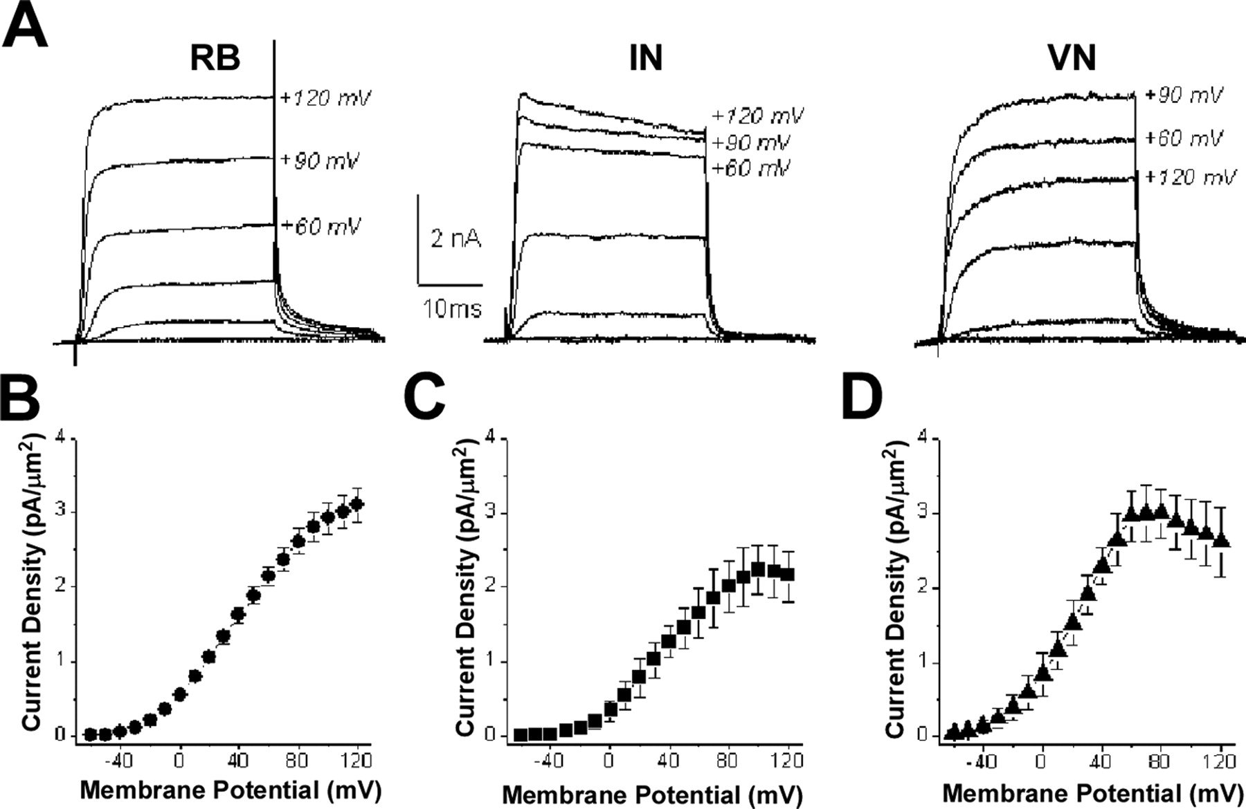
- Figure 9.
IKv recorded from RB cells, INs, and VNs in situ resemble those recorded from type I and type II neurons in culture. A, IKv values recorded from RB (left), IN (center), and VN (right) neurons in situ display different properties. B–D, Current–voltage curves for IKv values from RB cells (B; n = 6), INs (C; n = 7), and VNs (D; n = 5). On the basis of IKv properties, RB cells are type I neurons, whereas VNs are type II. INs contain a mixture of type I and II neurons, with ∼30% (2/7) being type I and the majority (5/7) type II.
- Figure 10.
RB cells, INs and VNs neurons have different firing properties. A, In response to brief stimulation (2.5 ms), RB cells (left), INs (middle), and VNs (right) fire single action potentials, as do type I and II neurons in culture (Fig. 5). Furthermore, after an action potential, VNs show a delayed depolarization (arrow), as do type II neurons in culture (Fig. 5). It is not known whether the delayed depolarization recorded in vivo from VNs was caused by synaptic input, as was the case for type II neurons in vitro (Fig. 5). B, In response to sustained current injections (150 ms), RB cells, INs, and VNs display different firing patterns. RB cells (left) fire a single action potential, as do type I neurons in culture (Fig. 5). In contrast, INs (middle) and VNs (right) fire repetitively, as do type II neurons in culture (Fig. 5). C, The frequency of action potential firing for different sustained levels of current injection differed for RB cells (circles; n = 4), INs (squares; n = 4), and VNs (triangles; n = 3). For current intensities >0.1 nA, RB cells and VNs had significantly different firing rates (*p < 0. 01; except for 1.0 nA, **p < 0.05). At 1.3 nA, VNs and INs also had significantly different firing rates (***p < 0.05).


Tables
Cell type Cap (pF) IR (MΩ) RMP (mV) 24 h Control Type I (10) 19 ± 5 360 ± 200 −56.8 ± 11.1 Type II (12) 16 ± 3 570 ± 310 −53.0 ± 10.5 48 h Control Type I (8) 20 ± 6 270 ± 160 −64.8 ± 8.6 Type II (8) 16 ± 3 580 ± 350 −51.6 ± 5.9 Acute I Type I (7) 19 ± 8 310 ± 210 −50.0 ± 13.6 Type II (10) 20 ± 6 410 ± 360 −48.8 ± 11.3 -
Values are means ± SD. n values are given in parentheses. One-way ANOVA determined that variations among means of capacitance, input resistances, and resting membrane potential values for type I or II neurons were not statistically significant. Cap, Cell capacitance; IR, input resistance; RMP, resting membrane potential.
-
Cell type Cap (pF) IR (MΩ) RMP (mV) RB (6) 19.1 ± 1.5 373 ± 85 −71 ± 5 IN (7) 13.9 ± 3.9 436 ± 138 −56 ± 18 VN (5) 16.9 ± 3.1 302 ± 73 −65 ± 12 -
Values are means ± SD. n values are given in parentheses. One-way ANOVA determined that variations among means of capacitance, input resistances, and resting membrane potential values for RB, IN, or VN neurons were not statistically significant. Cap, Cell capacitance; IR, input resistance; RMP, resting membrane potential.
-


