

Article Figures & Data
Figures
- Figure 1.
Structure and innervation of the lateral-line organ of a larval zebrafish. A , A schematic diagram of a zebrafish larva at 4 dpf depicts seven anteroposterior neuromasts (blue) and two dorsoventral neuromasts (green) of the PLL. Additional neuromasts, which are not shown, adorn the animal's head. The soma of a single afferent neuron (red) lies in the PLL ganglion immediately caudal to the developing ear. In this example, its peripheral axon runs in the PLL nerve and contacts hair cells in two neuromasts. The central axon bifurcates and synapses in the nascent octavolateralis nucleus along the length of the hindbrain. The diameters of the neuromasts, neuronal soma, and axons are exaggerated. B , Four hair cells occur at the center of a schematic depiction of a section through a single anteroposterior neuromast. Displacement of the gelatinous cupula by a hydraulic stimulus, in this instance directed toward the animal's posterior (red arrow), deflects the long kinocilia of the hair cells. When communicated to the stereocilia of the hair bundles, this movement depolarizes the posteriorly polarized hair cells (red) and hyperpolarizes the anteriorly polarized cells (blue). Supporting cells separate the hair cells; mantle cells outline the neuromast and contact the periderm cells of the larva's integument. The neuromast's innervation is not shown. The parallel dashed lines depict the plane of the parasagittal optical section shown in the following panel. C , A light micrograph of a neuromast's apical surface reveals the staining of filamentous actin by fluorescent phalloidin. The 20–30 stereocilia in each hair bundle form a crescent in whose concavity stands the unlabeled kinocilium. The dashed lines delineate the horizontal plane of section depicted in the preceding panel. A mitosis at the boundary between the mantle cells and supporting cells probably represents the division of an amplifying progenitor to form a mirror-symmetrical pair of hair cells. The two hair cells produced by an earlier mitosis remain immature: their hair bundles have yet to exhibit the polarization characteristic of mature hair cells. In this and all subsequent light micrographs, the animal's anterior is located to the left, and its dorsum is oriented upward. D , Labeling of hair cells in a living 6 dpf larva with 4-Di-2-ASP reveals 11 neuromasts in the PLL on the animal's left side. The neuromasts on the right side of the transparent larva appear out of focus. E , Efferent synaptic endings occur in a PLL neuromast in a living islet1:GFP fish at 3 dpf. F , Dual labeling with FM4-64 (red) demonstrates that one immature hair cell of this neuromast failed to take up the dye but was nevertheless innervated (arrowhead). G , GFP expression in the PLL nerve of a live HuC:GFP larva at 2 dpf documents the afferent innervation of two neighboring neuromasts. H , Labeling of the same specimen with FM4-64 reveals the hair cells (red). I , The expression of HuC:GFP in a single PLL afferent neuron reveals its soma in the PLL ganglion and its bifurcated axon reaching the hindbrain. An ascending fiber from the spinal cord is labeled as well (arrowheads). J , The peripheral projection of this neuron at 1.5 dpf features an actively migrating growth cone. D is a mosaic of several images; E–J are maximal-intensity projections of confocal Z-stacks. Scale bars: C , 5 μm; D , 1 mm; E–J , 20 μm.
- Figure 2.
In vivo imaging of afferent synaptogenesis in a developing neuromast. A , In a maximal-intensity projection of a Z-stack of an anteroposterior neuromast at 2.5 dpf, an mCherry-labeled afferent fiber (red) forms a putative synapse with the rostralmost of the hair cells expressing GFP (green). Two immature hair cells are only dimly labeled with GFP (arrowheads). B , A selected confocal section of the neuromast in A shows the extensive contact between the terminal and one hair cell as well as a substantially smaller contact with a second. C , A maximal-intensity projection of the same neuromast at 3.5 dpf illustrates extensive neuronal contact with the three posteriorly polarized hair cells. D , E , Selected optical sections of the neuromast depicted in C delineate the individual contacts. F , A maximal-intensity projection of the same neuromast at 4.5 dpf demonstrates five putative synapses, of which four occur with posteriorly polarized hair cells. G , H , Large boutons have formed on the three largest posteriorly polarized hair cells. I , A newly formed hair cell has been innervated (arrowhead) just as its hair bundle has begun to polarize posteriorly (see K ). J , One innervated hair cell of this neuromast (arrowhead) is of the opposite polarity with respect to the others (see K ). K , Staining of hair bundles in this neuromast with fluorescent phalloidin reveals the polarities of the hair cells at 4.5 dpf. The stereocilia in each bundle display a crescentic pattern of fluorescence surrounding a dark spot at the site of the kinocilium. A, Anterior; P, posterior; D, dorsal; V, ventral. Scale bars, 5 μm.
- Figure 3.
Statistical analysis of innervation bias and afferent neuronal receptive fields. A , In a plot of the weight of evidence for a biased model (W) against larval age, the ordinate represents the average weight of evidence contributed by a single neuromast at the given time. Summing the results over the ensemble of neuromasts yields a total weight of evidence of 375 db. B , Given that there is strong evidence for orientation selectivity, the parameter ω reflects the degree to which the neuron's choice of hair cells is biased. To illustrate the degree of bias as a function of larval age, the results have been expressed as means of the probability of |ω − 0.5| + 0.5, so that the ordinate reflects increasing bias. The error bars represent SDs. C , A histogram illustrating the fraction of a neuromast's hair cells innervated by the labeled fiber indicates that 84% of the neuromasts studied had 50% or fewer hair cells innervated. D , A plot of the number of neurons with the indicated receptive-field sizes demonstrates the preponderance of fibers innervating one or two neuromasts. E , The mean number of neuromasts innervated by a single afferent is essentially constant over the range of larval ages investigated. The error bars represent SEMs. F , The distribution of neuromasts per neuron demonstrates an excess of posteriorly biased (black) over anteriorly biased (gray) neurons.
- Figure 4.
Afferent connections of dorsoventral neuromasts. A , In a maximal-intensity projection of a confocal Z-stack, the mCherry-expressing afferent fiber turns ventrally from the lateral-line nerve to reach a dorsoventral neuromast. B , The hair cells in the same neuromast are labeled with GFP (green). C , D , This neuromast contains 10 hair cells, of which four receive bulbous synaptic endings. The most rostral is contacted by only a thin neurite (arrowhead). E , Labeling with fluorescent phalloidin indicates that these five hair cells have ventrally polarized hair bundles. Although the dorsally polarized hair cells are embraced by a thin, circular extension of the neuron ( A ), they lack synaptic boutons. Scale bars, 5 μm.
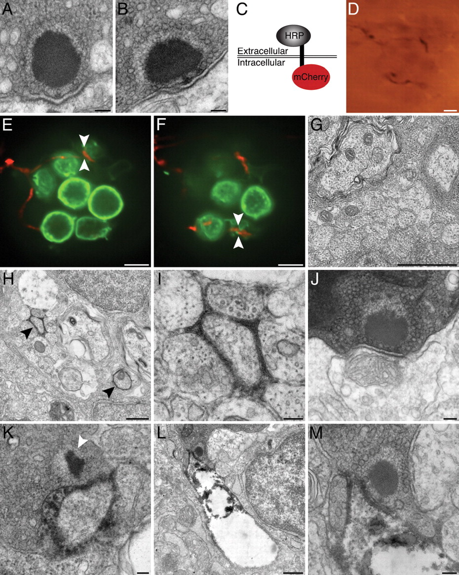
- Figure 5.
Correlative electron microscopy with HRP-mCherry. A , A ribbon synapse in a 2 dpf wild-type larva is indistinguishable from those in older animals. B , The synapse in a 5 dpf wild-type larva exhibits the characteristic features of a ribbon synapse, including a presynaptic dense body or ribbon, a halo of tethered synaptic vesicles, and prominent presynaptic and postsynaptic densities. C , Expression of the HRP-mCherry protein in the neurolemma places the fluorescent mCherry component intracellularly and the HRP moiety extracellularly. D , A bright-field micrograph depicts an afferent terminal expressing HRP-mCherry within a neuromast. The densely labeled fiber, which is also depicted in E , F , and H–M , is visible through the plastic resin in which the specimen has been embedded. E , An optical section through a neuromast of a living Brn3c:gap43-GFP larva features hair cells expressing a membrane-localized form of GFP (green). An afferent fiber labeled with HRP-mCherry (red) innervates three of the hair cells. The region bracketed by arrowheads is examined in greater detail in K . F , In an optical section through the basal region of the same neuromast, arrowheads bracket a site that was later explored under the electron microscope ( L , M ). G , A transverse section of the PLL nerve in a wild-type 5 dpf larva demonstrates several afferent axons. H , In a transverse section through a PLL nerve, two afferent fibers that express HRP-mCherry (arrowheads) produce prominent electron density in the surrounding extracellular space. The weakly labeled fiber in the lower right did not innervate the neuromast depicted in D–F and H–M . I , A higher-magnification view of the labeled neuron at the top left of H illustrates a localized precipitate that does not damage nearby cells. J , An unlabeled afferent neuron lacking electron density synapses with a hair cell of the neuromast. K , A synaptic ribbon (arrowhead) in the region of membrane contact denoted by arrowheads in E verifies that the membrane contact observed by light microscopy represents an afferent synapse. L , This ribbon synapse occurs at the site of membrane apposition bracketed by arrowheads in F . In this instance, the neuron has become distorted and exhibits poor preservation of intracellular organelles. M , Viewed at higher magnification, the ribbon synapse in L illustrates the typical attributes of hair-cell afferent synapses. Scale bars: A , B , I–K , M , 100 nm; D–F , 5 μm; G , H , L , 500 nm.
- Figure 6.
Reinnervation of regenerated hair cells. A , In an optical section through a 3 dpf neuromast before hair-cell elimination, the axis of planar cellular polarity (dashed line) can be inferred from the positions of the constituent hair cells. The afferent fiber has selectively synapsed with posteriorly polarized hair cells. B , In a maximal-intensity projection of the same neuromast 2 h after the application of 10 μm Cu2+, the hair cells have been eliminated, and the neuron has retracted its terminals. Note the presence in the lateral-line nerve of another labeled neuron that does not innervate this neuromast (arrowhead). C , After 6 h, the neuromast contains one weakly fluorescent progenitor that has not yet undergone mitosis to form two new hair cells. D , Twelve hours after Cu2+ treatment, the newly formed posteriorly polarized hair cell receives a small synapse. E , By 24 h, the synapse depicted in D has grown in size and in the extent of membrane contact. F , G , At 36 h after treatment, the neuron appears to contact two or three hair cells, but their polarities cannot be inferred because of the complex organization of the neuromast. It is likely, however, that the synapse depicted in G is identical to that in D and E . H–K , By 48 h, the neuromast has grown to encompass eight mature hair cells with polarized hair bundles (see L ). These four panels are ordered from the bases to the apices of the hair cells. H , A thin neurite reaches an anteriorly polarized hair cell (arrowhead). I , A larger bouton contacts the ventralmost of the posteriorly polarized hair cells (arrowhead). J , A synaptic contact blankets the basal surface of a posteriorly polarized hair cell (arrow), whereas only a tenuous process reaches an anteriorly polarized hair cell (arrowhead). K , The afferent neuron forms voluminous boutons on two posteriorly polarized hair cells. L , Staining with fluorescent phalloidin 48 h after treatment defines the polarities of the 10 hair bundles. Scale bars, 5 μm.
- Figure 7.
Hair-cell polarity in the absence of innervation. A , A maximal-intensity projection of a confocal Z-stack depicts immunolabeling for acetylated α-tubulin in the lateral line of a 5 dpf wild-type larva. The PLL nerve and superficial sensory neurons are labeled, as well as microtubules in the apices of hair cells. B , Immunolabeling of a neurogenin1 mutant sibling for acetylated α-tubulin illustrates the absence of a PLL nerve. Labeling persists in the microtubules of hair cells. C , Staining of a wild-type neuromast with fluorescent phalloidin (red) and immunofluorescent labeling of acetylated α-tubulin (green) reveal the polarities of the hair bundles in this anteroposterior neuromast. D , The hair-bundle polarities of a neurogenin1 mutant neuromast are unperturbed despite the lack of innervation. Scale bars: A , B , 30 μm; C , D , 5 μm.


Additional Files
Supplemental Data
Files in this Data Supplement:
- supplemental material - Supplemental Legend
- supplemental material - Supplemental Table


