

Article Figures & Data
Figures
- Figure 1.
Annexin V labels the hair cell apical surface after neomycin treatment. a–f, Wide-field fluorescence micrographs of basal-coil cochlear cultures. Culture in a–c was incubated in saline containing Alexa Fluor 488 annexin V for 30 min (a), 35 min (b), and 40 min (c). Culture in d–f was incubated in saline containing Alexa Fluor 488 annexin V for 30 min (d). Neomycin was then added to a concentration of 1 mm, and images were taken 5 min (e) and 10 min (f) later. Note the hair bundle labeling at 5 min after neomycin addition (e), and strong staining around the perimeter of the hair cell apex by 10 min (f). I, Inner hair cell row; O1, O2, and O3, outer hair cell rows 1, 2, and 3, respectively. Scale bar: (in f) a–f, 20 μm. The insets in e and f are 2× zooms of selected outer hair cells.
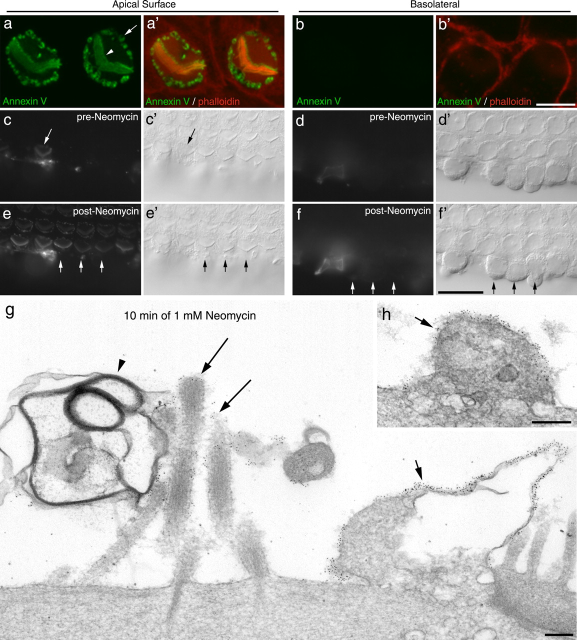
- Figure 2.
Annexin V labeling is restricted to the apical surface of the hair cell. a, b, Confocal images of hair cells from a culture that had been exposed to neomycin in the presence of Alexa Fluor 488 annexin V (green) for 10 min, fixed, and then double-labeled with phalloidin (red). Confocal images were taken at a level just above (a, a′) and just below (b, b′) the tight adherens junction. The arrowhead indicates the hair bundle, and the arrow points to the labeling at the perimeter of the hair cell apex. Scale bar: (in b) a, b, 5 μm. c–f, Wide-field fluorescence (c–f) and Nomarski interference contrast (c′–f′) images focused on the apical (c, e) and basolateral (d, f) surfaces of a cochlear culture in which the basolateral surfaces of the outer row of outer hair cells had been exposed by making an incision through the surface of the epithelium before the addition of Alexa Fluor 488 annexin V. Images were captured before (c, d) and 3 min after (e, f) the addition of 1 mm neomycin. The incision resulted in the labeling of one hair cell (arrow) before the neomycin treatment (c). The addition of 1 mm neomycin resulted in the rapid labeling of the apical surface of all the hair cells (e). Labeling of the exposed basolateral surfaces was not observed after addition of neomycin (f). The short arrows in e and f indicate the apical (e, e′) and basolateral surfaces (f, f′) of three adjacent outer hair cells. Scale bar: (in f) c–f, 20 μm. g, h, Immunogold labeling of annexin V after 10 min neomycin treatment. Gold particles are associated with the surface of peripheral membrane blebs (g, h, short arrows) and with the membranes of stereocilia (g, long arrows). The large membrane-filled bleb associated with the hair bundle (arrowhead) is only sparsely labeled. Scale bars, 200 nm.
- Figure 3.
Time course of annexin V binding in response to perfusion of neomycin. a, Confocal images captured at intervals (in seconds) after the onset of local perfusion with 1 mm neomycin. Labeling is first seen on the hair bundle, within 75 s of neomycin application. Pericuticular membrane staining is first detectable ∼150 s after the onset of neomycin application. Scale bar, 5 μm. b, Diagram of the organ of Corti indicating the region (gray box) that was analyzed. c, Time course of the change in fluorescence intensity (obtained by subtraction of the preapplication fluorescence signal, F0) from regions of interest centered on the hair bundle (blue) and the perimeter of the apical surface of the hair cell (red). The fluorescence signal from the hair bundle reaches a steady level at ∼250 s, whereas the signal in the perimeter region continues to increase. Data are mean ± SEM from six cells. Data are representative of four separate experiments. d, Normalized fluorescence signal measured from the hair bundle (circles) and the apical perimeter (triangles) of hair cells A (green symbols) and B (blue symbols) indicated by the arrows in a.
- Figure 4.
Neomycin-induced changes in whole-cell capacitance and effects of SQ225536, LY294002, brefeldin A, and ouabain. a, Transient capacitative currents (note timescale on x-axis) in response to a 10 mV hyperpolarizing voltage step (applied at t = 0 ms) from a holding potential of −84 mV for a representative outer hair cell in control conditions before (black line) and after (gray line) ∼2 min exposure to 1 mm neomycin. Traces are averaged from 10 repetitions of the voltage step. Whole-cell capacitance was calculated from the charge transfer during the voltage step, obtained by numerically integrating the area within the current transient. The measured whole-cell capacitances for these traces are 6.3 ± 0.2 pF (control) and 13 ± 1 pF (neomycin exposure). b–g, Fluorescence images of basal-coil cochlear cultures that were incubated in the presence of Alexa Fluor 488 annexin V for 30 min in 0.1% DMSO (b), 0.5 mm SQ225536 (c), 10 μm LY294002 (d), 50 μm brefeldin A (e), control solution (f), or 34 μm ouabain (g), before the addition of 1 mm neomycin. Images were captured 1 min before (b–g) and 10 min after (b′–g′) the addition of neomycin. Scale bar: (in g) b–g, 20 μm.
- Figure 5.
Effects of calcium chelation and PH domain expression. a–d, Wide-field images of apical (a, b) and basal (c, d) coil cochlear cultures that were incubated in saline (a, c) or saline containing 5 mm EGTA for 15 min, washed, incubated in saline for 45 min, and then exposed to 1 mm neomycin in the presence of Alexa Fluor 488 annexin V. Images were captured 10 min after the addition of neomycin. Scale bar: (in d) a–d, 20 μm. e, Confocal images of a pair of outer hair cells from a saline-treated (15 min) control culture that was fixed and double labeled with Texas Red-conjugated phalloidin. The cell on the right was transfected with PLCδ1-PH-EGFP. Scale bar, 5 μm. f, Live confocal images showing two PLCδ1-PH-EGFP transfected hair cells in a cochlear culture that had been exposed to 1 mm neomycin and 3 μm FM1-43 in the presence of Alexa Fluor 647 annexin V. The cell on the left (arrow) expresses a high level of PLCδ1PH domain and that on the right (arrowhead) is expressing at a lower level. The cell on the right has responded to neomycin with PS externalization, whereas that on the left has not. Both cells bleb and load with FM1-43. Scale bar, 10 μm. g, Confocal image of a pair of outer hair cells that were treated with 1 mm neomycin (15 min) in the presence of Alexa Fluor 647 annexin V, fixed, and double labeled with Alexa Fluor 350-conjugated phalloidin. The cell on the left expresses PLCδ1-PH-EGFP. PS externalization mainly occurs in areas that are not labeled by PLCδ1-PH-EGFP (arrows), and patches are observed that are not a labeled by either marker (arrowhead). Scale bar, 5 μm. h, Live confocal image from a culture that had been exposed to neomycin in the presence of Alexa Fluor 647 annexin V followed by the application of FM1-43. Note how the FM1-43 often partitions within membrane blebs (arrowheads) that are not labeled by annexin V (arrows). Image was obtained by spectral analysis of the fluorescence emission using the Zeiss Meta detector. Scale bar, 5 μm.
- Figure 6.
Effects of calcium on PS externalization. a, Sequence of images showing changes in Ca2+i in hair cells in response to the application of 1 mm neomycin. From left to right, the panels show the changes in fura-2 ratio (wide-field imaging) at the times indicated during brief exposure (40 s) to 1 mm neomycin. Times after application, and color scaling for ratio changes are indicated. The asterisk indicates a spontaneous Ca2+ event occurring in the greater epithelial ridge (Tritsch et al., 2007). b, Graph showing the change in fura-2 ratio in hair cells and Hensen's cells after application of 1 mm neomycin. Data are the averages from five cells of the recording in a and are representative of at least three different experiments. c, d, Fluorescence (c, d) and Nomarski interference contrast (c′, d′) images of a basal-coil cochlear culture that was incubated in 1 mm neomycin in nominally calcium-free saline (CFS) for 20 min in the presence of Alexa Fluor 488 annexin V (c, c′). After washout of both compounds, Alexa Fluor 488 annexin V was added in normal, calcium-containing saline and the culture was imaged after 10 min (d, d′). Blebs that formed in the absence of calcium, label with annexin V in the presence of calcium. e, f, Fluorescence (e, f) and Nomarski interference contrast (e′, f′) images of a basal-coil cochlear culture incubated in 5 μm ionomycin in the presence of Alexa Fluor 488 annexin V for 5 min. The image in e is focused on the apical surfaces of the outer hair cells (O1, O2, O3, outer hair cells in rows 1, 2, and 3, respectively), and the image in f is focused on the surfaces of the Hensen's cells (H). g–j, Fluorescence images of 2-d-old basal-coil cochlear cultures incubated for 10 min in Alexa Fluor 488 annexin V in saline with either 250 μm ATP (g), an additional 20 mm CaCl2 (h), an additional 20 mm MgCl2 (i), or 1 mm neomycin (j). k–m, Scanning electron micrographs of 2-d-old basal-coil cochlear cultures incubated for 15 min in saline (k), 20 mm CaCl2 (l), or 1 mm neomycin (m). Scale bars: (in j) c–j, 20 μm; (in m) k–m, 5 μm.
- Figure 7.
Ca2+i responses to application of high extracellular Ca2+ or ionomycin. a, Sequence of images showing changes in Ca2+i in hair cells in response to application of 20 mm Ca2+. Left panel, Fura 380 nm signal showing the cellular structure in the cochlear culture. The remaining panels show the changes in fura-2 ratio resulting from 20 mm Ca2+ application. Times after application, and color scaling for ratio changes are indicated. The asterisk indicates a spontaneous Ca2+ event occurring in the greater epithelial ridge (Tritsch et al., 2007). b, Data pooled from four different experiments showing the change in fura-2 ratio in hair cells and Hensen's cells after bath application of 20 mm Ca2+. c, Data showing the fura-2 changes in Hensen's cells and the hair cell region after bath application of 5 μm ionomycin. d, Quantification of the maximum change in Ca2+i after exposure to 20 mm Ca2+ (left) or 10 μm ionomycin (right). Dark bars, Hair cells (left) and for ionomycin, hair cell region (right); gray bars, Hensen's cells for both. e, Confocal imaging of changes in Ca2+i observed using Oregon Green BAPTA-488 before and after treatment with 5 μm ionomycin. Single confocal images at a focal plane above the nucleus of the hair cells are shown with the corresponding orthogonal projection below. Ionomycin causes a strong increase in the Ca2+i in the Hensen's cells (arrowhead) and the Deiters' cells (arrows), whereas the hair cells (asterisk) show little or no response. f, Traces from a confocal imaging series using Oregon Green BAPTA-488. The traces represent the responses of single outer hair cells and single Hensen's cells to local, sequential pipette application of 20 mm Ca2+, 100 μm ATP, and 10 μm ionomycin with intervals of ∼5 min (asterisk) between each application (traces are representative of four separate experiments). Scale bars: a, 50 μm; e, 10 μm. Pooled data shown are mean ± SEM.
- Figure 8.
Neomycin-induced PS externalization is reversible. a, b, Fluorescence (a, b) and Nomarski interference contrast (a′, b′) images of basal-coil cochlear cultures that were incubated in 1 mm neomycin for 15 min, washed in saline, and then incubated for 2 h at either 20°C (a, a′) or 37°C (b, b′) before the addition of Alexa 488 annexin V. Images were captured 5 min after the addition of annexin V. c–g, Orthogonal Z-projections of confocal image stacks of organs of Corti that, after neomycin-induced Alexa Fluor 488 annexin V labeling (green), had been allowed to recover for 0 min (c), 30 min (d), 60 min (e), or 120 min at 37°C (f), and 120 min at 20°C (g) before fixation and labeling with phalloidin (red). At 30 min, label is seen within the hair cells, and by 120 min labeling is no longer seen on the hair cell surface. Additional staining with DAPI (f) reveals that labeling with annexin V is located between the nucleus and the apical membrane. Internalization is not seen after 120 min recovery at 20°C (g). c–g show phalloidin labeling; c′–g′ show Alexa Fluor 488 annexin V labeling; c″–g″ are the merges. I, Inner hair cell; O1, O2, O3, outer hair cells in rows 1, 2, and 3, respectively. Scale bars: (in b, g) a–g, 20 μm.
- Figure 9.
Scanning electron microscopic and transmission electron microscopic analysis of recovery from neomycin-induced damage. Scanning (a–f) and transmission (g–j) electron micrographs of cochlear cultures that were incubated in saline for 5 min (a, d) or 1 mm neomycin for 5 min (b, e, g, i) or 30 min (c, f, h, j), and then either fixed immediately (a–c, g, h) or allowed to recover at 37°C for 2 h after neomycin washout (d–f, i, j) before fixation. Neomycin-induced blebs seen after 5 min (b, g) and 30 min (c, h) of neomycin treatment (b, c, arrowheads) are mostly no longer visible 2 h after neomycin washout (e, i, f, j). Scale bars: (in f) a–f, 5 μm; g–j, 200 nm.
- Figure 10.
Recovery from neomycin-induced damage in Myo6 mutants. a, b, Fluorescence (a, b) and Nomarski interference contrast (a′, b′) micrographs of basal-coil cochlear cultures from homozygous Sv/Sv mice that were incubated in 1 mm neomycin for 15 min, washed in saline, and then incubated in Alexa Fluor 488 annexin V immediately (a, a′) or after a 2 h recovery period at 37°C (b, b′). Images were captured 10 min after the addition of annexin V. Scale bar: (in b) a, b, 20 μm. c–f, Scanning electron micrographs of basal-coil cochlear cultures from homozygous Sv/Sv mice that were incubated in saline (c, e) or 1 mm neomycin (d, f) for 15 min, and then fixed immediately (c, d) or allowed to recover for 2 h at 37°C before fixation (e, f). Scale bar: (in f) c–f, 5 μm. g, h, Confocal Z-projections of hair cells in basal-coil cochlear cultures from heterozygous +/Sv (g) and homozygous Sv/Sv (h) mice that had been exposed to 1 mm neomycin in the presence of Alexa Fluor 488 annexin V for 15 min, followed by washout and recovery at 37°C for 2 h. The arrows indicate inner hair cells, and the arrowheads indicate outer hair cells. Scale bar: (in h) g, h, 10 μm.


Additional Files
Supplemental Data
Files in this Data Supplement:
- supplemental material - Supplemental Legend
- supplemental material - Supplemental Figure 1
- supplemental material - Supplemental Figure 2
- supplemental material - Supplemental Figure 3
- supplemental material - Supplemental Movie 1
- supplemental material - Supplemental Movie 2


