

Article Figures & Data
Figures
- Figure 1.
Glutamate-transporter mediated inward currents in cultured pinealocytes. A, Top, Family of currents activated by rapid application of 1 mm l-aspartate (left) or 1 mm l-glutamate (right) at membrane potentials between −80 and +40 mV in 20 mV steps. Solutions were applied to the lifted cell with exchange within a few milliseconds. Each trace is the mean of four records. All recordings were from the same cell with CsCl-based internal solution. Bottom, Current–voltage (I–V) relationships of peak currents evoked by l-aspartate (□) or l-glutamate (■) fitted with third-order polynomials. B, Average current density at V = −80 mV with several agonists of glutamate transporters and iGluRs applied at 1 mm, except AMPA and KA at 0.5 mm. Measured peak currents were divided by the capacitance of each cell. For l-, d-aspartate and l-glutamate, only cells showing current were included in the analysis. The NMDA application included 10 μm glycine, and external Mg2+ ions were omitted. C–E, Concentration dependence of currents activated by three excitatory amino acids normalized to the value with 10 mm in each cell. The averages were fitted with the Hill equation (see Materials and Methods). Half-maximal effective concentrations (Hill coefficient) are 89 μm (1.1), 73 μm (1.2), 306 μm (0.7) for l-aspartate, l-glutamate, and d-aspartate, respectively. Shown is the mean of three to four cells at −80 mV with CsCl-based internal solution. Error bars indicate SEM.
- Figure 2.
Steady inward currents require intracellular K+ ions. Currents were activated by 1 mm l-aspartate with CsCl (A)- or KCl (C)-based internal solutions at V = −80 mV (different cells; means of 2–3 records). The dashed lines represent the basal current level. B, D, I–V relationships of peak (■) and S-S currents (□; mean between 350 and 650 ms) in the same cells as A and C. The lines are fitted third-order polynomials. E, Currents were activated by 0.01 and 1 mm l-aspartate with KCl-based internal solution at V = −80 mV in the same cell. Traces are means of two to three records. The arrow indicates the peak current. F, Average current density evoked by 0.01 mm l-aspartate (n = 3) and 1 mm l-aspartate (n = 9). Error bars indicate SEM.
- Figure 3.
Sensitivity of current to intracellular anions and pharmacology. A, l-Aspartate (1 mm) evoked a large current of >50 pA with an SCN−-rich intracellular solution. The current was reduced by 300 μm DHK or TBOA present continuously. Traces from one cell at V = −80 mV. Full recovery of the current from DHK and TBOA was tested (data not shown). B, Average current density evoked by 1 mm l-aspartate with KCl (from Fig. 2F)- or KSCN (n = 11)-containing internal solutions at V = −80 mV. S-S current was averaged between 35 and 65 ms. C, Block of 1 mm l-aspartate-evoked currents by 300 μm DHK (n = 3) and TBOA (n = 3). Peak and S-S currents with KSCN-based internal solution were analyzed as in B and compared with those of control. V = −80 mV. Error bars indicate SEM.
- Figure 4.
Depolarization of the membrane potential by glutamate transporter-mediated currents. A, B, Membrane depolarizations evoked by 1 mm l-aspartate and 50 mm KCl (current-clamp mode). C, D, Membrane currents evoked by l-aspartate and KCl (voltage-clamp mode at V = −60 mV). All recordings were from the same cell in the perforated whole-cell configuration.
- Figure 5.
[Ca2+]i rise induced by glutamate transporters. Ratiometric calcium traces as agonists at 1 mm (except AMPA and kainate, 0.5 mm) were applied for 30 s using a local perfusion system (exchange within 0.5 s). A, B, Three transporter substrates evoked Ca2+ responses. d-Glutamate and two specific agonists of iGluR did not. C, D, TBOA (300 μm) and nimodipine (5 μm) reduced the [Ca2+]i rise induced by 1 mm l-aspartate. DHK at the same concentration were less effective (shown in F). E, Summary of experiments like A and B. F, Summary of experiments like C and D, normalized to l-aspartate alone; DHK (300 μm; n = 21), TBOA (n = 30), nimodipine (n = 14), 50 μm d-AP5 (n = 10), or 10 μm CNQX (n = 13). G, Dose–response relationship for l-aspartate evoked Ca2+ responses (n = 38–54 for each concentration). EC50 and Hill coefficient are 12 μm and 1.5. Error bars indicate SEM.
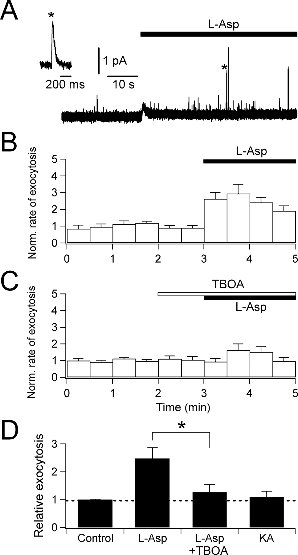
- Figure 6.
l-Aspartate-evoked exocytosis monitored by carbon fiber amperometry. A, An amperometric trace demonstrating increase in exocytotic events after 1 mm l-aspartate application (marked by a transient deflection of base line). Inset, One spike denoted by an asterisk is expanded to show a typical quantal release. B, Rate of exocytosis in 30 s bins normalized to the control (average of 11 cells). C, Inhibition by 300 μm TBOA (average of 19 cells). D, Summary of exocytosis evoked by 1 mm l-aspartate alone (n = 11), 1 mm l-aspartate in the presence of 300 μm TBOA (n = 19; *p < 0.01 compared with l-aspartate alone), and 1 mm KA (n = 3). Error bars indicate SEM.
- Figure 7.
l-Aspartate-evoked l-glutamate secretion monitored by HPLC. l-Glutamate release evoked by 100 μm l-aspartate in the absence and presence of different blockers: blockers of glutamate transporters (500 μm DHK and 100 μm TBOA), an L-type Ca2+ channel blocker (20 μm nimodipine), removal of extracellular Ca2+ (−Ca2+), a vesicular H+-pump inhibitor (1 μm bafilomycin A1), iGluR inhibitors (10 μm MK-801 and 50 μm CNQX), or an exocytosis toxin (10 nm BoNT/E, preincubated for 24 h). Shown are means of three to four independent experiments (*p < 0.01, **p < 0.001, compared with l-aspartate alone). Error bars indicate SEM.
- Figure 8.
Glutamate transporter-mediated currents in acutely prepared pineal slices. A, Whole-cell currents evoked by bath application of 1 mm l-aspartate. An intracellular solution containing KSCN was used to enlarge transporter currents. Holding membrane potential was −60 mV. Right, At the end of each experiment, the presence of transient outward K+ currents was confirmed by voltage steps from −60 to +60 mV in 20 mV intervals in the presence of internal and external TEA. B, Whole-cell currents evoked by 50 mm KCl (−60 mV). C, Whole-cell currents activated by 135 mm KCl (−60 mV).
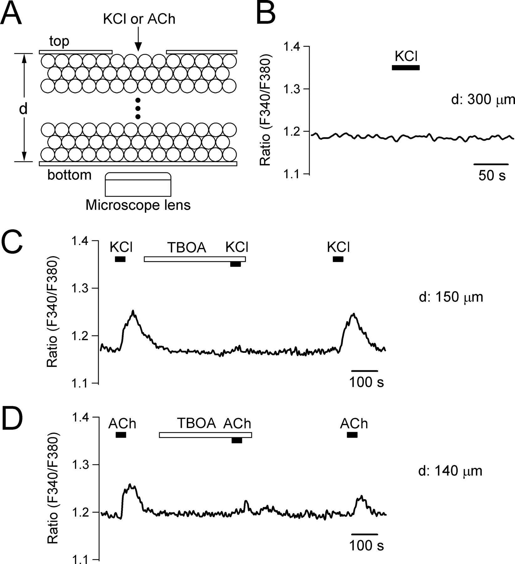
- Figure 9.
Propagation of [Ca2+]i signals via glutamate transporters in pineal slices. A, Schematic drawing of experimental setup. A solution containing 50 mm KCl was applied to cells on the top side of a pineal slice. [Ca2+]i response was monitored on the cells at bottom of the slice. See supplemental Figure 5 (available at www.jneurosci.org as supplemental material) for the detailed design of the chamber. “d” denotes the thickness of pineal slices. B, Ca2+ signal measured in the bottom cells of a thick slice (300 μm) on application of 50 mm KCl to the upper surface of the slice. C, The KCl stimulation evoked Ca2+ response at bottom of a thinner slice (150 μm). Preincubation of the slice with 600 μm TBOA reversibly abolished the Ca2+ signal. Shown is a representative record from eight similar experiments. D, Same experiment performed with a thin slice (140 μm) and 200 μm ACh. Shown is a single record from 11 similar experiments.
- Figure 10.
Effect of glutamate transporters on NE-evoked melatonin secretion. Pineal glands were treated with a combination of 10 μm NE, 500 μm l-aspartate, and 300 μm TBOA for 6 h at physiological 37°C, and released melatonin was measured (four independent experiments for each condition). Error bars indicate SEM. *p < 0.01.


Additional Files
Supplemental Data
Files in this Data Supplement:
- supplemental material - Supplemental Legend
- supplemental material - Supplemental Figure 1
- supplemental material - Supplemental Figure 2
- supplemental material - Supplemental Figure 3
- supplemental material - Supplemental Figure 4
- supplemental material - Supplemental Figure 5
- supplemental material - Supplemental Figure 6


