

Article Figures & Data
Figures
- Figure 1.
Spontaneous synchronous calcium oscillations are abolished by hAPP expression. A, B, F, G, Simultaneous traces of [Ca2+]i in three cells of a coverslip, measured with Fura-2 AM expressed as R/Rmean, were R is the ratio of F340/F380 and Rmean is the mean ratio value after 2 min recording. A, At 7 DIV, rat cortical neurons do not present calcium oscillations. B, After 13 DIV, neurons show spontaneous, synchronized calcium oscillations. C, In a neuronal network labeled by an anti-MAP-2 antibody (red), infection by AdAPPGFP allows APP and GFP (green) expression in a subset of neurons. D, Primary cortical neuron infected by AdAPPGFP, coexpressing GFP (green) and hAPP, labeled with hAPP specific monoclonal antibody, WO-2 (red). Scale bar, 20 μm. E, Western blot analysis of cell lysates (top panel) or culture media (bottom panel), from rat cortical neurons infected or not (NI) by Adβgal (βgal), AdAPPGFP (APP), or AdAPPswe (APPswe). Protein loading was controlled using rabbit actin antiserum. F, Neuronal network infected by Adβgal show spontaneous calcium oscillations, whereas in a network infected by AdAPPGFP or AdAPPswe, calcium oscillations are abolished (G, H).
- Figure 2.
hAPP expression increases the peak amplitude of mAHP. A, Representative trace for mAHP in a cultured cortical neuron. An action potential (AP) was elicited by a 2 ms depolarizing current injection, followed by an hyperpolarization phase corresponding to mAHP. The mAHP peak amplitude was measured 15 ms after the AP (arrow) (τ: time constant of decay). B, Resting membrane potential was similar in hAPP- or GFP-expressing neurons, whereas mAHP peak amplitude was higher in hAPP-expressing neurons when compared with GFP-expressing neurons (mean values ± SEM of n cells; *p < 0.05 was considered significant).
- Figure 3.
Long term L-type calcium channels stimulation inhibits calcium oscillations through activation of SK channels. A, Simultaneous traces of [Ca2+]i in three cells of a hAPP- expressing network show that cells recover synchronous calcium oscillations after bath application of apamin (200 nm), an SK channel inhibitor. B–C, A 60 min pretreatment of a βgal- expressing neuronal network by BayK6844 inhibits spontaneous calcium oscillations (B), which can be recovered by bath application of apamin (C).
- Figure 4.
hAPP expression increases L-type calcium channel activity in neurons. A, [Ca2+]i in a population of neurons. Response to Bayk6844 is higher in hAPP-expressing neurons, when compared with βgal-expressing neurons. B, Mean values ± SEM of [Ca2+]i in neurons expressing hAPP (n = 7) or βgal (n = 7); (**p < 0.01). C, D, Top, Representative traces of whole-cell calcium current elicited by a voltage step from −100 mV to + 10 mV, before and after nimodipine (5 μm) application. Bottom, I-V plot of whole-cell calcium currents (average of n cells; APP: n = 6; GFP: n = 5) before and after nimodipine (5 μm) application, where I/Imax is the current normalized to the maximal current of each cell. E, Saturation curve for specific binding of [3H]PN200–110 radioligand to intact cultured cortical neurons. Insert, Specific binding of [3H]PN200–110 is similar in APP (n = 15) or βgal- expressing neurons (n = 6; ns, not significant).
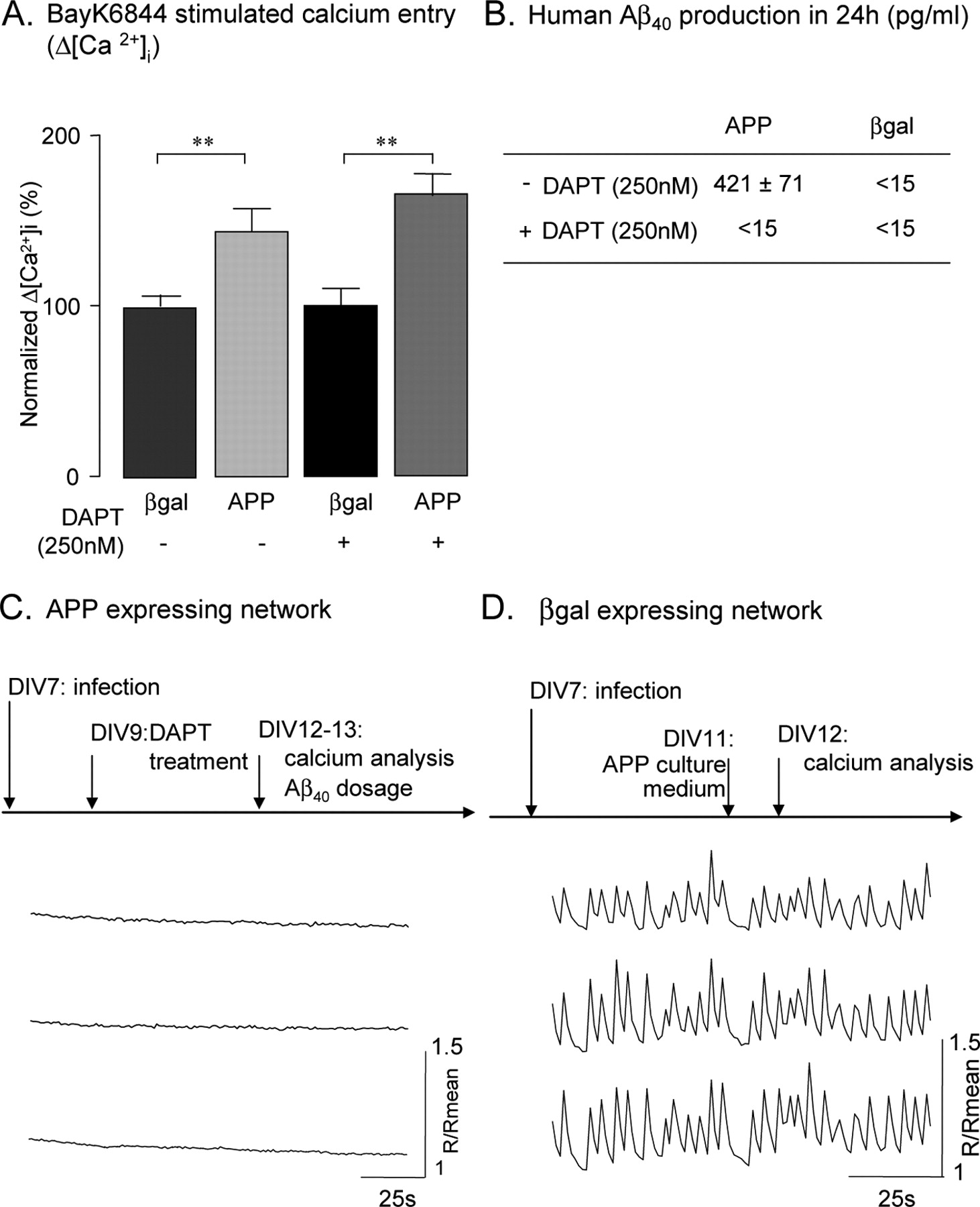
- Figure 5.
Inhibition of calcium oscillations by human APP is independent of γ-cleavage and extracellular APP metabolites. A, BayK6844 stimulated calcium entry was significantly higher in APP-expressing neurons, despite inhibition of γ-cleavage by DAPT treatment (as described in C). Error bars represent mean values ± SEM of normalized Δ[Ca2+]i where 100% is Δ[Ca2+]i in βgal-expressing neurons (βgal: n = 7; APP: n = 7; βgal+DAPT: n = 3; APP+DAPT: n = 3; **p < 0.01). B, Aβ40 production in 24 h (mean ± SEM; n = 4). C, Simultaneous traces of [Ca2+]i in three cells in a hAPP-expressing network show that cells do not recover synchronous calcium oscillations after DAPT treatment for 3–4 d. D, Simultaneous traces of [Ca2+]i in three cells in a βgal-expressing network show that conditioning cells in medium collected from hAPP-expressing network (for 24 h) does not abolish calcium oscillations.
- Figure 6.
Silencing endogenous APP affects spontaneous synchronous calcium oscillations. A, Western blot analysis of cell lysates from rat cortical neurons infected or not (NI) by APPshRNA (APPshRNA) or Neomycin (Neo) lentiviruses. Protein loading was controlled using rabbit actin antiserum. B, In a neuronal network labeled by an anti-MAP-2 antibody (red) and an anti-APPCterminal antibody (APPCter, green), infection by APPshRNA lentivirus (bottom) allows an efficient reduction of endogenous APP expression when compared with uninfected neurons. Scale bar, 20 μm. C, Simultaneous traces of [Ca2+]i in three cells in a Neo-expressing network show that calcium oscillations are not affected by lentiviral infection. D, Simultaneous traces of [Ca2+]i in three cells in a APPshRNA-expressing network show that reduction of endogenous APP expression increases frequency and decreases amplitude of calcium oscillations.


Additional Files
Supplemental Data
Files in this Data Supplement:
- supplemental material - Supplemental Legend
- supplemental material - Supplemental Figure 1
- supplemental material - Supplemental Figure 2


