

Article Figures & Data
Figures
- Figure 1.
Astrocytes express multiple γ-Pcdhs. In situ hybridization (ISH) using a Pcdh-γ C exon riboprobe (diagrammed in A ) revealed expression in cells of the white matter (arrowheads) as well as the gray matter in neonatal spinal cord ( B ). Double-labeling with a GFAP riboprobe indicated that many of these cells were astrocytes ( B , inset shows higher-magnification view). Immunofluorescence confirmed γ-Pcdh protein expression by GFAP-positive astrocytes in neonatal ( C ) and adult ( D ) spinal cords. Immunostaining of cultured astrocytes ( E ) and spinal cord sections ( F ) demonstrated that γ-Pcdh proteins colocalize with ezrin and Glt-1a, both markers of perisynaptic astrocyte processes. Reverse-transcriptase (RT)-PCR analyses detected expression of 20 of the 22 possible Pcdh-γ spliced transcripts in cultured spinal cord astrocytes ( G ). Scale bars: B , 100 μm (inset, 10 μm); C , D , 25 μm; E , 3 μm; F , 4 μm.
- Figure 2.
γ-Pcdh proteins localize to perisynaptic astrocyte processes. A , Schematic diagram of the Pcdh-γ fcon3 conditional mutant allele, in which a GFP-fused C exon 3 is flanked by loxP sites. The A–C subfamilies of V exons are in shades of blue, and the 3 C exons are in red. B , In Cre-ER; Pcdh-γfcon3/+ mice injected with tamoxifen ( B , right), Cre excision resulted in loss of the ubiquitous γ-Pcdh-GFP immunofluorescence observed in uninjected or Cre-ER-negative mice ( B , left). GFP-tagged γ-Pcdhs remained detectable, however, in a small number of GFAP-positive gray matter protoplasmic astrocytes ( B ). C , A higher-magnification image of a separate double-immunostained cryosection is shown. D , E , Confocal analysis of immunostained Cre-ER; Pcdh-γfcon3/+ spinal cord sections demonstrated that astrocytic γ-Pcdh puncta (stained for both GFP and γ-Pcdh; closed arrowheads; yellow in merged images) can localize directly adjacent to neuronal γ-Pcdh at synaptic puncta (stained for both bassoon and γ-Pcdh; open arrowheads, magenta in merged images). F , G , Immunostaining for GFP and presynaptic and postsynaptic markers confirmed that astrocytic γ-Pcdhs were frequently apposed to and/or wrapped around synaptic terminals. Large images are of single planes from z-stacks; perpendicular cross-sections through the entire z-stacks at locations indicated by white lines are shown at right and below. H , Tracings of several such perisynaptic contacts for the indicated boxes in F and G are presented. Scale bars: B , 100 μm; C , 6 μm; D , E , 1 μm; F , G , 2 μm.
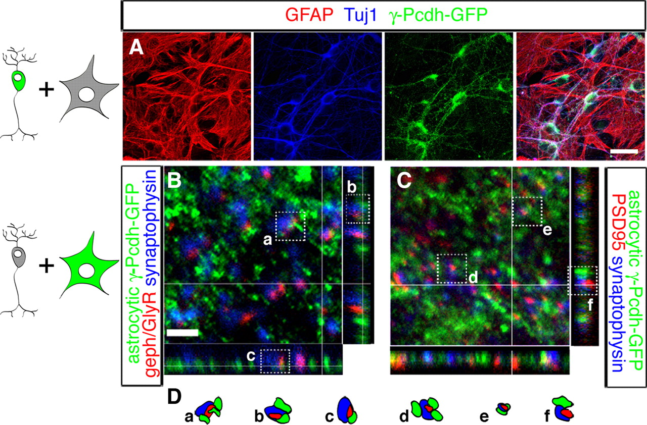
- Figure 3.
A–C , Cocultures of spinal cord interneurons and astrocytes of distinct genotypes. Cocultures of neurons from Pcdh-γ fusg mice with wild type astrocytes were immunostained with antibodies against the indicated proteins ( A ). Only TuJ1-positive neurons expressed GFP-tagged γ-Pcdhs, indicating that the coculture system allowed for effective separability of neuron and astrocyte genotypes with no carryover. Cocultures of wild type neurons with astrocytes from Pcdh-γ fusg mice stained for GFP and inhibitory ( B ) or excitatory ( C ) synaptic markers demonstrated that astrocytic γ-Pcdhs localized to many sites adjacent to synaptic terminals in vitro, as observed in vivo. Large images are of single planes from z-stacks; perpendicular cross-sections through the entire z-stacks at locations indicated by white lines are shown at right and below. D , Tracings of several such perisynaptic contacts for the indicated boxes in B and C are presented. Scale bars: A , 20 μm; B , C , 2 μm.
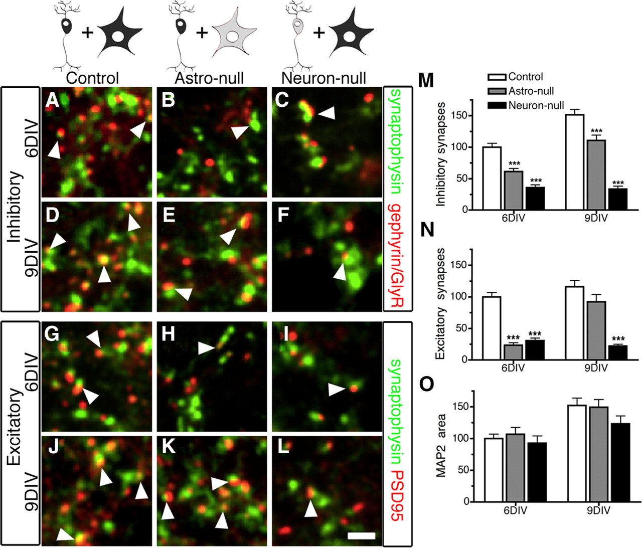
- Figure 4.
Astrocytic γ-Pcdhs are critical for synaptogenesis in developing neuronal cultures. Cocultures of embryonic spinal interneurons growing directly atop a confluent astrocyte monolayer were prepared such that either astrocytes (astro-null), neurons (neuron-null), or neither (control) were Pcdh-γ-null, and synaptogenesis monitored by quantifying the density of appositions of presynaptic and postsynaptic markers (i.e., the number of such synaptic contacts per a defined area) as neurons matured between 6 and 9 d in vitro (DIV). When astrocytes were mutant, the numbers of both excitatory and inhibitory synapses were significantly reduced at 6 DIV ( B , H , M , N ) compared with control cultures ( A , G , M , N ), although some recovery occurred by 9 DIV ( E , K , M , N ). When neurons were mutant, synapse density was drastically reduced at both 6 and 9 DIV ( C , F , I , L–N ). Neuronal differentiation and survival, monitored by quantifying the area of MAP2 immunostaining, did not differ across the three culture conditions ( O ). Data in M-O are graphed as percentage of control values. Graphs in M–O represent means ± SEM from 22 to 44 fields from 6 cultures per time point. *p < 0.05; ***p < 0.001. Scale bar (in L ): A–L , 2 μm.
- Figure 5.
Astrocytic γ-Pcdhs regulate synaptogenesis through a contact-dependent mechanism. Astrocyte-conditioned medium (ACM) was collected from astrocyte cultures derived from WT or Pcdh-γ del/del mice (KO). Western blot analysis of equal amounts of ACM showed no detectible difference between genotypes in the levels of thrombospondin-2 (TSP-2) secreted ( A ). Addition of concentrated WT or KO ACM to control cocultures over 6 DIV resulted in equally significant increases in the density of synapses (i.e., the number of synaptic contacts per a defined area) compared with control media ( B ); thus, WT and KO ACM are equivalent in their synaptogenic potency. Addition of concentrated WT ACM was unable to completely rescue the decrease in synapse density observed in astro-null cultures ( C ). Furthermore, the difference in synapse density between control and astro-null cocultures was maintained when the neurons were plated onto astrocytes that had been fixed with paraformaldehyde ( D ). Data are graphed as percentage of control values. Graphs represent means ± SEM from 12 fields from 3 cultures per time point.
- Figure 6.
Astrocyte-restricted Pcdh-γ mutation in vivo. In GFAP-Cre; Z/EG double transgenics ubiquitous expression of a β-galactosidase/neo fusion protein is replaced by that of GFP following Cre excision. GFAP-positive, but not NeuN-positive, cells express GFP, whereas NeuN-positive, but not GFAP-positive, cells express β-gal ( A , higher-magnification images of gray matter in B , C ), indicating that Cre is not expressed in neurons or their progenitors in the spinal cord. In the gray matter of GFAP-Cre; Pcdh-γfcon3/+ spinal cords, there is reduced colocalization of γ-Pcdh-GFP with Glt1a, confirming that Cre recombinase is able to efficiently excise the floxed allele in astrocytes in these mice ( D , E ). Scale bars: A , 100 μm; B , C , 25 μm; D , E , 10 μm.
- Figure 7.
Astrocytic γ-Pcdhs control synaptogenesis in the embryonic spinal cord in vivo. Quantification of immunostained synapses was performed on 50 μm circular fields imaged in spinal cord sections from GFAP-Cre; Pcdh-γ fcon3/fcon3 astrocyte-restricted mutants and controls at E15, E17 ( A–D ), and P0 (see supplemental Fig. S4, available at www.jneurosci.org as supplemental material). In A′–D′ , the yellow overlap between the red and green channels has been extracted and converted in Adobe Photoshop to black for clarity. E , Synapse density (i.e., the number of such synaptic contacts per 50 μm circle) was significantly reduced in GFAP-Cre; Pcdh-γ fcon3/fcon3 mutants (gray bars) compared with controls (white bars) at embryonic ages, but had recovered by P0, in contrast to Actin-Cre; Pcdh-γ fcon3/fcon3 mutants (black bars). Graphs represent means ± SEM from 18 fields from 3 animals per genotype per time point. *p < 0.05; **p < 0.01; ***p < 0.001.


Additional Files
Supplemental Data
Files in this Data Supplement:
- supplemental material - Supplemental Material


