

Article Figures & Data
Figures
- Figure 1.
Interaction between NR1 and importin α. A, Immunostaining of hippocampal neurons (21 DIV) with anti-NR1 (red) and anti-importin α (green) antibodies reveals partial colocalization in spines (arrowheads) of neurons silenced with TTX (1 μm). Following glutamate stimulation (40 μm, 15 min), the concentration of importin α is decreased in spines and dendrites and increased in the nucleus. Scale bars, 20 μm. B, Quantification of the percentage of NR1-immunopositive spines containing importin α immunoreactivity. *p < 0.05, **p < 0.01, one-way ANOVA F(3,17) = 4.73, p < 0.05. Post hoc least significant difference test was performed to compare all groups (*p < 0.05); n (number of cells) for each category is given below the graph. C, IB of adult rat forebrain homogenate (fb), synaptosome (syn), and PSD fractions (equal protein concentrations) with importin α and NR1 antibodies shows that both proteins are present in synaptosome and PSD fractions. Synaptophysin (p38), Synaptosome marker; PSD-95, PSD marker. D, NR1 and importin α co-IP from adult fb and syn. IPs were performed with monoclonal anti-NR1 antibodies or with polyclonal anti-importin α antibodies, and immunoblots with polyclonal anti-importin α antibodies or with monoclonal anti-NR1 antibodies, respectively. Control immunoprecipitates included monoclonal anti-GFP antibodies (for NR1) or pre-immune rabbit serum (for importin α). Anti-GAPDH antibodies were used as controls for IB. Experiments in C and D were replicated three times.
- Figure 2.
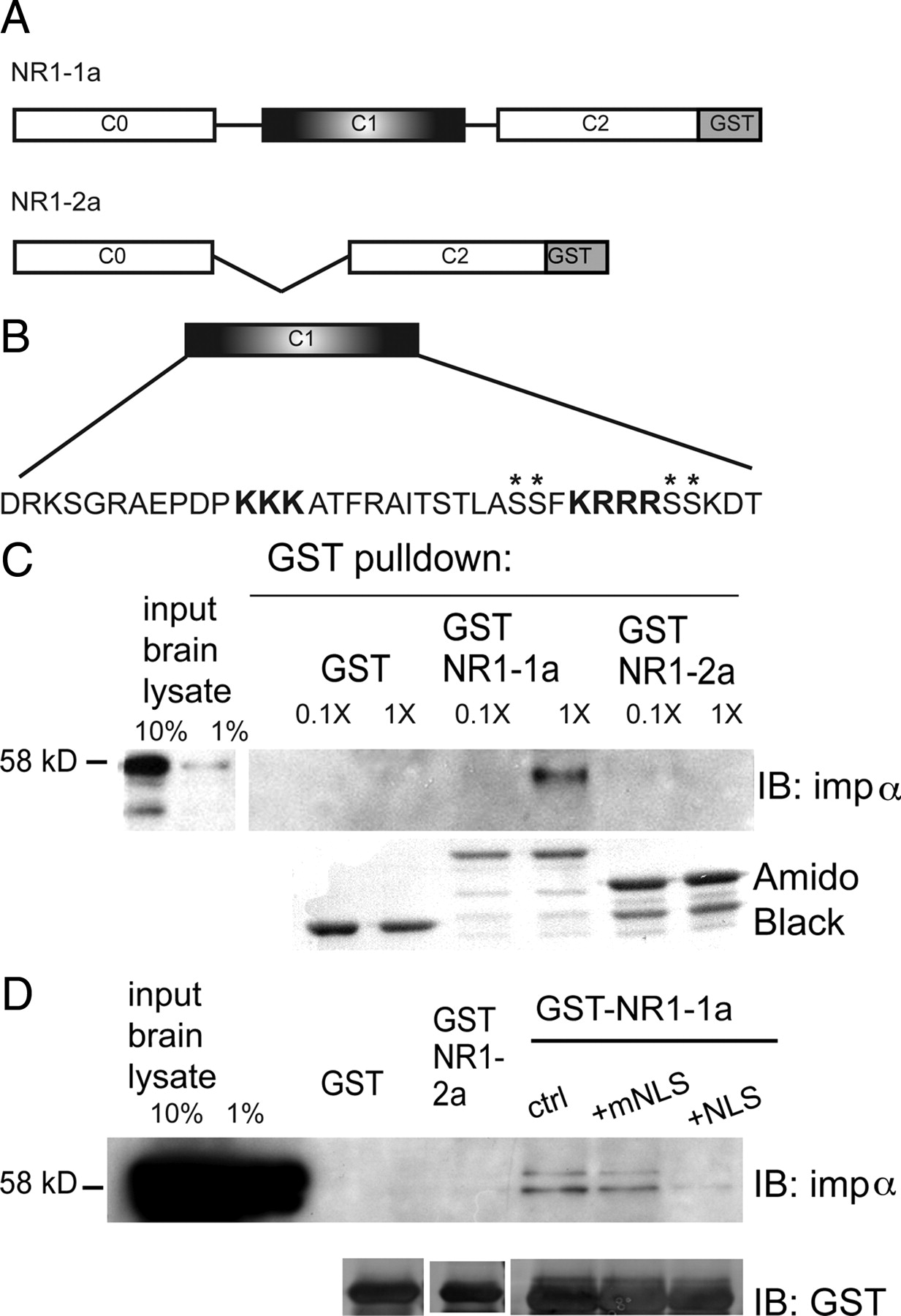
NR1-1a binds importin α via its NLS. A, The cytoplasmic tails of NR1-1a and NR1-2a were fused to GST. B, Amino acid sequence of the C1 cassette, with the NLS in bold; asterisks denote phosphorylation sites. C, Importin α binds GST NR1-1a but not NR1-2a or GST alone in adult rat forebrain lysates. Two input lanes show 10% and 1% of input used for pull-downs. The concentration of input lysate was 0.1× or 1× (2.3 mg), as indicated. Amido black shows amount of GST fusion proteins, which have distinct molecular weights. D, The interaction between importin α and GST-NR1-1a is inhibited by preincubation with 100-fold molar excess NLS but not mutant NLS (mNLS) peptide. Ctrl, No-peptide control. The presence of two bands is due to the fact that the importin α antibody recognizes importin α1 [molecular weight (MW) 61 kDa] as well as importin α2 (MW 58 kDa).
- Figure 3.
NR1-importin α binding is regulated by activity. A, 21 DIV cortical neurons were silenced with TTX (1 μm) for 6 h, incubated with 40 μm glutamate or vehicle for 5 min, lysed, and processed for IP with anti-NR1 antibodies, followed by IB for importin α (or NR1 as control). Importin α binds NR1 in TTX silenced neurons; binding is reduced following glutamate stimulation [TTX alone = 392.47 ± 67.58 arbitrary units (a.u.); glut = 92.07 ± 45.09 a.u., p < 0.002, independent-samples t test; n (n always represents number of independent experiments) = 3]. B, Hippocampal CA1 minislices were stimulated one or two times at 100 Hz (1 × 100, 2 × 100), lysed, and processed for IP with anti-NR1 antibodies followed by IB for importin α. Unstimulated (US) slices from the same chamber served as controls. Importin α is present in equal concentrations in NR1 immunoprecipitates from US slices and slices receiving 1 × 100 Hz stimuli, but is reduced following stimulation with 2 × 100 Hz tetani (US = 1.00 ± 0.07 a.u.; 1 × 100 Hz = 1.08 ± 0.05 a.u.; 2 × 100 Hz = 0.42 ± 0.17 a.u.; 1 × 100 Hz vs US p > 0.05, ns; 2 × 100 Hz vs US p < 0.04, independent-samples t test; n = 4). IB with NR1 antibodies reveals equal NR1 concentrations in stimulated and US slices. Additional controls include IP with GFP antibodies and IB with PSD-93 antibodies. C, The reduction in importin α binding to NR1 is blocked by preincubation with the NMDA receptor antagonist APV (100 μm) (APV alone = 2.14 ± 0.19 a.u.; APV + 2 × 100 Hz = 2.00 ± 0.21 a.u.; p > 0.05, ns, independent-samples t test; n = 4).
- Figure 4.
PKC phosphorylation of NR1 disrupts importin α binding. A, GST-NR1 C-terminal constructs were phosphorylated in vitro with recombinant PKC (−PKC, GST-NR1-1a incubated with ATP but without PKC). Unphosphorylated GST-NR1-1a (ctrl and −PKC) pulls down importin α; this interaction is abrogated by PKC phosphorylation of GST-NR1-1a (+PKC = 0.378 ± 0.131 a.u.; −PKC = 0.940 ± 0.133 a.u.; p < 0.006, independent t test; n = 4). IB with NR1 antibodies shows equal concentration of GST-NR1 constructs in each condition (GST-NR1-2a smaller due to lack of C1 cassette). B, GST-NR1-1a constructs were phosphorylated in vitro with recombinant PKA (−PKA, GST-NR1-1a incubated with ATP but without PKA). Both unphosphorylated GST-NR1-1a (−PKA) and PKA phosphorylated GST-NR1-1a pull-down importin (+PKA = 1.33 ± 0.03 a.u.; −PKA = 1.43 ± 0.04 a.u.; p > 0.05, ns, independent t test; n = 3). IB with NR1 antibodies shows equal concentration of GST-NR1 constructs in each condition.
- Figure 5.
PKC is necessary for importin α dissociation from NR1 with glutamate stimulation. A, Immunostaining of hippocampal neurons (21 DIV) with anti-NR1 (red) and anti-importin α (green) antibodies reveals partial colocalization in spines of neurons silenced with TTX (1 μm), with decreased colocalization after glutamate stimulation (“glut,” 40 μm, 8 min). When preincubated for 30 min with specific inhibitors of PKC (“glut+chel”, 5 μm chelerythrine), glutamate does not decrease the colocalization of importin α and NR1. B, Quantification of the percentage of NR1-immunopositive spines containing importin α immunoreactivity (one-way ANOVA F(7,84) = 5.66, p < 0.001). Post hoc Dunnett's test was performed to compare all groups to glutamate (*p < 0.05; n, number of cells). Scale bars, 20 μm. C, TTX-silenced cortical neurons (21 DIV) were incubated with 20 nm phorbol esters (phor) to activate PKC or vehicle (TTX, 1 μm) for 10 min, lysed, and processed for IP with anti-NR1 antibodies followed by IB with anti-importin α antibodies. More importin α is present in NR1 immunoprecipitates from control cultures (TTX) than in cultures in which PKC is activated by phorbol esters (phorbol esters = 83 ± 4.7% of TTX control; p < 0.05, 1-sample t test; n = 4). NR1 IB reveals equal concentration of NR1 in all conditions.
- Figure 6.
In our model, importin α binds the NLS in the cytoplasmic tail of NR1. Upon stimulation, importin α is released from the NMDA receptor, binds synaptically localized soluble NLS-containing cargoes, and together with importin β1, transports them to the nucleus. Synaptic stimulation activates PKC, which phosphorylates residues flanking the NLS in NR1-1a, thereby disrupting importin α binding. Enlargement of the NR1 subunit shows the amino acid sequence of exon 21/cassette C1 containing the NLS (orange), and residues flanking the NLS that are PKC (blue)/PKA (brown) targets of phosphorylation.


Additional Files
Supplemental Data
Files in this Data Supplement:
- supplemental material - Supplemental Material


