

Article Figures & Data
Figures
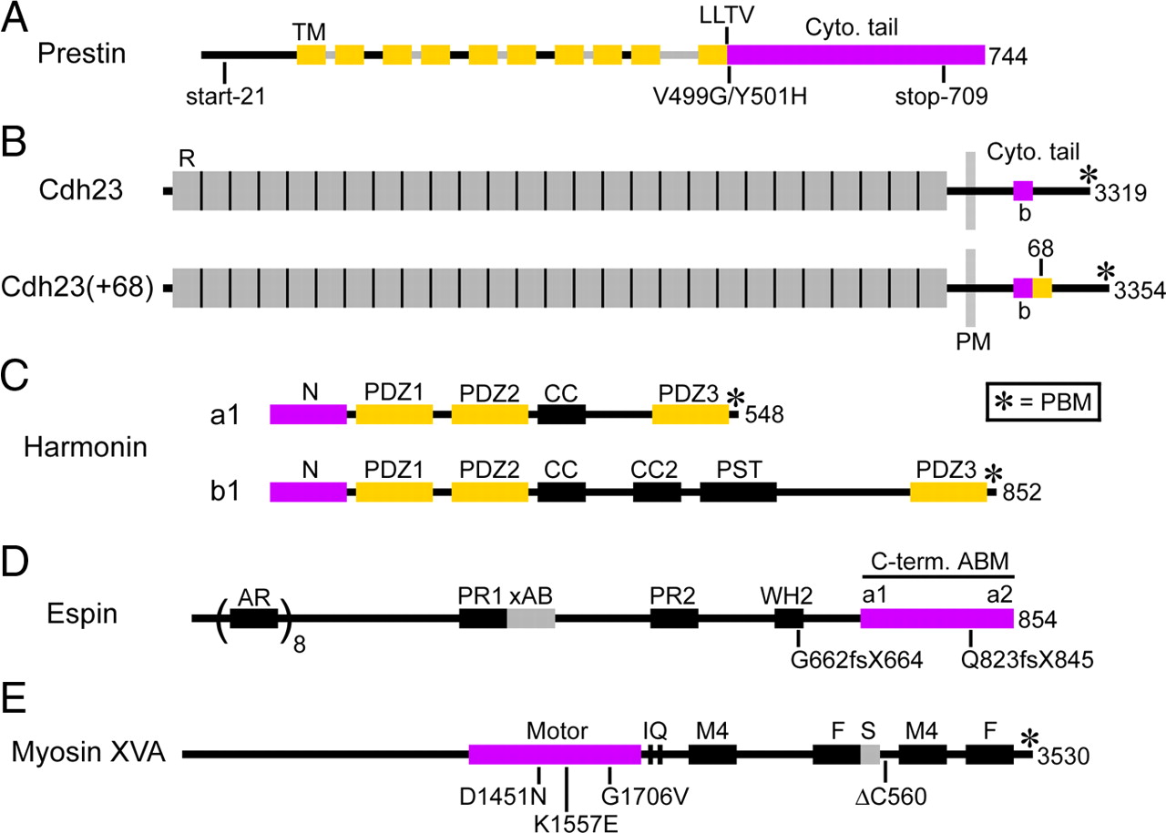
- Figure 1.
Stick-figure diagrams of the proteins examined, highlighting selected domains and mutations. Proteins are shown from N terminus (left) to C terminus (right), and the number of amino acids is shown at the right. For the Cdh23 proteins, the number of amino acids is for the precursor with the N-terminal signal peptide. Asterisk indicates the C-terminal PBM. A, For prestin, predicted transmembrane (TM) domains are gold, and the cytoplasmic tail (Cyto. tail) is purple. Upstream of the C-terminal transmembrane domain, predicted cytoplasmic segments are black, and predicted extracellular segments are gray. B, For Cdh23 and Cdh23 with exon 68 [Cdh23(+68)], the cadherin ectodomain repeats (R) are gray, the 35-residue exon 68 peptide (68) is gold, and the 35-residue harmonin binding site (b) in the cytoplasmic tail (Cyto. tail) is purple. The FLAG-tagged Cdh23 proteins examined are estimated to contain 11 full cadherin ectodomain repeats. PM, Plasma membrane. C, For harmonins a1 and b1, the N domain (N) is purple, and the three PDZ domains (PDZ1, PDZ2, and PDZ3) are gold. CC, Predicted coiled-coil domain; PST, domain rich in proline, serine, and threonine. D, For espin, the 116-residue C-terminal actin-bundling module (C-term. ABM) is purple and has putative F-actin binding sites (a1, a2) disposed at opposite ends. The GFP-tagged espins examined are splice-isoform 3A, which begins approximately midway between xAB and PR2, 59 aa upstream of PR2. AR, Ankyrin-like repeat; PR, proline-rich domain; xAB, additional F-actin binding site; WH2, Wiskott-Aldrich homology 2 domain. E, For myosin XVa, the motor domain is purple. The GFP-tagged myosin XVa proteins examined are isoform 2a, which is missing the N-terminal extension upstream of the motor domain. IQ, Two IQ domains; M4, MyTH4 (myosin tail homology 4) domain; F, FERM (band 4.1, ezrin, radixin, and moesin) domain; S, SH3 (Src homology 3) domain.
- Figure 2.
Domain-specific plasma membrane targeting of prestin and Cdh23. Cells are CL4 unless indicated otherwise in top right corner. A–C, CL4 cell grow in islands (A) and undergo epithelial differentiation, as revealed in part by Texas Red-X phalloidin, which labels F-actin in the junctional belt and short brush border microvilli (B, C). D–F′, Prestin–GFP (prestin) accumulates in cytoplasmic puncta in HEK293 cells (HEK) (D, D′) but in the basolateral plasma membrane of CL4 cells (E, E′) and LLC–PK1 cells (LLC) (F, F′). G–H′, FLAG–Cdh23 (Cdh23) accumulates in irregular, filopodium-like surface projections of HEK293 cells (HEK) (G, G′) but in the microvillar (apical) plasma membrane of CL4 cells (H, H′) (also see Fig. 3). I, I′, A cotransfected CL4 cell accumulates prestin–GFP (prestin; green) and FLAG–Cdh23 (Cdh23; red) in the basolateral and microvillar (apical) plasma membrane, respectively. J–L, Prestin–GFP with the V499G/Y501H (VY) double mutation accumulates in the basolateral plasma membrane (J), but prestin–GFP with the start-21 (K) or stop-709 (L) truncation mutation accumulates in intracellular puncta. M–O′, Pendrin–V5 (pendrin) accumulates in the microvillar (apical) plasma membrane (M, M′), prestin–V5 (prestin) accumulates in the basolateral plasma membrane (N, N′), and a pendrin–prestin–V5 chimera (chimera), in which the prestin cytoplasmic tail substitutes for the pendrin cytoplasmic tail, accumulates in the basolateral plasma membrane (O, O′). Orthogonal sections: x,z (D′–F′, H′, M′); y,z (G′, I′, N′, O′). Scale bars, 10 μm (scale bar in D applies to D–O′).
- Figure 3.
Live-cell surface immunolabeling of microvillar FLAG–Cdh23 at 0–1°C and colocalization with the plasma membrane marker GFP-PH. A, A′, Live-cell labeling of CL4 cell expressing FLAG–Cdh23 with FLAG antibody and Texas Red-labeled secondary antibody followed by fixation, all at 0–1°C to restrict internalization, reveals intense microvillar staining. A′, y,z orthogonal section. B, C, CL4 cell expressing FLAG–Cdh23 with a severely truncated extracellular domain (Cdh23ΔEx), which accumulates in intracellular puncta, shows no such live-cell labeling with FLAG antibody and Texas Red-labeled secondary antibody at 0–1°C (B), but this intracellular FLAG–Cdh23 construct is revealed after a second labeling with FLAG antibody followed by fluorescein-labeled secondary antibody after brief permeabilization (perm.) at 4°C with 0.1% (v/v) Triton X-100 (C). D–F, Live-cell anti-FLAG labeling at 0–1°C, as in A, of a triply transfected CL4 cell expressing FLAG–Cdh23, untagged espin (to elongate microvilli), and the plasma membrane marker GFP–PH. Note that the surface-immunolabeled FLAG–Cdh23 is present along espin-elongated microvilli (A) and colocalizes with GFP–PH (B), as evident in the merged image (C). Scale bar: A–F, 10 μm.
- Figure 4.
Colocalization of GFP–harmonin a1 with microvillar FLAG–Cdh23. A, A′, When expressed without FLAG–Cdh23, GFP–harmonin a1 (harm) accumulates in the nucleus and cytoplasm but not in microvilli. B–D′, When coexpressed with FLAG–Cdh23 (Cdh23), a large fraction of GFP–harmonin a1 (harm) colocalizes with FLAG–Cdh23 in microvilli, suggestive of binding between the two expressed proteins. E–G, Elimination of the four-residue C-terminal PBM from harmonin a1 (harmΔC4) does not decrease GFP–harmonin a1 (harm) construct colocalization in microvilli (compare with B–D). H–J, Elimination of the four-residue C-terminal PBM from FLAG–Cdh23 (Cdh23ΔC4) does not decrease GFP–harmonin a1 (harm) colocalization in microvilli (compare with B–D). K–P, Although FLAG–Cdh23 with the exon 68 peptide [Cdh23(+68)] shows approximately threefold lower levels of accumulation than FLAG–Cdh23 without the exon 68 peptide (compare L with C; and see Results), elimination of the four-residue C-terminal PBM from FLAG–Cdh23 with the exon 68 peptide [Cdh23(+68)ΔC4] does not decrease GFP–harmonin a1 (harm) colocalization in microvilli (compare N–P with K–M). Scale bar (in A): A–P, 10 μm.
- Figure 5.
Colocalization of harmonin b1 with microvillar Cdh23 and mapping the harmonin a1 binding site on the Cdh23 cytoplasmic tail using the colocalization assay. A–C, When expressed without FLAG–Cdh23, GFP–harmonin b1 (harm b) does not accumulate in microvilli but becomes colocalized with F-actin in a belt-like accumulation at the lateral margin and in a lacy cytoplasmic network (prominent in left half of cell), as revealed by labeling with Texas Red-X phalloidin (B). Dashed line in A, Plane of x,z orthogonal section shown in A′–C′. A′–C′, A 1.5× enlarged x,z orthogonal section through the right half of the transfected cell shown in A–C (along dashed line in A) highlighting the accumulation of GFP–harmonin b1 and F-actin in what appears to be an exaggerated version of the F-actin-containing junctional belt normally present near the apical end of the lateral domain. D–F, When coexpressed with FLAG–Cdh23 (Cdh23), a large fraction of GFP–harmonin b1 (harm b) colocalizes with FLAG–Cdh23 in microvilli, suggestive of binding between the two expressed proteins, and is no longer prevalent in a lateral belt-like accumulation or lacy cytoplasmic network. G–I, Elimination of the four-residue C-terminal PBM from FLAG–Cdh23 (Cdh23ΔC4) does not decrease GFP–harmonin b1 (harm b) colocalization in microvilli (compare with D–F). J–L, Elimination of the 108 residues from the C terminus of FLAG–Cdh23 (Cdh23ΔC108) does not decrease GFP–harmonin a1 (harm) colocalization in microvilli (compare with Fig. 4B–D). M–O, Elimination of 143 residues from the C terminus of FLAG–Cdh23 (Cdh23ΔC143) drastically reduces colocalization of GFP–harmonin a1 (harm) in microvilli. Instead, the GFP–harmonin a1 returns to the nucleus and cytoplasm (compare M to Fig. 4A), and the merged image changes from yellow to red–orange (O). Scale bars, 10 μm (scale bar in D applies to D–O).
- Figure 6.
Mapping the harmonin a1 binding site on the Cdh23 cytoplasmic tail using the in vitro binding assay. A, A Coomassie blue-stained SDS gel shows the results of representative in vitro binding assays between purified 6xHis–harmonin a1 protein (harm) and the designated purified GST–Cdh23 cytoplasmic tail protein, or GST alone, immobilized on glutathione–Sepharose 4B beads. GST–Cdh23 cytoplasmic tail constructs either include [Cdh23(+68)] or exclude (Cdh23) the exon 68 peptide and are missing the designated number of amino acid residues from their C terminus (ΔC). Lanes i–k contain control samples that exclude harmonin protein to illustrate that bands comigrating with harmonin are not present in any of the GST–Cdh23 tail proteins. Lane l contains 30% of the harmonin protein input into the binding assay. The numbers at left indicate the positions of apparent molecular mass markers in kilodaltons. B, Graph showing results of three independent in vitro binding assays like that shown in A. The mean levels of harmonin (harm) bound to the designated constructs are plotted as a percentage of the amount of harmonin added to the binding assay. Error bars represent +1 SD. The Tukey-Kramer multiple comparisons test indicated that the means are not significantly different from each other (p > 0.05) except during comparison with Cdh23ΔC143 (*p < 0.001). Note that elimination of the four-residue C-terminal PBM of ΔC4 does not diminish harmonin binding with or without the exon 68 peptide, GST–Cdh23 tail constructs with and without the exon 68 peptide bind similar levels of harmonin, deletion of up to 108 residues (ΔC108) does not diminish harmonin binding, and deletion of 143 residues (ΔC143) drastically reduces binding to near the background level observed with GST alone.
- Figure 7.
Mapping the Cdh23 binding site on harmonin a1 using the colocalization assay. A–C, GFP–harmonin a1 (harm) missing the N domain and PDZ1 domain (PDZ2+PDZ3) does not colocalize with FLAG–Cdh23 (Cdh23) in microvilli. D–F, A large fraction of GFP–harmonin a1 (harm) missing the PDZ2 and PDZ3 domains (N domain + PDZ1) colocalizes with FLAG–Cdh23 (Cdh23) in microvilli (compare with Fig. 4B′–D′). G–I, Elimination of the PDZ1 binding pocket and mutation of histidine-147 to alanine (ΔBP + HA) does not decrease GFP–harmonin a1 (harm) colocalization with FLAG–Cdh23 (Cdh23) in microvilli (compare with Fig. 4B–D). J–L, Elimination of the N domain (ΔN-domain) drastically reduces colocalization of GFP–harmonin a1 (harm) with FLAG–Cdh23 (Cdh23) in microvilli. Instead, the GFP–harmonin a1 construct returns to the nucleus and cytoplasm (compare J with Fig. 4A), and the merged image changes from yellow to red–orange (L). Scale bars, 10 μm (scale bar in G applies to G–L).
- Figure 8.
Examining human espin and myosin XVa deafness mutations using espin-mediated microvillar elongation. A–I are labeled with Texas Red-X phalloidin to reveal F-actin and highlight the actin bundles of CL4 cell microvilli. A, Wild-type GFP–espin (wt espin) is efficiently targeted to microvillar actin bundles and causes pronounced microvillar elongation (average microvillar length, 7 ± 1 μm for this cell). B, GFP–espin with the c.2469delGTCA human deafness mutation (Q823fsX845 espin) retains only weak microvillar targeting and elongation activity, evident mainly near the cell periphery. This mutated espin accumulates in the junctional belt, nucleus, and cytoplasm. C, GFP–espin with the c.1988delAGAG human deafness mutation (G662fsX664 espin) neither targets microvilli nor causes microvillar elongation and instead accumulates in the nucleus. This localization does not reflect recent cell division, because individual transfected cells look the same. D, GFP–myosin XVa (myosin XVa) accumulates at the tips of brush border microvilli, but this is difficult to resolve because the microvilli are small. E, When coexpressing GFP–myosin XVa and untagged espin (myosin XVa + espin), GFP–myosin XVa accumulates at the tips of the espin-elongated microvilli. Compared with the situation without espin (D), tip accumulation is much easier to discern. F–I, The transfected cells are coexpressing GFP–myosin XVa with the designated mutation and untagged espin (+espin). Accumulation at the tips of espin-elongated microvilli is observed for GFP–myosin XVa missing the C-terminal 560 residues (ΔC560; F) and GFP–myosin XVa with the G1706V human deafness mutation (I) but is not observed for GFP–myosin XVa with the D1451N (G) or the K1557E (H) human deafness mutation. The latter two constructs are concentrated below, in the cytoplasm (G, H). Average microvillar length is similar for the transfected cells shown in E–I (8 ± 1–2 μm). Scale bar (in A): A–I, 10 μm. J, A representative GFP antibody-labeled Western blot of SDS extracts prepared from CL4 cells cotransfected with the untagged espin construct and the designated GFP–myosin XVa construct shows that there is no change in apparent molecular mass or major difference in expression levels or integrity among the GFP–myosin XVa protein bands (GFP-XVa) examined in E and G–I: wild-type (lane W), D1451N (lane D), K1557E (lane K), G1706V (lane G). When applied to the results of three independent Western blotting experiments, the Dunnett's multiple comparisons test indicates that mean band intensities for the mutated GFP–myosin XVa proteins are not significantly different from those of the wild-type control (p > 0.05). The bands other than the GFP–myosin XV band (GFP-XVa) are background bands, which are also present in SDS extracts prepared from mock-transfected cells (lane M) and cells transfected with the untagged espin construct alone (lane E). The numbers at left indicate the positions of apparent molecular mass markers in kilodaltons.


