

Article Figures & Data
Figures
- Figure 1.
Passive steady-state properties of layer 6 (L6) pyramidal neurons. a, L6 pyramidal neurons reconstructed from biocytin-filled neurons after the experiment or sketched from fluorescent-dye-filled neurons during the experiment (red electrodes, dendritic recording sites; black electrodes, somatic recordings). The number below each neuron indicates the distance of the dendritic electrode from the soma (in μm). The neuron shaded in gray is the cell for which data are shown in b. b, One-second-step current injections (top) to the dendrite (b1) and to the soma (b2) and resulting voltage responses (middle, red traces, dendrite; black traces, soma). Bottom, The steady-state I–V relationships at dendrite and soma for both dendritic and somatic current injection. b3, Voltage recordings with current injection at the reciprocal site for both somatic and dendritic current injection. c, Local membrane time constant (τ membrane) [black, soma, 9.1 ms; red, dendrite (Dend), 5.7 ms]. d, Steady-state voltage attenuation from soma to dendrite (black) and from dendrite to soma (red) for recordings from different cells at different distances along the apical dendrite. e, Input resistance (Rinput) at the dendrite versus soma (red circles) as a function of distance from soma. f, Sag at dendritic recording as a function of distance from soma. g, Ratio of sag at the dendrite to the soma.
- Figure 2.
Backpropagation of single somatic APs. a, Schematic diagram showing the experimental setup with the distance between the somatic (black) and the dendritic (red) electrode for different recording sites. A short (2 ms) current injection near threshold (bottom trace, Isoma) evoked an AP at the soma (black trace) that propagated back into the dendrite (red trace). b, Amplitude of single bAPs in the dendrite plotted as a function of distance from soma. c, Ratio of somatic AP to dendritic bAP amplitude as a function of distance from soma. d, e, Half-width (d) and latency (e) of bAPs in the dendrite as a function of distance from soma.
- Figure 3.
Back propagation of action potentials in a train. a, Representative examples from a range of dendritic distances from the soma illustrating dendritic responses to long (1000 ms) somatic current injections at twice rheobase, which evoked a train of APs (black traces) that backpropagated into the dendrite (red trace) with variable amplitude. Insets show enlargement of the first three to five APs. b, Dendritic amplitude of bAPs with the smallest (open circles) and the largest (filled circles) bAP amplitude in the train as function of distance from soma. c, Ratio of dendritic amplitude of smallest to biggest bAP of the train of somatic APs. d, e, Half-width (d) and latency (e) of smallest (open circles) and largest (filled circles) dendritic bAP amplitudes as a function of distance from soma. Data were fit with a single exponential where possible (dashed line, smallest bAP; solid line, biggest bAP).
- Figure 4.
Abrupt increase in bAP amplitude with dendritic current injection dependent on dendritic Na+ channels. a, Experimental setup with the dendritic electrode in red and the somatic electrode in black. The dendritic membrane potential was controlled by dendritic current injection for 300 ms with varying amplitude (red traces) starting 100 ms before a 2 ms current injection at the soma (black trace) that evoked an AP. b, bAPs recorded 457 μm from the soma (red traces) at different dendritic membrane potentials (Vm_dend). c, Example of bAP amplitude as a function of Vm_dend in one cell. Dashed lines indicate average values before and after the abrupt increase in amplitude (low and high, respectively). d, High (black circles) versus low (gray circles) amplitudes of bAPs in all cells recorded as a function of distance from soma. e, Relative amplification of bAP amplitude with respect to the low level as a function of distance from soma. f, Left, Experimental setup for local blockade of dendritic Na+ channels. Dendritic and somatic recording electrodes are in red and black, respectively, and the TTX puffing pipette is in blue. Right, bAPs recorded 340 μm from soma at different Vm_dend in control conditions (red traces) and during the local application of TTX (blue traces). g, bAP amplitude as a function of Vm_dend of the same cell in control conditions (red circles), after application of TTX (blue circles), and after washout (pink circles). h, bAP amplitude in percentage at low and high Vm_dend, in control conditions (red bars), and during local application of TTX (blue bars). Asterisk indicates statistical significance using a paired, one-tailed t test (n = 3; p = 0.004).
- Figure 5.
Ca2+ influx along the apical dendrite due to bAPs. a, Reconstructed L6 pyramidal neuron showing the regions of interest used for measuring Ca2+ influx (green squares) along the apical dendrite (numbers to the right indicate the distance from the soma). A somatic recording electrode (green) was used to fill the cell with the calcium indicator OGB-1 (100 μm) and evoke a single bAP. A second extracellular stimulation electrode was placed close to the dendrite (<∼10 μm) at the border of L5 and L6. b, Example of bAP (top trace) that evoked fluorescence increases (lower traces) for the first 200 μm along the apical dendrite under control conditions. Diagonal slashes in top trace indicate truncation of the somatic AP. c, bAP-evoked fluorescence measurements along the same dendrite with simultaneous synaptic stimulation. d, Synaptic stimulation (top trace) alone showing no detectable Ca2+ influx (bottom traces). e, Fluorescence profile for all distances measured in the cell shown in a–d. f, Average fluorescence profile for five cells. Error bars indicate the SEM. Asterisks indicate statistical significance between the control condition versus synaptic stimulation (n = 5; p = 0.007, 0.02, 0.007, 0.006, 0.024, in order from left to right).
- Figure 6.
Critical frequency for dendritic electrogenesis. a, Dual somatic (black trace) and dendritic (red trace) recordings from a L6 pyramidal neuron. Responses to brief trains of APs at 10 (top) and 140 Hz (bottom) evoked by three brief somatic current injections (2 ms each). The resting membrane potential at the soma and at the dendrite was −77 and −82 mV, respectively. The soma was 1540 μm from pia and the dendritic recording pipette was localized 354 μm from soma. b, Overlay of the last AP in the train of three APs at different frequencies of 40 to 150 Hz for dendritic (top) and somatic (bottom) recording. The peak amplitude (and width) of the last AP recorded at the dendrite increased in a nonlinear manner. This was also reflected in an increase in depolarization after the last AP (ADP) at the soma (time point indicated by the dashed line). c, Measurements of [Ca2+]i in a region of interest (green box) in the apical dendrite following low (top) and high (bottom) frequency trains of APs as in a and b. d, Dendritic and somatic potentials measured at the time point indicated with dashed lines in a and b shown as a function of AP frequency. e, Somatic after-depolarizing potential and fluorescence measurements from c shown as a function of AP frequency. f, Histogram of critical frequency measured from 46 neurons. g, Histogram of somatic after-depolarizing potential values from the same neurons as in f.
- Figure 7.
Dendritic electrogenesis evoked by long current injection to the distal dendrite and its interaction with somatic action potentials. a, Long (1 s) dendritic current injections near threshold evoking different somatic AP response patterns depending on the distance of dendritic injection site from the soma (electrode placement shown schematically on the left). b, Width of dendritic potentials (DP), including bAPs with dendritic current injection (see Materials and Methods). b–e, Black squares represent the average somatic value. c, Variability of AP firing pattern (coefficient of variation of interspike interval over the entire duration of current injection) due to steady-state current injection as a function of the distance of injection site from the soma. d, Threshold current injection to evoke one or more APs. In some cases, the first AP was initiated at the soma (filled circles) and in other cases at the dendrite (open squares). e, Threshold current required for generating somatic APs with long current injection at the dendrite (D) in proportion to threshold for APs with long current injection at soma (S).
- Figure 8.
Dendritic spikes generated by EPSC-like current injection to the dendrite. a–d, Dual recording from the soma and dendrite (at a dendritic distance of 383 μm). Dendritic EPSC waveform current injections (bottom red traces) evoked voltage deflections in the dendrite (top red traces, Vdend) and soma (top black traces, Vsoma). a, Subthreshold EPSC waveform current injection (bottom) evoked similarly shaped voltage deflections in the dendrite (top, red trace). b, Dendritic spike with a fast initial component generated with a peak EPSC amplitude of 550 pA. The extrapolated passive response at suprathreshold EPSC amplitudes is shown in gray in b to d (Vextr). This was used to calculate the active component of the response (Vdiff) by subtracting Vextr from Vdend (middle, dark gray line) c, Dendritic spike with two components generated with increased current injection (750 pA) in the same cell as b. d, Threshold for a somatic AP in the same cell reached with a 950 pA dendritic current injection. e, Diagrammatic representation of the locations of the dendritic current injection that evoked single- and two-component dendritic spikes. The second component could only be generated with current injection more distal than 250 μm from soma, which typically corresponds to the region of the dendrite reaching into L5 and L4. f, Threshold in nA for the single-component (gray circles) and the two-component (red circles) dendritic spike as a function of distance from soma. Linear fits to the data show the progressive decrease in threshold for initiation of single- and two-component dendritic spikes. g, Ratio of the thresholds for the generation of dendritic spikes (Idend) versus somatic APs (IsomaAP) via dendritic current injection. The intersection of the linear fit to the data and the dotted line (IsomaAP/Idend = 1) indicates the approximate position along the apical dendrite where dendritic spikes tend to precede somatic APs or occur in isolation. h, An example of a two-component dendritic spike (red) evoked by EPCS waveform current injection to the dendrite that was reduced to a one-component dendritic spike (gray) by the bath application of Ni2+ (100 μm) and Cd2+ (50 μm). Application of TTX (1 μm; black) blocked the remaining component. i, Enlarged view of the region inside the dashed box in h. j, Average amplitudes of the first and second components before and after application of drugs (n = 3).
- Figure 9.
Facilitation of dendritic spikes in L6 neurons with backpropagating APs. a1, EPSC current injection just below and at threshold to the dendrite (383 μm from the soma) recorded at the dendrite (red trace, Vdend) and soma (black trace, Vsoma). a2, The subthreshold dendritic EPSP current injection 5 ms following a somatically evoked backpropagating AP produced a local dendritic spike. b, Average dendritic current threshold for a dendritic spike with the injection of dendritic current alone (left, no AP) and following a backpropagating AP (right, 5 ms). Asterisk indicates statistical significance (p = 0.017; n = 6).
- Figure 10.
AP-5-sensitive spikes evoked in the distal apical dendrites of L6 pyramidal neurons. a, Experiment setup with a somatic recording electrode (gray) and a distally located extracellular stimulating electrode close (<∼5 μm) to the apical dendrite. Gabazine (0.1 μm) was added to reduce the inhibitory transmission. b, Two extracellularly evoked EPSPs at 50 Hz with progressively increasing stimulus strength up to 200 μA (red traces). AP-5 (50 μm; black traces) blocked the large increase in amplitude and duration of the second EPSP but had only a small effect on the first EPSP. c, Integral of the second EPSP shown in b as a function of stimulus strength. d, Average integral of the second EPSP for five cells. “Low” and “High” refer to response below and above threshold (dashed gray line in c), respectively, for plateau-like responses. Asterisk indicates statistical significance (n = 5; p = 0.013).
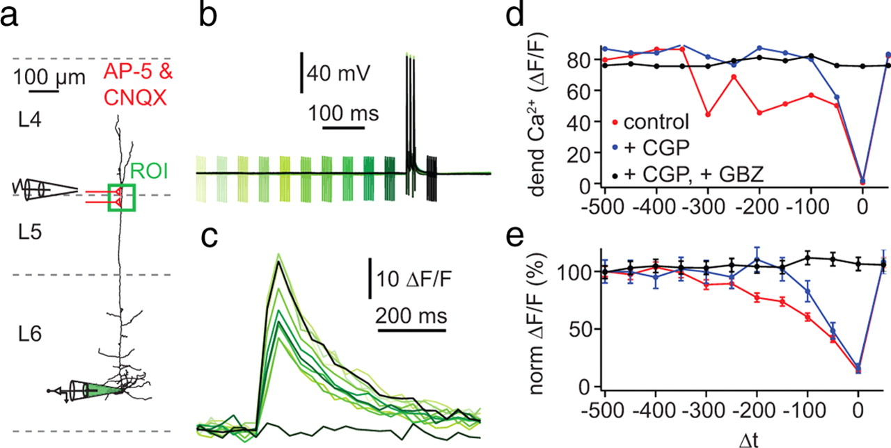
- Figure 11.
Activation of distal inhibitory inputs induces a long-lasting blockade of dendritic Ca2+ electrogenesis. a, Experimental arrangement: somatic whole-cell recordings were made with a pipette containing OGB-1 (100 μm; bottom, green) while monitoring ΔF/F at an ROI on the apical dendrite ∼400 μm from soma (green box). An extracellular bipolar electrode was placed in L4 ∼150 μm lateral to the apical dendrite to evoke inhibitory input (represented schematically in red). CNQX (10 μm) and AP-5 (50 μm) were included in the extracellular bathing solution to prevent excitatory synaptic transmission. b, Electrical recordings from the soma (black trace) while evoking a train of three APs at 120 Hz with somatic current injection (2 ms pulses of 1 nA; critical frequency of the cell shown in this example was 84 Hz). The compound IPSP was concurrently evoked by five pulses at 200 Hz. The time of the extracellular stimulation was altered in steps of 50 ms from 500 before to 50 ms after the train of action potentials (green to black traces). c, Gradual blockade of distal Ca2+ transients recorded in the distal ROI. d, Peak amplitudes of ΔF/F as a function of the time interval between the extracellular stimulation and the train of somatic APs (Δt) for the example shown in a–c. Measurements were obtained in control conditions (red) and in the presence of the GABAB antagonist CGP 52432 (1 μm; blue) and after further addition of the GABAA antagonist gabazine (GBZ; 3 μm; black). e, Average inhibition curve for five cells as in d.
- Figure 12.
Anatomical comparison of apical dendrites of different pyramidal neurons in the neocortex. a, Neurolucida reconstructions of pyramidal neurons from layers 2/3, 5, and 6 of the somatosensory cortex in rats. L6 cells were reconstructed from cells used in this study. The L2/3 cells were taken from our unpublished recordings and the L5 cells are those used in the figures of Larkum et al., (2001) for which dendritic and somatic recordings were published (used with permission). b, Table showing the average data for five neurons from L2/3, L5, and L6 neurons from L6 shown in a. Measurements were made for the entire length of the apical tree, i.e., the path from the cell body to the dendritic endpoint furthest from the cell body and also for the apical shaft, i.e., the path from the cell body to the main bifurcation point on the apical dendrite. The terms “effective length constant” and “effective electrotonic length” refer to λeff as defined in the main text and the analogous concept of Leff = l/λeff (where l is the physical length). These are empirically derived distances for the attenuation of steady-state signals traveling along the apical dendrite and should not be confused with the λ and L used for modeling. They are used here for comparison between cell types. †, from Larkum et al. (2007); ‡, from Williams (2004); used with permission.


Tables
- Table 1.
Summary of passive properties in the soma and apical dendrite of L6 pyramidal neurons
Soma Dendrite Resting Vm (mV) −68.37 ± 1.71 −72.23 ± 1.53 Rin (MΩ) 114.36 ± 6.38* 153.86 ± 8.83* τ (ms) 9.09 ± 0.46* 5.75 ± 0.34* CAR 102.37 ± 14.73* 97.82 ± 13.77* Sag (%) 7.48 ± 0.71* 15.39 ± 1.40* Reobase (pA) 184.21 ± 12.90* 265.00 ± 24.55* AP threshold (mV) −46.26 ± 1.57† −20.99 ± 2.22* AP amplitude (mV) 88.86 ± 2.00† 47.69 ± 5.51† AP half width (ms) 0.881 ± 0.051† 5.922 ± 1.433† Recording distance from soma (μm) 0 336 ± 126 - Table 2.
Comparison of the dendritic properties of neocortical pyramidal neurons in different lamina
Layer 2/3 Layer 5 Layer 6 Na+ channels  12,3
12,3Ca2+ channels 12,3,4,5,6K+ channels ( )17,8( )Active bAPs 12Critical frequency 1209 9810 96 Local Na+ spike 93,11Local Ca2+ spike ( ) short92,3,4 shortLocal NMDA spike (basal)1211,13BAC firing 914Parentheses indicate that only indirect evidence is available.
↵12Gordon et al. (2006);


Supplemental Material
Files in this Data Supplement:
- supplemental material - Supplemental Figure


