

 Cellular/Molecular

TRPA1 Channels Respond to CO2 Elevation
Yuanyuan Y. Wang, Rui B. Chang, and Emily R. Liman
(see pages 12958–12963)
Many types of CO2 receptors are present in the nervous system. Brainstem nuclei that regulate respiration and vascular tone express chemoreceptors that respond to changes in blood CO2 levels. Environmental CO2 is detected by neurons innervating the nasal and oral cavities; these underlie the stinging sensation produced by carbonated beverages. Wang et al. report that a subset of mouse trigeminal neurons is sensitive to CO2 elevation and that the transient receptor potential channel TRPA1 confers this sensitivity. Although CO2-sensitive neurons also expressed TRPV1 channels, only TRPA1 blockers diminished CO2 responses. Like other CO2 receptors, TRPA1 channels are gated not by molecular CO2, but by changes in pH, which directly follow changes in CO2, as CO2 combines with water to generate bicarbonate and free protons. Because CO2 freely diffuses through membranes, it can lower intracellular pH and activate channels that bind protons on the intracellular face. This is what opens TRPA1 channels in trigeminal neurons.
Development/Plasticity/Repair

Sodium Influx Triggers Regeneration of Tadpole Tails
Ai-Sun Tseng, Wendy S. Beane, Joan M. Lemire, Alessio Masi, and Michael Levin
(see pages 13192–13200)
Young Xenopus tadpoles regenerate severed tails, with muscles, nerves, and vasculature. A regeneration bud appears 24 hr after amputation and the new tail is constructed within 7 d. Many factors are required for regeneration, including early activity of a proton pump that modulates membrane potential, and later upregulation of several genes that drive development. Tseng et al. discovered that sodium influx and subsequent activation of salt-inducible kinase (SIK) are required to initiate regenerative processes. Expression of the voltage-sensitive sodium channel Nav1.2 increased in the regeneration bud starting 18 hr after amputation in young tadpoles, but not in older tadpoles that do not regenerate tails. SIK expression also increased. Inhibition or knockdown of Na]v1.2 abolished sodium influx, reduced cell proliferation, prevented upregulation of developmental genes, and inhibited regeneration. Knockdown of SIK also prevented regeneration. Restoring sodium influx with a sodium-specific ionophore rescued regeneration, even in older tadpoles, indicating that sodium influx is necessary and sufficient to trigger regeneration.
Behavioral/Systems/Cognitive

Claustrum Neurons Are Unimodal
Ryan Remedios, Nikos K. Logothetis, and Christoph Kayser
(see pages 12902–12907)
The claustrum is a sheet of neurons lying between the insula and the putamen. Because of its thin, irregular shape, recording from the claustrum is difficult, and therefore its function is not well understood. But neuroanatomical studies showed that the claustrum receives inputs from and projects to most cortical regions, and functional imaging suggested that neurons in the claustrum receive inputs from multiple sensory modalities, leading to the hypothesis that it helps bind diverse inputs into a unified experience. Because of its low spatial resolution, however, functional imaging cannot determine whether sensory inputs to the claustrum overlap. Using single-unit recordings, Remedios et al. found that most neurons in one region of primate claustrum responded only to sound, whereas neurons in another region responded only to visual stimuli. Presenting simultaneous auditory and visual stimuli did not alter neurons' responses. Furthermore, few bimodal neurons were found, arguing against extensive sensory integration in the claustrum.
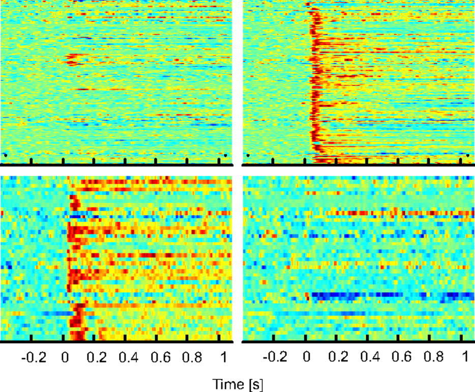
Neurons in the central claustrum (top) responded to sound (right), but not video (left), whereas neurons in the ventral claustrum responded to video but not sound. See the article by Remedios et al. for details.
Neurobiology of Disease

Weight Gain Is Linked to Reduced Striatal Responses to Food Intake
Eric Stice, Sonja Yokum, Kenneth Blum, and Cara Bohon
(see pages 13105–13109)
Like most behaviors, eating is regulated by reward pathways involving dopamine signaling in the striatum. A human polymorphism in the dopamine D2 receptor gene that leads to decreased expression in the striatum is associated with an increased risk of obesity. This suggests that obesity sometimes results from hyposensitivity to reward, which compels people to consume more to become sated. Ironically, however, rats given free access to highly palatable food not only consumed more and gained weight, but also showed reduced striatal expression of D2 receptors and elevated reward thresholds. Stice et al. report that a similar phenomenon may occur in humans. Functional imaging of striatal activity in women after consumption of a milkshake showed that reduced activation was correlated with weight gain over a 6-month interval. These results support the hypothesis that overconsumption of palatable food engages a positive feedback loop involving decreased activation of reward pathways, which promotes further overconsumption.


