

Article Figures & Data
Figures
- Figure 1.
Kinetic properties of VGSCs in nucleated patches from MSO neurons. A, A nucleated patch pulled from the soma of a MSO principal neuron, viewed with DIC microscopy and 160× magnification. B, Nucleated patch recordings of somatic Na+ currents in response to voltage steps between −70 and +40 mV from a −120 mV prepulse. Na+ currents isolated pharmacologically (see Materials and Methods). Bottom, Na+ currents were blocked by 1 μm TTX. C, Current versus voltage plots of Na+ currents produced in control and 1 μm TTX (Control n = 12, 1 μm TTX n = 3). D, Normalized mean conductance versus voltage plot of VGSC activation. Line represents average Boltzmann fit to data: V1/2 = −30.9 ± 1.3 mV and k = 7.5 ± 0.3.
- Figure 2.
VGSC steady-state inactivation and recovery from inactivation. A, Currents recorded using the nucleated patch (left) or cell attached (right) configuration. Currents were elicited by voltage steps between −70 and +40 mV from a −120 mV (75 ms) prepulse. (Currents isolated as detailed in Materials and Methods.) B, There was no significant difference in steady-state inactivation between nucleated patch (V1/2 = −77.4 ± 1.4 mV, k = −7.4 ± 0.5, n = 6) and cell-attached (V1/2 = −76.9 ± 1.3, k = −6.9 ± 0.5, n = 8) configurations (p = 2.4). The cell attached curve was shifted by the average difference between estimated membrane potential and that of the measured membrane potentials (2.8 mV). Activation curve recorded from nucleated patches (light gray) illustrates an unusually small window current. C, Example traces showing the recovery time course of sodium currents recorded in nucleated patches using a –120 mV holding voltage and –10 mV inactivating and recovery pulses (10 and 30 ms, respectively). D, Plots of current recovery as a function of interstimulus interval (Δt) for holding voltages of –120 and –70 mV, fit with dual exponentials. Sodium currents recovered more quickly at –120 mV (τfast = 0.59 ms, τslow = 4.7 ms, n = 5) than –70 mV (τfast = 3.0 ms, τslow = 9.7 ms, n = 6). Inset shows time course over the first 5 ms.
- Figure 3.
Density of Na+ currents decreases along the dendrites of MSO neurons. A, Na+ currents recorded in cell-attached patches from the soma and dendrites of MSO principal neurons using a step pulse to −100 mV from a presumed −120 mV prepulse. After each recording the membrane was ruptured and the resting potential immediately measured (−52.8 ± 0.27 mV). B, Scatter plot of somatic (n = 18) and dendritic (n = 30) cell-attached recordings illustrating the overall decrease in current density as a function of distance from the center of the soma. Filled square indicates somatic average (−6.42 ± 1.82 pA).
- Figure 4.
VGSC-dependent amplification of subthreshold EPSPs occurs at the soma but not in the dendrites. A, Dual somatic and dendritic (40 μm, lateral) recordings from a single neuron (P17; Vrest = −60 mV). Responses to a series of EPSCs injected and recorded at the soma (upper traces, 0–1700 pA, 200 pA steps) or injected and recorded at the dendritic site (lower traces, 0–1800 pA, 200 pA steps). Bath application of 1 μm TTX revealed VGSC amplification of somatic but not dendritic EPSPs. B, Plot of EPSP amplitude measured from rest with respect to EPSC amplitude. Open and closed symbols indicate responses before and after the application of 1 μm TTX, respectively. AP thresholds (dotted lines) were set at control values for both control and TTX conditions. The percentage reduction of control EPSP amplitude by 1 μm TTX was determined at the current producing a just-subthreshold response. C, Mean values for the percentage reduction in EPSP amplitude at somatic (open bars) and dendritic (30–55 μm; closed bars) recording sites by the application of TTX reveal much less influence of TTX in the dendrite than in the soma (left; n = 5; p < 0.005). TTX application substantially increased the current required to reach the amplitude of a just-subthreshold EPSP at the soma but not in the dendrite (right; n = 5; p < 0.05). *Statistically significant difference (paired t test), with a criterion level of 0.05. D, Ipsilateral and contralateral stimulating electrodes evoked independent synaptic responses. Stimuli were delivered at the arrows (upper left diagram). The response to paired ipsilateral and contralateral stimulation was similar to the arithmetic sum of the responses to each stimulus delivered individually (lower left panel, right panel). Histograms show means + SEM.
- Figure 5.
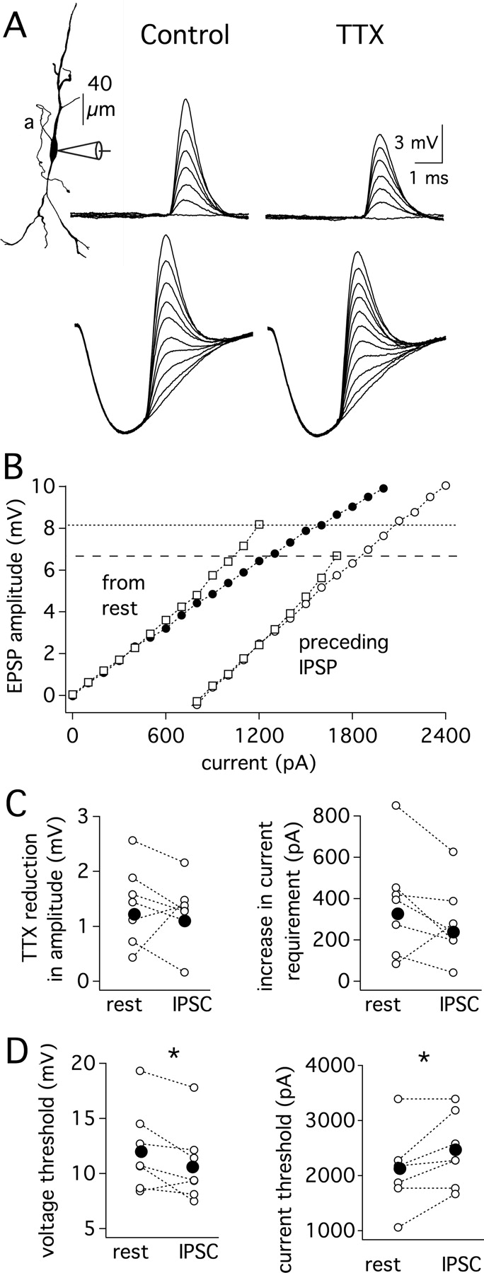
Leading inhibition alters subthreshold gain and lowers the threshold for action potential initiation, but the magnitude of just-subthreshold EPSP amplification by VGSCs remains relatively constant. A, Somatic recordings from a single neuron (P16; Vrest = −65 mV). Responses to a series of EPSCs delivered at rest (upper traces, 0–2.4 nA, 0.4 nA steps) or delivered 2.5 ms after the start of an IPSP (lower traces, 0–1.7 nA, 0.2 nA steps, 8.7 mV IPSP). With preceding IPSPs, peak amplitude measurements were limited to EPSPs large enough to have clear peaks. Bath application of 1 μm TTX reveals a similar magnitude of EPSP amplification in each condition. AP thresholds were set at control values for both control and TTX conditions. B, Plot of EPSP amplitude measured from rest versus EPSC amplitude. Squares and circles indicate responses before and after the application of 1 μm TTX, respectively (closed symbols, from rest; open symbols, from IPSP). Dotted lines indicate threshold for each condition. C, The reduction of EPSP amplitude by TTX determined at the current producing a just-subthreshold response in control conditions (left; p > 0.1). The TTX-induced increase in current required to reach the amplitude of a just-subthreshold EPSP (right; p < 0.1). D, The IPSP-induced decrease in action potential voltage threshold was significantly different with respect to control conditions (left; p < 0.002). The IPSP-induced increase in the current threshold for action potential generation was significantly greater after a preceding IPSP (right; p < 0.05). Mean, closed symbols; individual responses, open symbols (n = 7).
- Figure 6.
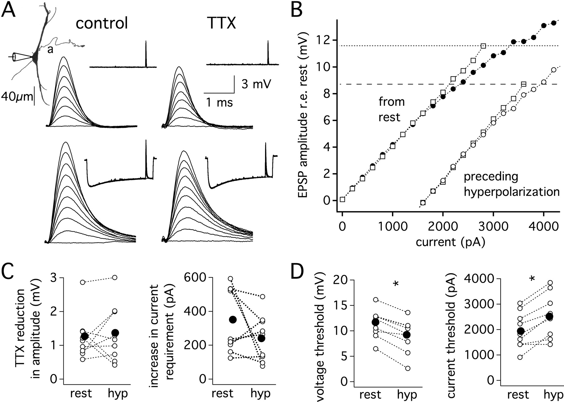
Prolonged membrane hyperpolarization lowers the voltage threshold for action potential initiation and shifts resting conductances, but VGSCs provide the same magnitude of just-subthreshold EPSP amplification as in resting conditions. A, Somatic recordings from a single neuron (P16; Vrest = −62 mV). Responses to a series of EPSCs delivered at rest (upper traces, 0–2.8 nA, 0.4 nA steps) or delivered 80 ms into a 100 ms hyperpolarizing step pulse (lower traces, 0–3.6 nA, 0.4 nA steps). Bath application of 1 μm TTX revealed similar amplitudes of just-subthreshold EPSP amplification in each condition. B, Plot of EPSP amplitude measured from rest versus EPSC amplitude. Square and circle symbols indicate responses before and after the application of 1 μm TTX, respectively. Closed symbols, from rest. Open symbols, from hyperpolarization. Dotted lines indicate AP threshold in the control condition (normal ACSF). C, The reduction of EPSP amplitude by TTX determined at the current producing a just-subthreshold response is similar from rest and from a hyperpolarizing pulse (left; n = 9; p > 0.1). The TTX-induced increase in current required to reach the amplitude of a just-subthreshold EPSP is similar in each condition (right; n = 9; p > 0.1). D, Hyperpolarization decreased action potential voltage threshold (left; n = 9, p < 5 × 10⁻⁵). Hyperpolarization increased the current threshold for action potential generation (right; n = 9, p < 0.05). Mean, closed symbols, and individual responses, open symbols. *Statistically significant difference (paired t test), with a criterion level of 0.05.
- Figure 7.
Subthreshold amplification of EPSPs by somatic VGSCs in a computational model of an MSO neuron. A, Membrane properties of a model neuron with gK-LVA, gleak, and gH, but no gNa. Step pulses (5 ms, −0.2 to 0.2 nA in 0.05 nA steps) were injected at the soma (Vrest = −61 mV). Left: A somatic response to a −200 pA step has a time constant of 565 μs. Right: The slope of a plot of maximal voltage responses versus current amplitudes indicates a somatic input resistance of 12.3 MΩ. B, C, For the same model cell as in A, four simulation configurations are indicated using a 2-dimensional morphological representation of the cell. In all cases, an EPSG-like waveform at the soma evoked responses. The electrode indicates the location, soma or the distal end of the axon initial segment) at which responses were recorded. Red indicates gNa insertion at either the soma (B) or axon initial segment (C). Sample EPSP traces show responses to EPSGs in 5 nS steps to threshold; a plot of EPSP amplitude versus EPSG amplitude (0–50 nS, 5 nS steps) is shown below. The amount of gNa is indicated by line or trace color (S/cm2). Threshold was set at a somatic potential of 12 mV to match the average somatic EPSP amplitude required for action potential generation. Spikes occurring at voltages <12 mV are indicated by asterisks. With all gNa absent (black traces), there is a sublinear relationship between peak EPSP amplitude and the underlying conductance both at the soma (left panels) and the axon initial segment (right panels). Somatic but not axonal gNa amplifies somatic EPSPs (compare B and C left panels). Both somatic and axonal gNa amplify depolarizations in the initial segment (right panels). *Statistically significant difference (paired t test), with a criterion level of 0.05.
- Figure 8.
Restriction of VGSCs to the perisomatic region reduces variability of EPSP amplitude with changes in synapse location. A, Stimulus configuration and VGSC distribution of the computational model used in B and C. EPSG-like waveforms were injected 50 μm from the soma, either into the lateral or the medial dendrite, and the resulting EPSP was recorded at both the dendrite and soma. VGSCs were added incrementally, and distributed either in a restricted manner at the soma (red) or throughout the somatodendritic compartment (blue). The amount of gNa is reported as the total gNa added throughout the cell. Threshold was set at a somatic potential of 12 mV above rest to match the average somatic EPSP amplitude required for threshold action potential generation in MSO neurons. B, Upper left panel: traces of near-threshold EPSPs recorded in the lateral dendrite in a model with 50 S of gNa restricted to the soma (red traces) or throughout the soma and dendrites (blue traces). These EPSPs are contrasted with those evoked by the same synaptic conductance but in the absence of VGSCs (black traces). Right panel: the percentage amplification of just subthreshold EPSPs recorded at the synapse is plotted as a function of whole-cell gNa for medial and lateral synapses. Dendritic VGSCs lead to substantially more EPSP amplification at the synapse than somatic VGSCs. C, Left panel: traces of the same near-threshold EPSPs as in B, but recorded at the soma. Right panel: the percentage amplification of just subthreshold EPSPs at the soma is plotted as a function of whole-cell gNa for medial and lateral synapses. Both somatically located and somatodendritically located VGSCs amplified EPSPs at the soma. ∼45 S of somatic gNa provides a similar amount of somatic EPSP amplification as that found in MSO neurons. D, Top: schematic showing synapse location. All other aspects of the model were the same as in A-C. For the bilateral conditions, synapses at 50 or 100 μm from the soma were stimulated on opposite dendrites, whereas for the unilateral conditions, the 2 synapses on one dendrite were stimulated. Bottom: a scatter plot shows the percentage amplification of just subthreshold EPSPs recorded at the soma for each synaptic condition when 45 S of gNa was added either to the soma or throughout the soma and dendrites. When gNa was restricted to the soma, somatic EPSPs were amplified ∼20%, regardless of synapse location. Somatic EPSP amplification was not consistent when gNa was distributed throughout the somatodendritic compartment.
- Figure 9.
Restricting VGSCs to the soma enhances MSO neurons' sensitivity to bilateral versus unilateral coincident inputs. A, Stimulus configurations used in the computational model. Unilateral simulations (left schematics) were run with two synapses on either the lateral or medial dendrite (configurations 1 and 2 respectively), at positions 50 and 100 μm from the soma. Bilateral simulations (right schematics) were also run with two synapses at 50 and 100 μm, but these synapses were located on opposite dendrites (configurations 1 and 2 have proximal and distal positions reversed). B, The minimum synaptic conductance required for reaching action potential threshold, for bilateral (red) and unilateral (black) synapses plotted as a function of whole-cell gNa. Simulated EPSGs were injected at each synapse simultaneously, and the summed EPSPs were recorded at the soma. EPSG amplitude was increased until somatic EPSP amplitude reached action potential threshold, defined as 12 mV above rest at the soma (the average voltage threshold for action potential generation in MSO neurons). This procedure was repeated with different whole-cell levels of VGSCs, with channels either restricted to the soma (top graph) or distributed uniformly throughout the soma and dendrites (bottom graph). Insets show expanded views of thresholds for whole-cell gNa between 40 and 60 S, the range of gNa that closely reproduced EPSP amplification observed in experiments (Fig. 4). When gNa was restricted to the soma, threshold synaptic conductances were lower for bilateral than unilateral synapses at all levels of gNa. In contrast, when gNa was distributed throughout the soma and dendrites, the thresholds for the unilateral and bilateral conditions overlapped.


Additional Files
Supplemental Data
Files in this Data Supplement:
- supplemental material - Supplemental Legend
- supplemental material - Supplemental Figures


