

Article Figures & Data
Figures
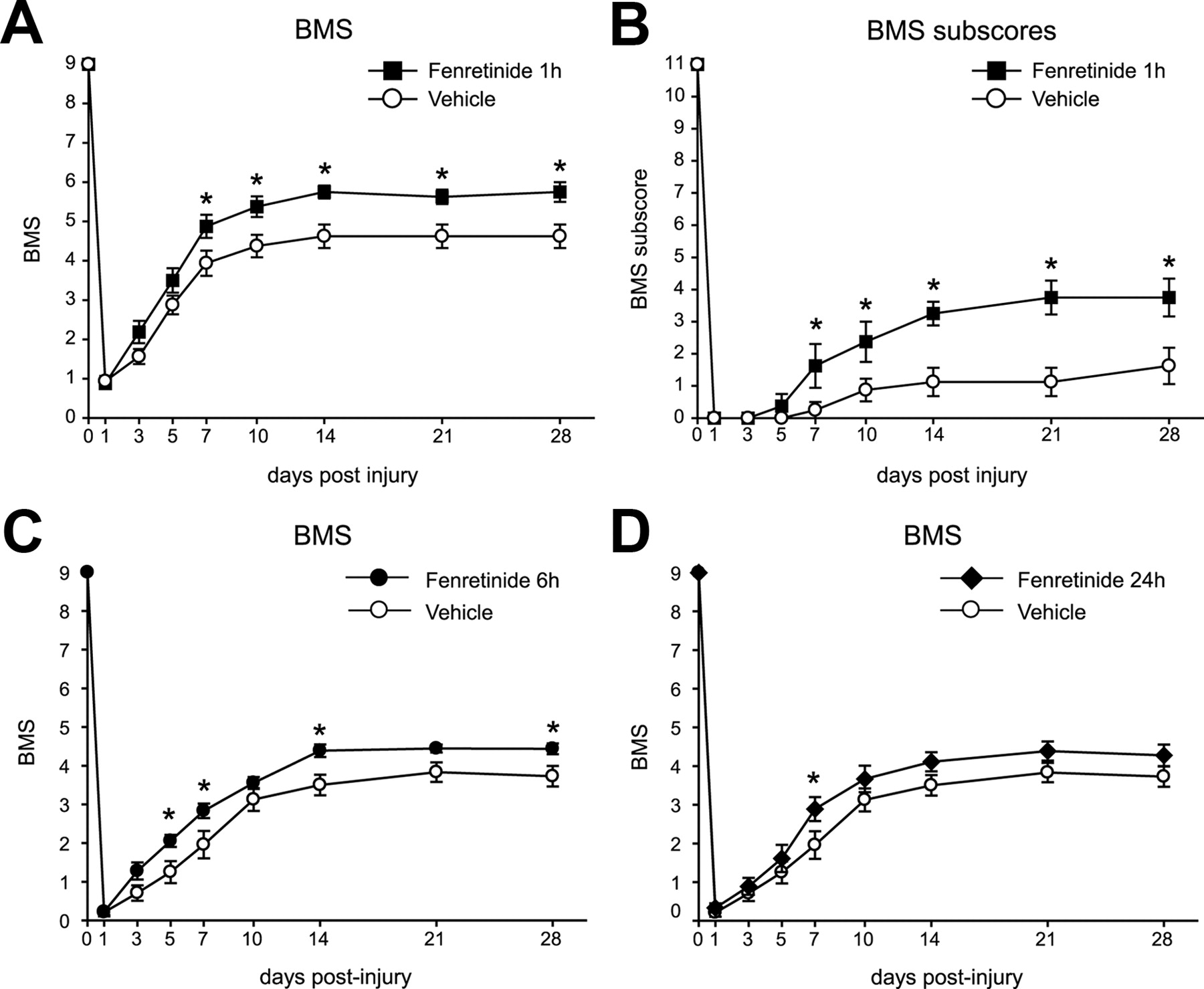
- Figure 1.
A, B, Time course of locomotor recovery in mice treated with fenretinide starting 1 h after SCI. Evaluation was done using the 9-point BMS (A) and the 11-point BMS subscores (B), which evaluate finer aspects of locomotor control. Note that animals treated with fenretinide show significantly enhanced locomotor recovery starting from day 7 after SCI in the BMS scores and in the BMS subscores (*p < 0.05; n = 8 for each group). C, BMS scores of locomotor skills show that fenretinide treatment started after a delay of 6 h after SCI also promoted significant improvement in locomotor recovery (fenretinide group, n = 9; vehicle group, n = 11). D, After a 24 h delay in the start of treatment, there was a significant improvement in locomotor score early after injury (1 week), but this effect was not sustained (fenretinide group, n = 9; vehicle group, n = 11) (*p < 0.05). Error bars indicate SEM.
- Figure 2.
A, Quantification of histological sections stained with anti-GFAP at different distances from the lesion epicenter (0) show that tissue sparing was significant improved at the epicenter and areas rostral and caudal in mice treated with fenretinide (*p < 0.05). B, C, Representative micrographs showing GFAP immunostaining at the epicenter of the injury in vehicle (B)- and fenretinide (C)-treated mice at 28 dpi. Note the greater tissue sparing in mice treated with fenretinide. D, Graph showing that mice treated with fenretinide have greater neuron survival at distances of 300 and 500 μm rostral and 500 μm caudal to the lesion epicenter (*p < 0.05). Quantification done on tissue sections stained with NeuN. E, F, Micrographs showing NeuN staining of neurons in the ventral horn 500 μm rostral to the lesion epicenter in mice treated with vehicle (E) and fenretinide (F) at 28 dpi. Note the marked increased in neuronal profiles in mice treated with fenretinide. G, Mice treated with fenretinide display significantly greater serotonergic innervation in the ventral horns 1 mm caudal to the lesion epicenter (*p < 0.01). H, I, Representative micrographs showing 5-HT-immunoreactive fibers in the ventral horn at a distance 1000 μm caudal to the injury site in vehicle (H)- and fenretinide (I)-treated mice. A marked increased in serotonergic fibers is seen in the ventral horns of mice treated with fenretinide compared with vehicle-treated injured mice (n = 8 for each group). Scale bars: B, C, 500 μm; E, F, H, I, 100 μm. Error bars indicate SEM.
- Figure 3.
Representative micrographs showing NF immunostaining at the epicenter of the injury in vehicle (A)- and fenretinide (B)-treated mice at 28 dpi. Note the greater axonal sparing in mice treated with fenretinide (arrows). The area outlined in the box is shown in higher magnification in C and D. Higher magnification images show greater sparing of axons in the dorsal white matter after fenretinide treatment (D) compared with vehicle-treated controls (C). Scale bars: B, 500 μm; D, 100 μm.
- Figure 4.
Quantification of AA and DHA levels in plasma and spinal cord tissue after SCI. A, Treatment with fenretinide led to a significant reduction in plasma AA levels compared with vehicle-treated mice from days 1 to 28 after SCI (n = 5). B, DHA levels in plasma were significantly higher from 3 to 28 dpi in mice treated with fenretinide (n = 5). Treatment with fenretinide also caused a significant decrease in AA (C) and a significant increase in DHA (D) in spinal cord tissue taken 3 d after SCI (n = 3) (*p < 0.05). Error bars indicate SEM.
- Figure 5.
A, B, The mRNA expression of iNOS, IL-1β, TNF-α, and MCP-1 by RT-PCR (A) and Q-PCR (B) at 24 h after SCI. Quantitative real-time PCR analyses revealed that mice treated with fenretinide had a significant reduction in the mRNA expression of TNF-α and iNOS compared with vehicle-treated mice (*p < 0.05). C, D, Spinal cords from mice treated with fenretinide also showed a significant reduction in the levels of MDA (C) and 3-nitrotyrosine (D) at 3 d after SCI (*p < 0.01; n = 3). Error bars indicate SEM.
- Figure 6.
Effects of fenretinide treatment on TNF-α release from microglial cell cultures stimulated with LPS (10 ng/ml). Fenretinide induced a significant reduction in TNF-α release from activated microglia (*p < 0.05; n = 3). Error bars indicate SEM.


Additional Files
Supplemental Data
Files in this Data Supplement:
- supplemental material - Supplemental Figure


