

Article Figures & Data
Figures
- Figure 1.
Localization of RPE65 protein within the cone OS. A, Frozen sections prepared from 4-week-old C57BL/6 (top) and BALB/c (middle) eyecups and 2-week-old Rpe65−/− eyecups (bottom) were costained for RPE65 (PETLET antibody), S-opsin, and nuclei (DAPI). B, Analysis of sequential single-plane images acquired as a z-series (1–3) and the merged image (M) from a single-cone OS is displayed, showing the distribution of RPE65. C, C57BL/6 eyecup section costained for RPE65 and GS, showing the absence of RPE65 in the Müller glia. D, Visualization of IS in C57BL/6 mouse eyecup using propidium iodide (PI) allows for additional analysis of RPE65 localization within the cone, demonstrating that the majority of RPE65 is localized at the base of the cone OS. E, FITC-conjugated PNA was used to counterstain the glycoprotein sheath surrounding C57BL/6 mouse cone OS and IS, and a single-plane image was acquired to show the glycoprotein sheath surrounding RPE65 immunostaining within the OS. ONL, Outer nuclear layer; OPL, outer plexiform layer; INL, inner nuclear layer; IPL, inner plexiform layer; GCL, ganglion cell layer.
- Figure 2.
Specificity of anti-RPE65 antibodies. Immunostaining of the 8B11 anti-RPE65 antibody was evaluated with 4-week-old C57BL/6 retinas and eyecups. A, The 8B11 antibody failed to detect RPE65 within cones of the flat-mounted retina. B, Evaluation of eyecup sections provided confirmation, with 8B11 immunostaining detected only within the RPE. C, Immunoblot analysis of whole eyecup homogenates prepared from 2-week-old C57BL/6 and Rpe65−/− mice with the PETLET antibody showed detection of a 61 kDa major band corresponding to RPE65 protein in the C57BL/6 sample but not in the Rpe65−/− sample. The 8B11 antibody also detected RPE65 protein in the C57BL/6 sample but none in the Rpe65−/− sample. Blots were stripped and reprobed for β-actin, shown as insets. Arrows represent location of RPE65 band; M, marker; DIC, differential interference contrast; ONL, outer nuclear layer.
- Figure 3.
Variation of amount of cone RPE65 among mouse strains/lines. Whole eyecup homogenates were prepared from 4-week-old eyecups. A, RPE65 protein per eyecup was quantified from immunoblots for six animals (n = 6) from each group using densitometry analysis. Errors are shown as ±SD. *p < 0.05. B, RPE65 immunofluorescence was quantified for cones (n = 10) from each indicated group, background fluorescence was subtracted, and values are plotted on a logarithmic scale in red as fold of amount quantified for C57BL/6 cones (C57BL/6 = 1.00 ± 0.22; BALB/c × Rpe65−/− = 0.31 ± 0.07; BALB/c = 0.08 ± 0.02) with the amount of RPE65 per eyecup calculated from A displayed in blue. *p < 0.001. Errors are represented as ±SD. C, Retinas (2 week-old Rpe65−/− mice; 4-week-old for all other strains/lines) from each group are costained for RPE65 and S-opsin and flat mounted for analysis. Status of the amino acid at position 450 of RPE65 is indicated for each group. Images are taken from the central region of the retina immediately ventral to the optic nerve head. D, RPE65 is detected within cones of albino C57BL/6 mice. ONL, Outer nuclear layer.
- Figure 4.
Effects of exogenous 11-cis-RAL on cone and rod function. Six-week-old animals (n = 5 per group) were intraperitoneally injected with 0.375 mg of 11-cis-RAL (treated) or saline (untreated) 12 h before ERG recordings. A, Summaries of cone responses are shown for mice exhibiting high (C57BL/6), intermediate (BALB/c × Rpe65−/−), and undetectable (BALB/c) levels of RPE65 within cones (Fig. 3B). An approximate threefold increase in the sensitivity of cone b-waves is observed for BALB/c and BALB/c × Rpe65−/− mice. B, No significant differences were observed in rod responses between treated and untreated mice. C, D, Sample cone (C) and rod (D) ERG traces are shown for treated and untreated BALB/c mice. Errors are represented as ±SD.
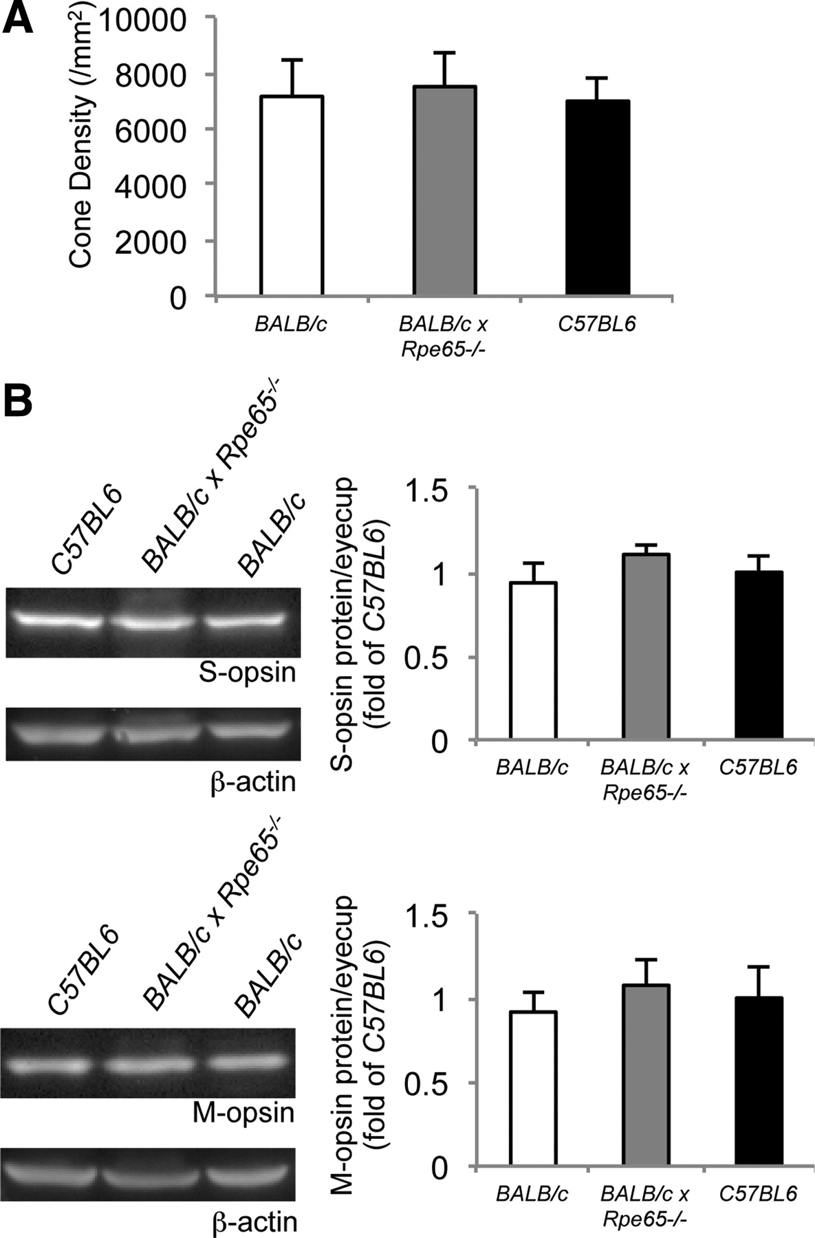
- Figure 5.
Comparable cone levels in retinas of C57BL/6, BALB/c × Rpe65−/−, and BALB/c mice. A, Cone densities measured by S-opsin staining from the central region of BALB/c (p = 0.82; n = 5) and BALB/c × Rpe65−/− (p = 0.46; n = 5) retina flat mounts did not differ significantly from that of C57BL/6 mice (n = 5). B, This was further confirmed from quantification of both M- and S-cone opsin proteins per eyecup using immunoblot analysis, indicating the amounts of both M- and S-opsins from BALB/c (n = 5) and BALB/c × Rpe65−/− (n = 5) eyecups did not differ significantly from that of C57BL/6 (n = 5) eyecups after normalization to β-actin. Errors are represented as ±SD.


