

Article Figures & Data
Figures
- Figure 1.
RB membrane potential varies with background illumination. In darkness, VM = −54 ± 0.2 mV (n = 23). As background increases, VM depolarizes by up to ∼15 mV.
- Figure 2.
The strength of transmission at the RB–AII synapse is reduced by tonic presynaptic depolarization. A, The presynaptic RB is stepped to prepulse potentials between −60 and −42 mV for 750 ms before a test pulse: a 10 ms depolarization of +10 mV relative to the prepulse potential. Averaged (n = 4 responses at each potential) presynaptic Ca currents (middle) and evoked EPSCs (bottom) are illustrated. Some inactivation of the presynaptic Ca current during the prepulse is evident at −48 and −42 mV. EPSCs have been separated by a vertical offset. B, Ca currents and EPSCs are illustrated at higher temporal resolution. EPSCs are not offset. C, Summary of n = 7 experiments. Ca currents (integrated) and EPSCs (quantal contents) recorded during the test pulse from each RB–AII pair were normalized to the largest currents recorded in that pair. The magnitude of Ca influx was relatively constant following each prepulse, but evoked exocytosis was inhibited significantly by the more depolarizing prepulses. Error bars indicate SEM. D, Examples of EPSCs recorded in the 100 ms before the test pulse demonstrate both the increased exocytosis evoked by tonic depolarization (colors as in A) and our ability to resolve and evaluate individual synaptic events (denoted as vertical lines in the traces above the EPSCs). E, The EPSC amplitudes do not vary with interevent interval: amplitudes are normalized to the mean quantal EPSC amplitude (recorded in the absence of presynaptic stimulation), and data are binned in 3 ms bins. The quantal content of the EPSCs is larger than one (average mEPSC, 27.6 ± 3.1 pA), reflecting the multivesicular release that is known to occur at this synapse. The average interevent intervals at −54, −48, and −42 mV were 37.2 ± 33.8, 10.3 ± 8.7, and 4.9 ± 1.8 ms, respectively. Events were exponentially distributed, indicating that they arose from a Poisson process. The average interevent interval in the absence of stimulation was 208.9 ± 38.0 ms (n = 11 paired recordings).
- Figure 3.
Presynaptic Ca currents inactivate at depolarized membrane potentials in the physiological range. A1, The RB was stepped to potentials between −60 and −42 mV for 750 ms. Then, the membrane potential was ramped at 1 mV/ms to −10 mV. A2, The maximal Ca current is reduced by inactivation. B, Summary data from n = 16 RBs illustrate the reduction in maximal current (to ∼75% of control) induced by inactivation. C, Comparison of the conductance–voltage relationships recorded following prepulses to −60 (no prepulse) or −42 mV illustrates a small rightward shift in half-maximal activation potential (from −37.1 to −33.4 mV as determined by a Boltzmann sigmoid fit to the averaged data) that accompanies inactivation.
- Figure 4.
Linear–nonlinear model for characterizing the RB–AII synapse. A, Schematic representation of the synaptic transfer function between presynaptic membrane potential and postsynaptic current. In both cases, synapse is partly rectifying (zero for potentials negative to rest) and otherwise linear (left) or nonlinear (right). Both a high-gain (GHIGH) and low-gain (GLOW) condition are shown; gain in the GLOW condition was scaled by 0.5 (i.e., ordinate values are plotted against abscissa values multiplied by 2). Response to a +5 mV test pulse from baseline (0 mV) in the low-gain (RLOW) and high-gain (RHIGH) conditions are shown. Measuring gain by the ratio RLOW/RHIGH yields a correct 0.5 change in the linear case (A1) but an incorrect 0.25 change in the nonlinear case (A2); measuring the complete input–output function using the L-N analysis enables a scaling procedure to reveal the correct gain change in both cases (see Materials and Methods). B, The RB was stimulated with a voltage command (48 ± 3 mV) that included both repeated sequences (350 ms) and a unique sequence (4 s). A single trial is represented schematically, and a representative sample of the repeated stimulus is illustrated below. Trials were separated by 20 s. The L-N model was generated from the unique sequences and tested on the repeat sequences (i). The unique response is convolved with a linear filter (ii) to generate a linear prediction of the AII output (given in arbitrary units). This prediction is passed through a static nonlinearity (iii), characterized by strong inward rectification, to generate the L-N model of AII output. The predicted L-N output (iv) resembles the measured synaptic current (AII Isyn; averaged across six repeats; r2 = 0.57 ± 0.07 for n = 5 recorded pairs). The nonlinearity shows the raw data (gray points, downsampled to 1 kHz), the binned data (black points, 200 bins), and the fit compared with the averaged response to the repeated stimulus (red line). C, The measured filter is approximated by a delayed DOE after applying a 250 Hz cutoff (fitted parameters: td = 2.0 ms; κ1 = 2.6; σ1 = 1.0 ms; κ2 = 0.05; σ2 = 26.6 ms). The entire L-N analysis was restricted to frequencies <250 Hz, and the measured filter was used to generate the linear prediction in B. D, Six responses to the repeated stimulus are shown to illustrate the variability of the synaptic responses. This variability gives rise to the scatter evident in the raw data (gray points) binned to generate the nonlinearity in B. The gray bars highlight responses to relatively large depolarizations; these are reproduced with considerable variability from trial to trial.
- Figure 5.
Depolarizing the rod bipolar cell membrane potential reduced synaptic gain. A, B, The static nonlinearity and linear filter (insets) are shown for conditions in which the mean rod bipolar command potential (Vcom_mean) was either −51 (A) or −45 mV (B); in both cases, the SD of the white noise input was 3 mV. The nonlinearities were fit (red line) simultaneously by allowing a scale factor to differ between conditions. C, The nonlinearity of the depolarized mean condition could be aligned with the nonlinearity of the hyperpolarized mean condition by adding a constant (9.3 pA for this recording and 5.5 ± 2.4 pA for n = 5 paired recordings; B, orange arrow) and scaling the x-axis (by 0.36 for this recording and 0.32 ± 0.04 for n = 5 paired recordings; B, green arrows). To maintain a constant L-N model output, the filter was multiplied by the same scaling factor (inset). Depolarizing the RB reduced output gain to 36% of the gain in the hyperpolarized condition (32 ± 4% for the population of n = 5 paired recordings). The red dashed line shows the unscaled fit in the depolarized condition in B. Depolarizing the RB reduced synaptic gain. D, Three responses to repeated stimuli with means of −51 mV (left) and −45 mV (right) illustrate the variability inherent in the data. The L-N prediction is better correlated with the averaged response at −51 mV than with the averaged response at −45 mV because of the increased amount of uncorrelated release at depolarized potentials as well as the reduction in the size of the responses correlated with the stimulus. The average response in the −45 mV condition is shown both at the same scale as the −51 mV condition and on an expanded scale. E, Pooling data from n = 5 recorded pairs improves the predictive power of the L-N model. Gray trace, Response to repeated stimulus averaged across all recorded pairs; red trace, L-N prediction derived from model constructed with pooled data. r2 = 0.86 and 0.72 for −51 and −45 mV, respectively.
- Figure 6.
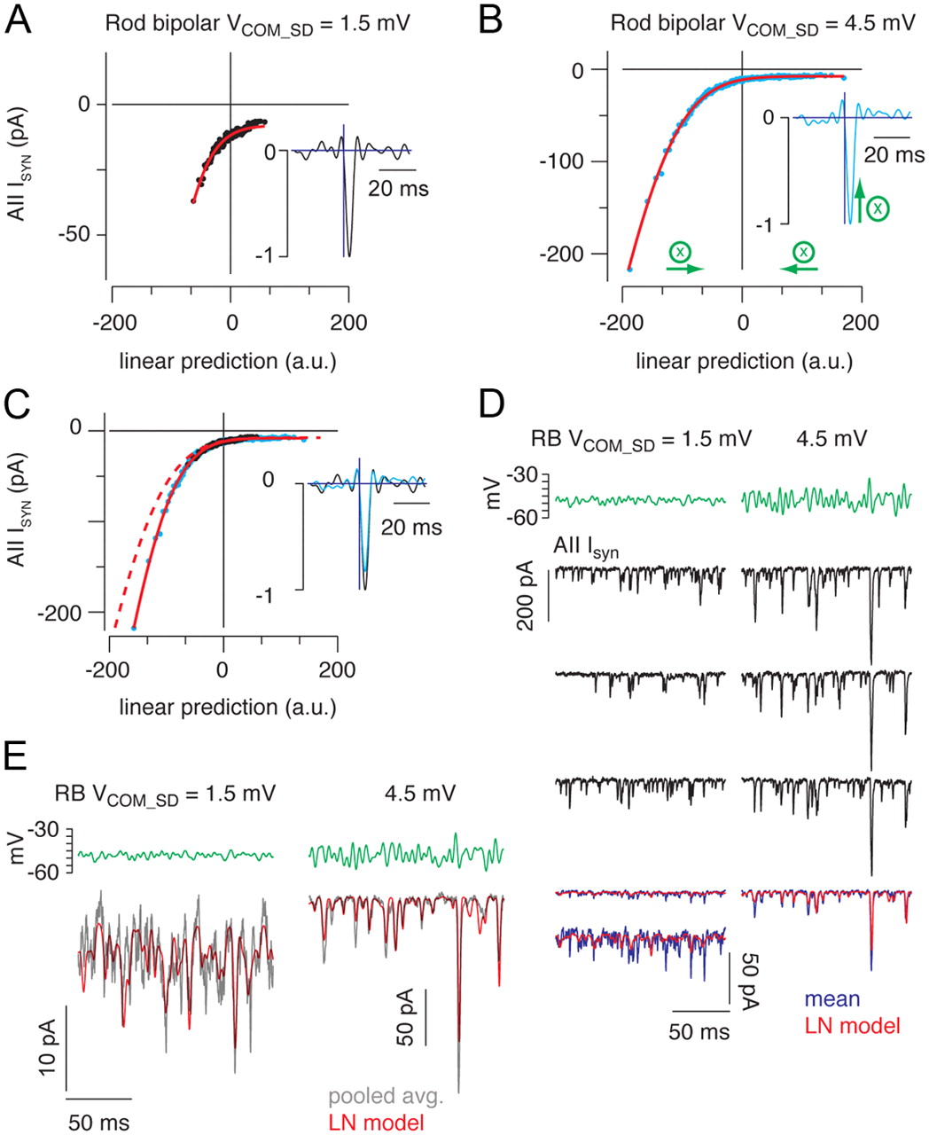
Increasing SD of the rod bipolar cell membrane potential reduces synaptic gain. A, B, The static nonlinearity and linear filter (insets) are shown for conditions in which the mean RB command potential was −48 mV, and the SD of the command potential (Vcom_SD) was either 1.5 (A) or 4.5 mV (B). The nonlinearities were fit (red line) simultaneously by allowing a scale factor to differ between conditions. C, The nonlinearity of the high SD condition could be aligned with the nonlinearity of the low SD condition by scaling the x-axis (by 0.83; B, green arrows). To maintain a constant L-N model output, the filter is multiplied by the same scaling factor (inset). Increasing the SD of the rod bipolar reduced output gain to 83% of the gain in the low SD condition. The red dashed line shows the unscaled fit in the high SD condition in B. D, Three responses to repeated stimuli with SD of 1.5 mV (left) and 4.5 mV (right) illustrate the variability inherent in the data. The L-N prediction is better correlated with the averaged response when the modulated voltage is larger (i.e., when the proportion of release events correlated with the stimulus is higher). The average response in the low SD condition is shown both at the same scale as the high SD condition and on an expanded scale. E, Pooling data from n = 5 recorded pairs improves the predictive power of the L-N model. Gray trace, Response to repeated stimulus averaged across all recorded pairs; red trace, L-N prediction derived from model constructed with pooled data. r2 = 0.70 and 0.88 for SD of 1.5 and 4.5 mV, respectively.
- Figure 7.
A phenomenological model of the RB–AII synapse. A, A presynaptic voltage command (top) is applied to the model to generate a simulated synaptic response (red trace, bottom). The simulated response (red) resembles the experimentally observed postsynaptic response to the same presynaptic stimulus (black; response averaged from n = 5 recorded RB–AII pairs). B, The linear filter (i) and static nonlinearity (ii) extracted from L-N analysis of the simulated output of the synapse (red) matches closely the linear filter and static nonlinearity describing the experimentally measured input–output relationship (black). C, E, The phenomenological model of the RB–AII synapse reproduces experimentally observed adaptation to stimulus mean and variance. Stimuli (i, iii) and responses (ii, iv) (black, experiment; red and blue, model) for each condition are illustrated. Experimental responses are averages from n = 5 paired recordings for each condition. D, F, Static nonlinearities derived for experimental (dots) and simulated (lines) conditions. Increasing stimulus mean from −51 to −45 mV reduced gain by 63%; raising stimulus SD from 1.5 to 4.5 mV reduced gain by 15%. For simplicity, the scaled nonlinearities are not illustrated (see Materials and Methods).
- Figure 8.
The RRP at the RB–AII synapse is reduced by tonic presynaptic depolarization. A, The presynaptic RB is stepped to prepulse potentials between −60 and −42 mV for 700 ms before a 5 ms step to −10 mV. Presynaptic Ca currents (middle) and evoked EPSCs (bottom) are illustrated. EPSCs have been separated by a vertical offset. B, Ca currents and EPSCs are illustrated at higher temporal resolution. EPSCs are not offset. C, Summary of n = 7 experiments. Ca currents (integrated) and EPSCs (quantal contents) recorded during the test pulse from each RB–AII pair were normalized to the largest currents recorded in that pair. D, Simulated synaptic currents generated by our phenomenological model (stimulus as in A). The inset illustrates a small, steady-state component of release at depolarized potentials. E, Simulated responses to the depolarization to −10 mV at higher temporal resolution. F, Comparison of the output of the model to the experimentally measured responses. Here, the error bars on the experimental data points are ±SD.
- Figure 9.
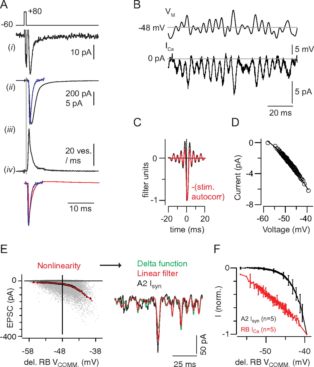
A, A Ca tail current (i) evoked by a 1 ms step from −60 to +80 mV evokes a large, fast EPSC (ii, black trace) that is slower than a quantal miniature EPSC (ii, blue trace) because of the asynchrony inherent in the release process. The extended time course of release is illustrated by deconvolving the miniature EPSC from the evoked response (iii). The gray area highlights the synaptic delay. The waveform of the quantal miniature EPSC (blue) resembles strongly the DOE (black) approximating the true linear filter of the synapse (iv). B, The Ca current elicited by the quasi-white noise stimulus. C, The linear filter exhibits a delay of ∼0.8 ms because of the time required for channel activation (black). In red, The autocorrelation of the stimulus, shifted by +0.8 ms. D, For the recording illustrated, the relationship between Ca current and voltage (delayed by 0.8 ms) was relatively linear. E, The linear prediction is generated by convolving the stimulus with a delayed Dirac delta function, in effect time-shifting (delaying) the stimulus by a constant value. The nonlinearity is represented as a function of the delayed stimulus, and the predicted output generated here (green) is almost identical with that derived from the conventional L-N analysis (B) (red; r2 = 0.55 ± 0.06; n = 5 recorded pairs). F, A comparison of the Ca current and postsynaptic current nonlinearities averaged from n = 5 recordings in each case. Error bars are ±SD.


Tables
VMean ≤−54 VMean = −51 VMean = −48 VMean = −45 VMean = −42 V1/2∞(VMean) −41 −39.5 −38 −36.5 −35 h∞(VMean) 1 1 0.87 0.48 0.18 B(VMean) 0 0 0.003 0.055 0.25 Values are expressed in millivolts.


