

Article Figures & Data
Figures
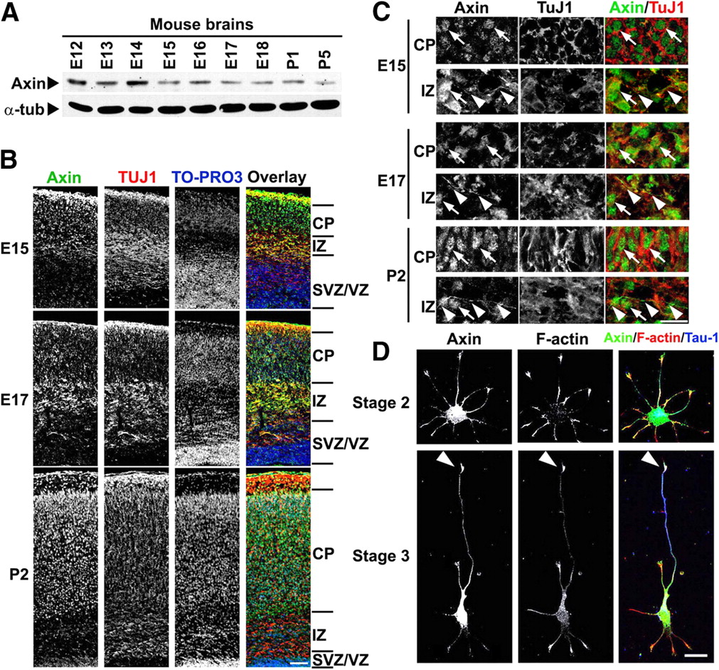
- Figure 1.
Axin expression is developmentally regulated during axon formation. A, Axin is expressed in the mouse brain during embryonic and early postnatal development. α-Tubulin (α-tub) served as a loading control. E, Embryonic day; P, postnatal day. B, Expression of Axin protein during development of cortical plate (E15–P2). Typical layers of the cerebral cortex were shown. Mouse brain sections were stained with anti-Axin (green), TuJ1 (red) antibodies and TO-PRO3 iodide (nuclei; blue). SVZ, Subventricular zone; VZ, ventricular zone. Scale bar, 100 μm. C, Axin protein is detected in the soma and axons of neurons. Axin protein was in the nucleus (arrows) and cytoplasm of neurons, which was marked by TuJ1. Low level of Axin was also detected in the axons (arrowheads). Scale bars, 20 μm. D, Axin is preferentially enriched at the tips of nascent axons. Cultured hippocampal neurons at stage 2 or at stage 3 were stained with anti-Axin (green), anti-Tau-1 (axon; blue) antibodies together with rhodamine-phalloidin (for F-actin; red). F-actin was concentrated at the peripheral area of growth cones, labeling the tips of neurites or axons. Arrowheads depicted the axon tips. Scale bar, 20 μm.
- Figure 2.
Axin is required for axon formation during mouse neocortical development. A–C, Endogenous Axin expression in neurons was silenced by pSUPER–Axin shRNA (shAxin) construct. A, Cultured hippocampal neurons at 0 DIV were transfected with shAxin construct. Protein lysates were collected at 3 DIV after transfection and subjected to Western blot analysis for Axin. B, Mouse brains were in utero electroporated with shAxin construct and GFP expression construct at embryonic day 14.5. Brain sections were collected 2 d later and were stained with Axin antibody. Transfected cells were outlined in purple. C, Cultured hippocampal neurons at 0 DIV were transfected with shAxin construct. Immunostaining was performed at 3 DIV. Scale bar, 20 μm. D–F, Knockdown of Axin inhibits axon formation in developing mouse brains. Expression of Axin protein in cerebral cortical neurons was suppressed using in utero electroporation. shAxin construct was coelectroporated with GFP expression construct into mouse brains at E13.5 or E14.5. After allowing in vivo development, brain sections were obtained from mouse pups at E15.5 (D), E17.5 (E), or P2 (F). For each condition, at least three independent experiments were performed and at least six brains were examined. TAG1, Axonal maker. TO-PRO3 iodide stained the nuclei. Arrowheads indicated the axonal tracts. Green arrows indicated the trailing processes/axons of control neurons. Scale bar, 50 μm. G, Quantification of the percentage of neurons (∼80 individual neurons for each condition) with trailing processes/axons. **p < 0.01, n = 3, Student's t test, mean ± SEM.
- Figure 3.
Axin directs axon formation in cultured hippocampal neurons. A–F, Axin is required for axon formation. Dissociated hippocampal neurons were transfected with shAxin construct, together with GFP expression construct, and fixed at 3 DIV (A, B), 4 DIV (C, D), or 5 DIV (E, F) after transfection. Neurons were costained with antibodies against GFP, axonal markers Tau-1 (green), smi-312 (green), or synapsin I (Syn I; green), and dendritic marker MAP2 (red) as indicated. The axon was defined as Tau-1, smi-312, or synapsin I positive, longer than 100 μm, and at least twice as long as the other processes. Arrows indicated the axons; arrowheads indicated the non-axon neurites. Scale bar, 20 μm. B, D, F, Quantitative analyses of axonal phenotypes. The axonal phenotypes of transfected hippocampal neurons were classified into three groups: NA (no-axon; negative for Tau-1, smi-312, or Syn I but positive for MAP2), SA (single-axon), and MA (multiple-axon) neurons. More than 300 neurons were examined and quantified for each condition. Data were presented as mean ± SEM; **p < 0.01, n = 3, shAxin versus control (Student's t test). G–I, Expression of shRNA-resistant Axin restores axon formation in Axin-deficient neurons. G, Expression of shRNA-resistant Axin (AxinRes–WT) in HEK 293T cells. HEK 293T cells were transfected with expression constructs encoding wild-type (Axin–WT) or shRNA-resistant Axin (AxinRes–WT) together with shAxin construct as indicated. H, shRNA-resistant Axin construct restores the expression of Axin protein in shAxin-transfected hippocampal neurons. I, Expression of shRNA-resistant Axin rescues axon formation in Axin-deficient neurons. More than 300 neurons were examined and quantified for each condition. Data were presented as mean ± SEM; **p < 0.01, n = 3, one-way ANOVA. J, Knockdown of Axin does not cause axon degeneration or a transition of axon to dendrite. Cultured neurons were cotransfected with shAxin construct and GFP expressing construct at 3 DIV. The neurons were then fixed at 3 d after transfection and stained with antibodies for Tau-1 and MAP2. Arrows indicated the magnified axonal distant ends, which is positive for Tau-1 (red) but negative for MAP2 (blue). Scale bar, 50 μm.
- Figure 4.
Cytosolic Axin regulates the microtubule stability through inhibitory phosphorylation of GSK-3β. A, B, Axon formation is attributable to the cytosolic pool of Axin in cultured hippocampal neurons. Expression of the shRNA-resistant wild-type Axin (AxinRes–WT) or its mutant AxinRes–NLSm, but not the AxinRES–NESm, restored the axon formation in Axin-deficient neurons. Scale bar, 20 μm. More than 200 neurons were examined and quantified for each condition. Data were presented as mean ± SEM; **p < 0.01, n = 3. C, Axin-directed axon formation is independent of the canonical Wnt signaling. Cultured hippocampal neurons were transfected with vector (Control), Axin–WT, or AxinΔβ-cat (Axin mutant lacking the binding domain for β-catenin) as indicated. More than 300 neurons were analyzed and quantified for each condition. *p < 0.05, **p < 0.01, n = 3, Student's t test, mean ± SEM. D, Axin interacts with GSK-3β during axon formation in cultured cortical neurons at 2.5 DIV. Rabbit or mouse IgG (rIgG or mIgG) was used as negative control. E, Knockdown of Axin increases activity of GSK-3β. Silencing Axin expression in cultured hippocampal neurons led to a reduction of the phospho-GSK-3β at Ser9 (p-GSK-3β; ∼40% reduction, p < 0.05) and an increase of phospho-CRMP-2 at Thr514 (p-CRMP-2; ∼2.4-fold increase, p < 0.05), which resulted in impaired microtubule stability. F, Knockdown of Axin reduces the level of Ace-α-tub and dephosphorylated tau (Tau-1) in cultured neurons, indicating the impaired microtubule stability. G, Loss of Axin does not affect aPKC and Akt activity in cultured neurons. p-aPKC, phospho-aPKC (at Thr410/403); p-Akt, phospho-Akt (at Ser473). H, Knockdown of Axin abolishes accumulation of phospho-GSK-3β (Ser9) at the tip of the axon or the longest neurite. Neurons cotransfected with shAxin and GFP constructs were fixed and costained with anti-phospho-GSK-3β antibody (green) and rhodamine–phalloidin to indicate the growth cones (F-actin; red). Magnified views of the longest neurite tips (asterisks) were shown. Scale bar, 20 μm. I, J, Knockdown of Axin leads to the mislocalization of GSK-3β from the tip of the axon or the longest neurite. Magnified views of the longest neurite tips (asterisks) were shown (insets). CMRA is a cytosolic marker, labeling the neuronal soma and processes. Scale bar, 20 μm.
- Figure 5.
The specific interaction between Axin and GSK-3β is required for GSK-3β activity regulation and axon formation. A, Mutation of Axin at Leu392 site to proline abolishes the interaction of Axin and GSK-3β. HEK 293T cells were transfected with Axin–WT or Axin–L392P. Protein lysate was coimmunoprecipitated with GSK-3β antibody. Western blot analysis for Axin was performed. B, The Axin–GSK-3β interaction is essential for inhibiting GSK-3β activity in cultured neurons. *p < 0.05; ns, not significant; n = 3, Student's t test, mean ± SEM. C, Axin–GSK-3β interaction in neurons is important for maintaining the level of Ace-α-tub and dephosphorylated tau (Tau-1). D, Axin–GSK-3β interaction is required for the proper localization of GSK-3β. Scale bar, 20 μm. E, The specific Axin–GSK-3β interaction is required for axon formation during mouse brain development. Note that a small proportion of neurons expressing Axin–L392P were found in the IZ or the lower CP, indicating that the migration of these neurons was slightly affected. TAG1, Axonal maker. Arrowheads indicated the axonal tracts, whereas green arrows depicted the trailing processes/axons. Scale bar, 50 μm. F, Quantification of the percentage of neurons (∼60 individual neurons for each condition) with trailing processes/axons. **p < 0.01, n = 3, Student's t test, mean ± SEM. G, H, Axin–GSK-3β interaction is essential for axon formation in cultured hippocampal neurons. Although expression of shRNA-resistant Axin restored axon formation in Axin-deficient neurons, Axin–L392P expression failed to rescue axon formation. Big arrow indicated the axon; small arrow indicated the neighboring untransfected axon; arrowheads indicated non-axonal neurites. More than 300 neurons were analyzed and quantified for each condition. *p < 0.05; **p < 0.01, n = 3, one-way ANOVA, mean ± SEM. Scale bar, 20 μm.
- Figure 6.
Cdk5 phosphorylates Axin in cultured neurons and in developing mouse brains. A, The putative Cdk5 phosphorylation sites of Axin in different species [mouse (m), rat (r), and human (h)]. B, Cdk5 phosphorylates Axin at Thr485 in HEK 293T cells. Axin and its mutants were coexpressed with p35 as indicated. Total Axin protein was immunoprecipitated by specific antibody, and phospho-Axin level was examined using commercial p-Ser/Thr antibody. C, Axin is directly phosphorylated by Cdk5 at Thr485 using in vitro phosphorylation assay. Axin proteins were overexpressed in HEK 293T cells and immunoprecipitated by anti-Axin antibody. The pulled down Axin proteins were then incubated with and phosphorylated by the active Cdk5/p25 complex. p-Axin was detected by our custom phospho-specific antibody targeting Axin at the Thr485 site. D, CIP assay confirms the specificity of the phospho-specific antibody. E, Inhibition of Cdk5 activity in neurons substantially reduced the level of p-Axin. F, p-Axin level was barely detected in cdk5−/− cultured neurons. G, Knockdown or knock-out of p35, the activator of Cdk5, markedly suppressed Axin phosphorylation at Thr485 in cultured neurons. H, Axin phosphorylation was detected both in the cytosol and nuclei of cultured neurons. α-Tubulin was used as the cytosolic protein marker.
- Figure 7.
Cdk5-dependent phosphorylation of Axin is required for axon formation through regulating Axin–GSK-3β interaction and thus GSK-3β inhibition. A, Cdk5 activity is required for Axin–GSK-3β interaction. Ros was used to inhibit Cdk5 activity. B, Cdk5-dependent phosphorylation of Axin is essential for the interaction of Axin and GSK-3β. Wild-type Axin or its phosphorylation point mutants were overexpressed in HEK 293T cells as indicated, and the interaction of Axin with endogenous GSK-3β was examined by coimmunoprecipitation using GSK-3β antibody. C, GSK-3β activation did not affect Axin–GSK-3β interaction. Treating neurons (3 DIV) with Wort, a PI3K inhibitor, upregulated GSK-3β activity, shown by the reduction of phospho-GSK-3β (at Ser9). Wort treatment did not regulate the level of p-Axin, indicating that Axin was not phosphorylated by GSK-3β at Thr485 in neurons. D, Cdk5-dependent phosphorylation of Axin is required for inhibiting GSK-3β activity. shRNA resistant constructs of Axin WT or its phosphorylation deficient mutant (T485A) was coexpressed together with the shAxin construct in cultured neurons. Western blot analysis for p-GSK-3β and p-CRMP-2 was performed. *p < 0.05; ns, not significant; n = 3, Student's t test, mean ± SEM. E, Cdk5-dependent phosphorylation of Axin was required for proper localization of GSK-3β in cultured neurons at 2.5 DIV. Magnified views of the longest neurite tips (asterisks) were shown (insets). Scale bar, 20 μm. F, Phosphorylation of Axin at Thr485 was required for axon formation during mouse brain development. TAG1, Axonal maker. Arrowheads indicate the axonal tracts, whereas green arrows indicated the trailing processes/axons. Scale bar, 50 μm. G, Quantification of the percentage of neurons (∼60 individual neurons for each condition) with trailing processes/axons. *p < 0.05, **p < 0.01, n = 3, Student's t test, mean ± SEM. H, I, Phosphorylation of Axin at Thr485 is essential for axon formation in cultured hippocampal neurons. Whereas expression of shRNA-resistant Axin restored axon formation in Axin-deficient neurons, Axin–T485A failed to rescue axon formation. The arrow indicated the axon; arrowheads indicated non-axonal neurites. Scale bar, 20 μm. More than 300 neurons were examined and quantified for each condition. *p < 0.05, **p < 0.01, n = 3, one-way ANOVA, mean ± SEM.
- Figure 8.
Axin phosphorylation by Cdk5 mediates the neurotrophin-stimulated GSK-3β inhibition through Axin–GSK-3β interaction. A, Axin is required for neurotrophin-induced inhibition of GSK-3β. The p-GSK-3β level was enhanced by BDNF and NT-3 treatment, but this increase was abolished by Axin depletion. *p < 0.05, **p < 0.01, n = 3, Student's t test, mean ± SEM. B, Axin phosphorylation at Thr485 is induced by treatment of neurotrophin (BDNF or NT-3). Cultured neurons at 4 DIV were starved for 1 h and then treated with neurotrophins (50 ng/ml) at 37°C for 30 min. C, Transient neurotrophin stimulation enhanced the Axin–GSK-3β interaction remarkably. D, Axin phosphorylation mediates the Axin–GSK-3β association and the inhibitory phosphorylation GSK-3β induced by neurotrophins. Levels of both p-Axin and p-GSK-3β were substantially reduced by inhibition of Cdk5. E, Cdk5-dependent phosphorylation of Axin transduces signals from the activated cell surface neurotrophin receptors to the GSK-3β-dependent microtubule dynamics during axon formation. Although basal interaction of Axin and GSK-3β can be detected in neurons, this specific interaction increases significantly in a Cdk5-dependent manner upon stimulation with neurotrophins, extracellular cues for inducing axon formation. Importantly, the Axin–GSK-3β interaction is critically required for an inhibitory regulation of GSK-3β activity and reduced phosphorylation of GSK-3β downstream effectors (such as CRMP-2) that are important for axon formation.


