

Article Figures & Data
Figures
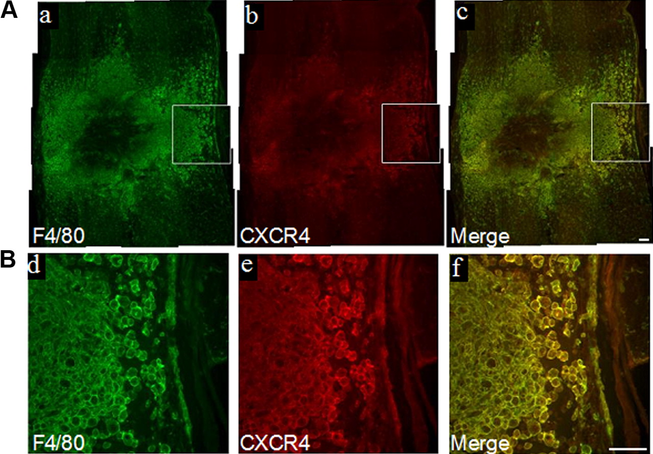
- Figure 1.
SDF-1 and CXCR4 are upregulated in the injured cord. Double immunostaining at 7 d after injury reveals abundant F4/80+ cells that coimmunoexpress CXCR4 (A). Enclosed areas in a–c are shown at higher magnification in d–f (B). Scale bars, 100 μm (a–f).
- Figure 2.
Macrophages express gelatinolytic activity in the injured spinal cord. In situ zymography reveals prominent gelatinase activity within the epicenter that is localized to F4/80+ macrophages in MMP-9 WT mice at 7 d after injury (A, B). Gelatinase activity appeared markedly reduced in the injured cord of the MMP-9 KO and there is no colocalization in F4/80+ macrophages (C, D). Higher magnification of the epicenter reveals localization of gelatinase activity in F4/80+ macrophages in WT mice (E–G). Scale bars: 50 μm (A–D), 100 μm (E–G).
- Figure 3.
Infiltration of bone marrow-derived GFP+ macrophage-like cells into the injured spinal cord. Irradiated WT mice, transplanted with GFP-expressing bone marrow cells (GFP chimeric mice), were subjected to SCI. GFP+ cells were visualized by fluorescence microscopy in longitudinal sections of the spinal cord, centered over the site of injury. At 7 d after injury, the infiltration of GFP+ cells was prominent in the injured cord (A). The majority of infiltrated GFP+ cells exhibited a macrophage-like phenotype (enclosed box in A). Higher magnification shows these cells are round (20–30 μm in diameter) with no processes (B). These GFP+ macrophage-like cells expressed MMP-9 (C–E). Double immunolabeling reveals colocalization of GFP (green in F), with F4/80 (red in G). Scale bars: 250 μm (A, B), 100 μm (C–H).
- Figure 4.
MMP-9-expressing BMDMs show enhanced cell motility. Zymography of supernatants from BMDMs stimulated with inflammatory cytokines for 18 h. While MMP-9 expression is prominent, there appears to be no change in MMP-2 (A). In vitro migration assays show greater cell motility in MMP-9-expressing WT BMDMs compared to MMP-9 KO BMDMs. Synthetic MMP inhibitors GM6001 and MMP-9 inhibitor I attenuate migration of WT BMDMs, but have no effect on KO BMDMs (B). ***p < 0.001.
- Figure 5.
SDF-1α-induced transendothelial migration of BMDMs is enhanced in the presence of MMP-9. Cultured BMDMs when stimulated with LPS, immunoexpress CXCR4 (A). There was a twofold increase in the migration of BMDMs across uncoated Transwells in the presence of SDF-1 in the bottom well. This migration, however, was effectively reduced in the presence of the CXCR4 inhibitor AMD3100 (B). SDF-1α induced a similar magnitude of migration in MMP-9 KO and WT BMDMs. The migration across uncoated Transwells was not affected by MMP-9 inhibitors (C). Transendothelial migration of WT and MMP-9 KO BMDMs was first studied across activated (TNF-α and IFN-γ) and unactivated endothelial cells with growth medium (10% FCS) in the bottom chamber. Transmigration of MMP-9 KO and WT BMDMs across unactivated endothelial cells is relatively modest. While transmigration of WT BMDMs across activated endothelial cells is markedly increased, no differences in transmigration are seen in MMP-9 KO BMDMs (D). When SDF-1 α was added to the bottom chamber as a chemoattractant, MMP-9 WT BMDMs showed greater migratory ability compared to MMP-9 KO BMDMs. No differences in transmigration are seen in MMP-9 KO BMDMs with or without SDF-1 α as the chemoattractant in lower chamber (E). AMD3100, a CXCR4 inhibitor (F), and an MMP-9 inhibitor (G) completely blocked SDF-1α-induced transendothelial migration. Results are expressed as percentages of transmigrated cells that are normalized to WT control. **p < 0.01, ***p < 0.001. Scale bar, 100 μm (A).
- Figure 6.
F4/80+ macrophages are reduced in MMP-deficient mice after SCI. Treatment with the broad-spectrum MMP inhibitor GM6001 from 4 to 6 d after SCI, when macrophage infiltration was most prominent, resulted in reduced axial distribution of F4/80+ macrophages relative to vehicle-treated mice at 7 d after injury (A). The proportional area of F4/80+ macrophages was reduced at the epicenter at 7 d after injury in MMP-9 KO relative to WT mice (B). Scale bars: 200 μm (A), 200 μm (B).
- Figure 7.
Adoptive transfer confirms MMP-dependent migration of myeloid cells into the injured cord. Interestingly, there are very few GFP+ cells in the meninges in the vehicle-treated group (A), whereas there was an abundance of these cells in the meninges in the SB-3CT-treated group (B). At 24 h after adoptive transfer, most of the infiltrated GFP+ cells were CD11b+Gr1+ cells, which are characteristic of immature myeloid cells (C, D). However, by 48 h after transfer, many of the infiltrated GFP+ cells were Ly6G− (E). At 72 h after transfer, the cell bodies of the infiltrated GFP+ cells assumed a larger phenotype relative to those seen at 24 h after transfer and were F4/80+ (F). The presence of infiltrated GFP+ cells within the injured site was confirmed by immunostaining with anti-GFP antibody 24 h after transfer (G–J). Treatment with the MMP inhibitor SB-3CT or CXCR4 inhibitor AMD3100 reduced the number of adoptively transferred bone marrow-derived myeloid cells that were recruited to the injured cord to 70%, and further reduced to 55% when treated with SB-3CT and AMD3100 together (N = 5/group) (K). *p < 0.05, **p < 0.01. Scale bars: 50 μm (A, B, G–J), 100 μm (C–F).


