

Article Figures & Data
Figures
- Figure 1.
Identification of PIIs in the DG and CA1. A, Confocal image stack projection of a PII in the DG. Cells were filled with biocytin during whole-cell patch-clamp recordings and visualized with Alexa647-conjugated streptavidin. Note the dense axonal arbor in the PCL. Insets show the fast action potential firing response of the cell to 600 pA depolarizing current injection and the expression of PV in the cell's dendrite. Scale bar, 10 μm. B, A PII in CA1. Inset shows firing response to 600 pA current injection. sto, stratum oriens.
- Figure 2.
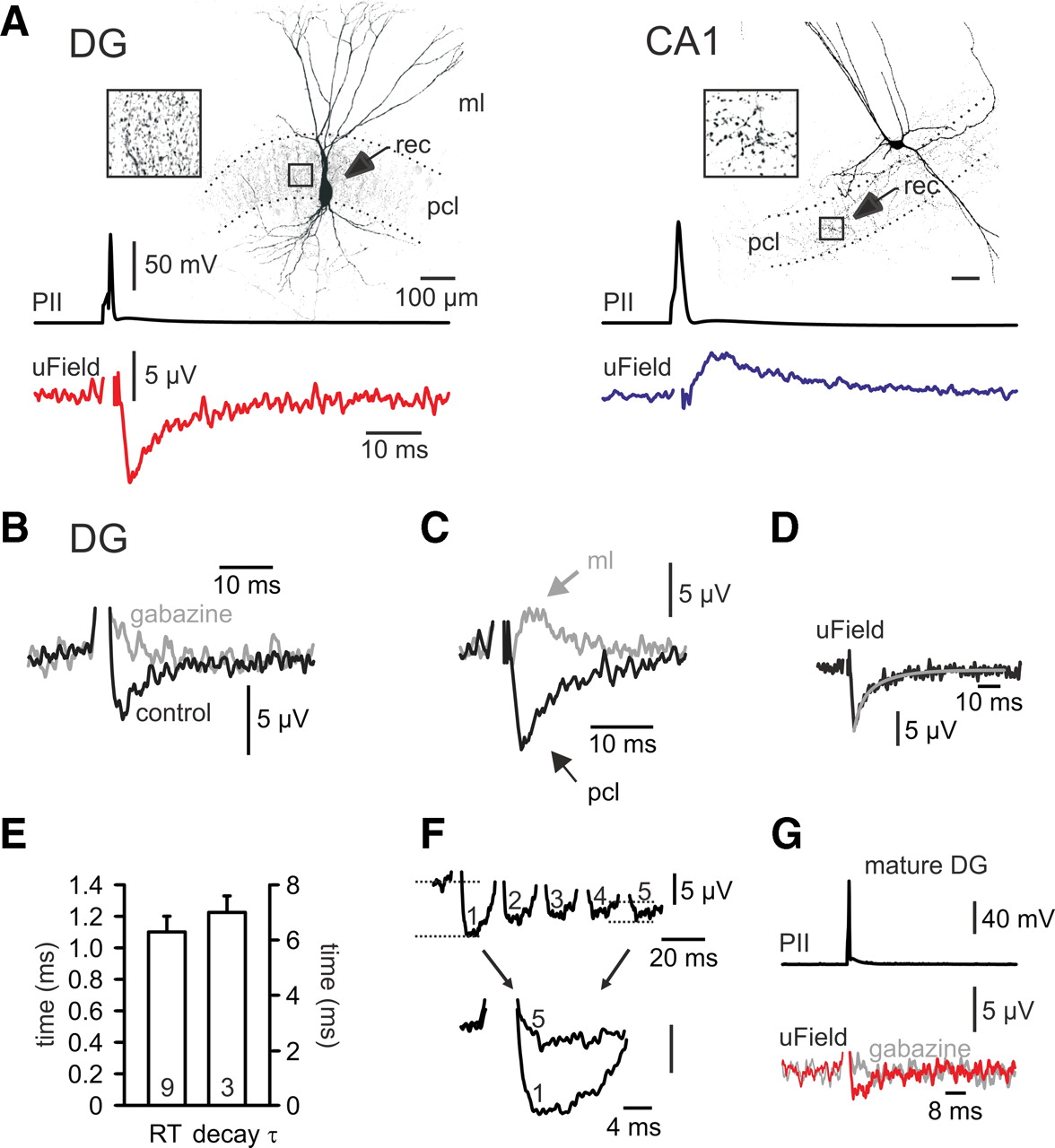
Depolarizing versus hyperpolarizing nature of perisomatic inhibition in the DG and CA1. A, Left, Single action potentials evoked in a DG PII generate negative uFields in the PCL. Right, Single action potentials evoked in a PII located in CA1 give rise to positive uFields. Boxes show the PII axon in the PCL at higher magnification. B, Gabazine (5 μm) blocks uFields, confirming their GABAA receptor-mediated nature. C, When the recording pipette (rec) is moved to the ML, uFields reverse their polarity. D, Biexponential fit (black) to the decay phase of an average uField recorded in the DG reveals its fast time course. E, Summary plot of the 20–80% rise time (RT; left scale) and the decay time constant (decay τ; right scale) of average uFields. F, Multiple-pulse depression of uFields in response to a presynaptic train of five action potentials at 50 Hz. Bottom, The first and the fifth uField of the train are superimposed. G, uFields in the DG of mature rats (P57–P61) are negative in amplitude and can be blocked by gabazine.
- Figure 4.
Dendritic GABA-signaling is depolarizing in the DG. A, Confocal image of a DII with axon in the ML (arrows). B, Top, Adapting action potential firing pattern in response to a 500 pA depolarizing current injection. Bottom, DIIs do not express PV (13 cells tested). C, Example of an EGABA measurement of DII-mediated IPSPs. Signals were recorded at various Vhold. D, IPSPs are plotted as a function of Vhold to reveal EGABA. E, Summary graph of dendritic and perisomatic EGABA in relation to action potential threshold (thres) and resting potential (rest). F, Stimulation in the DG ML (gray) elicits slower IPSPs than stimulation in the PCL (black), indicating that they are of dendritic origin. Traces are amplitude-scaled perforated-patch recordings from the same PC. **p < 0.001; n.s., not significant; stim, stimulation.


