

Article Figures & Data
Figures
- Figure 1.
Unilateral 6-OHDA lesion increases Gαolf levels in the dorsal striatum. Immunoreactivity was detected using Odyssey–LI-COR system (A–D) or confocal microscope (E, F). A, B, D, Serial coronal brain sections from a mouse that received a unilateral 6-OHDA injection into the right striatum. A, TH immunofluorescence (TH IF). B, pAcH3 immunofluorescence (pAcH3 IF) showing the extent of signaling hypersensitivity in the dorsolateral part of the lesioned striatum. C, Plot of Gαolf immunofluorescence quantification with Odyssey–LI-COR, as a function of the proportion of Gαolf protein in a dot blot assay. The samples were obtained by mixing various proportions of protein extracts from striatum (Str) expressing Gαolf and cortex (Cx) devoid of Gαolf. The immunofluorescence values were determined in parallel in two regions of the striatum, in two brain sections (i.e., non-homogenized striatal slices; right, Striatal regions). These values were within the linear range of the standard curve. D, Gαolf immunofluorescence (Gαolf IF). Quantification of mean immunofluorescence intensity (IF) on the unlesioned (UL) and lesioned (6-OHDA) sides is shown in A, B, and D in the dorsolateral part of the striatum (circled region of interest drawn in A). Data are means ± SEM (n = 11–13). Paired two-tailed Student's t test: TH, t = 4.57; pAcH3, t = 12.8; Gαolf, t = 4.00. Scale bar, 1 mm. E, F, Single confocal sections in the dorsolateral striatum showing TH (E) and Gαolf immunofluorescence (F). Data are means ± SEM (n = 8–12). Paired two-tailed Student's t test: TH, t = 4.47; Gαolf, t = 3.19. Scale bar, 100 μm. **p < 0.01, ***p < 0.001. a.u., Arbitrary units.
- Figure 2.
The increase in striatal Gαolf levels correlates with dyskinetic behavior. A, Quantification of Gαolf immunofluorescence (IF) in the dorsal striatum of sham-operated mice treated with l-DOPA for 10 d (Sham; n = 6), 6-OHDA-lesioned mice treated with a single injection of l-DOPA (Acute; n = 21), or daily for 10 d (Chronic; n = 44). Data are expressed as percentage of the unlesioned (UL) side in each group and are means ± SEM. One-way ANOVA, F(2,68) = 4.16, *p < 0.05. Post hoc comparison (Bonferroni's test): *p < 0.05 versus sham. B, Comparison of Gαolf immunofluorescence in the unlesioned and 6-OHDA-lesioned striata of mice with low (Low Dysk, top row) and high (High Dysk, bottom row) scores of LID. Scale bar, 100 μm. C, Comparison of Gαolf levels in the 25% 6-OHDA-lesioned chronically l-DOPA-treated mice in A that developed the weakest (total AIM score < 12.5; Low Dysk; n = 11) and strongest (>33.5; High Dysk; n = 11) dyskinesia. Data are means ± SEM. Unpaired two-tailed Student's t test: t = 2.89, **p < 0.01. D–F, Correlation between Gαolf levels and total AIMs (D) (r = 0.40, F(1,42) = 7.96, p < 0.01), LOC AIMs (E) (r = 0.41, F(1,42) = 8.56, p < 0.01), and ALO AIMs (E) (r = 0.34, F(1,42) = 5.38, p < 0.05).
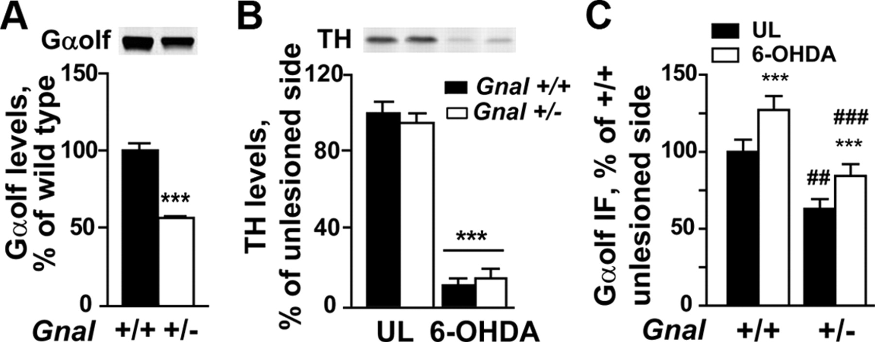
- Figure 3.
Gαolf and TH levels in 6-OHDA-lesioned Gnal+/− and control mice. A, Quantification of Gαolf levels in mice heterozygous for a null mutation of Gnal gene (+/−) and their wild-type littermates (+/+) by immunoblotting. Data are expressed as percentage of the mean in wild-type mice and are means ± SEM (n = 4–6). Unpaired two-tailed Student's t test: Gαolf t = 10.93, ***p < 0.0001. B, Quantification of TH levels by immunoblotting. Data are expressed as percentage of the means of unlesioned (UL) side of Gnal+/+ mice and are means ± SEM (n = 7–8). Two-way ANOVA: effect of lesion, F(1,24) = 357, p < 0.0001; effect of genotype, F(1,24) = 0.01, not significant; interaction, F(1,24) = 0.90, not significant. Post hoc comparison (Bonferroni's test): ***p < 0.001, 6-OHDA versus unlesioned (UL) for each genotype. C, Quantification of Gαolf mean immunofluorescence (IF) intensity in 6-OHDA-lesioned Gnal+/− and wild-type control (+/+) mice chronically treated with l-DOPA. Gαolf immunofluorescence was measured in the dorsolateral striatum by confocal microscopy in three independent experiments for each mouse and normalized to the mean values in wild-type mice for each experiment. Two-way ANOVA analysis effect of the lesion, F(1,18) = 133, p < 0.0001; effect of the genotype, F(1,18) = 13.97, p < 0.01; interaction, F(1,18) = 1.81, not significant. Post hoc comparison (Bonferroni's test): ***p < 0.001, 6-OHDA-lesioned versus unlesioned; ##p < 0.01 and ###p < 0.001, Gnal+/− versus Gnal+/+.
- Figure 4.
LID is not altered in Gnal heterozygous mice. Heterozygous (+/−) Gnal mutant and wild-type (+/+) mice were lesioned and treated with l-DOPA and benserazide during 10 d. A, Forelimb use was determined using the cylinder test in the 6-OHDA-lesioned mice before (−) and after (+) administration of l-DOPA on the first day of treatment. Data are means ± SEM (n = 7–8). Repeated-measure ANOVA (with the within-subjects factor of treatment and the between-subjects factor of genotype): effect of treatment, F(1,26) = 56, p < 0.0001; effect of genotype, F(1,26) = 0.49, not significant; interaction between genotype and treatment, F(1,26) = 0.09, not significant. Post hoc comparison (Bonferroni's test): ***p < 0.001, before versus after l-DOPA. B, Sum of ALO AIMs scored during 140 min period after l-DOPA. Comparison between +/+ (n = 18) and +/− (n = 23) mice. Data are means ± SEM. Unpaired two-tailed Student's t test: t = 0.62, not significant. C, Sum of LOC AIMs scored during 140 min period after l-DOPA in the same animals. Data are means ± SEM. Unpaired two-tailed Student's t test: t = 0.61, not significant. D, Time course of total AIMs (sum of ALO and LOC AIMs) scored every 20 min over a period of 140 min after the last l-DOPA administration. Repeated-measures two-way ANOVA (with the within-subjects factor of time and the between-subjects factor of genotype): effect of genotype, F(1,234) = 0.18, not significant; effect of time, F(6,234) = 71, p < 0.0001; interaction, F(6,234) = 0.43, not significant. E, Total AIMs (sum of ALO and LOC AIMs) scored during 140 min period after l-DOPA on day 5 of l-DOPA treatment. Data are means ± SEM. Student's t test, t = 0.181, not significant.
- Figure 5.
l-DOPA-induced PKA-dependent phosphorylation is markedly impaired in Gnal heterozygous mice. A, Single confocal section showing phospho-PKA substrate immunofluorescence in the unlesioned (UL) and 6-OHDA-lesioned dorsal striatum of Gnal+/+ (top row) and Gnal+/− (bottom row) mice, 30 min after the last injection of l-DOPA + benserazide. Scale bar, 50 μm. B, Number of phospho-PKA substrate (pPKA substrate)-positive cells in the dorsolateral striatum of wild-type and heterozygous mice. Data are means ± SEM of positive cells in 375 × 375 μm confocal images (n = 8–11). Repeated-measures two-way ANOVA (with the within-subjects factor of lesion and the between-subjects factor of genotype): effect of the lesion, F(1,17) = 54.5, p < 0.001; effect of the genotype, F(1,17) = 7.04, p < 0.02; and interaction, F(1,17) = 6.49, p < 0.05. Post hoc comparison (Bonferroni's test): **p < 0.01and ***p < 0.001, 6-OHDA versus unlesioned; ##p < 0.01, Gnal+/− versus Gnal+/+. C, Immunoblot analysis using antibodies against phospho-Ser845-GluA1 (pSer845) and total GluA1, phospho-Thr34–DARPP-32 (pThr34), and total DARPP-32 in the unlesioned (UL) and 6-OHDA-lesioned striatum of Gnal+/+ and Gnal+/− mice, 30 min after the last injection of l-DOPA + benserazide. D, Quantification of pSer845 normalized to total GluA1, expressed as percentage of unlesioned (UL) striatum of Gnal+/+ mice. Data are means ± SEM (n = 9–15). Two-way ANOVA: effect of the genotype, F(1,48) = 6.02, p < 0.02; effect of the lesion, F(1,48) = 10.8, p < 0.01; interaction, F(1,48) = 6.8, p < 0.05. Post hoc comparison (Bonferroni's test): ***p < 0.001, 6-OHDA versus unlesioned; ##p < 0.01, Gnal+/+ versus Gnal+/−. E, Same representation for pThr34 normalized to total DARPP-32. Data are means ± SEM (n = 5–9). Two-way ANOVA: effect of the genotype, F(1,22) = 1.13, not significant; effect of the lesion, F(1,22) = 15.5, p < 0.001; interaction, F(1,22) = 10.1, p < 0.01. Post hoc comparison (Bonferroni's test): ***p < 0.001, 6-OHDA versus UL; #p < 0.05, Gnal+/+ versus Gnal+/−.
- Figure 6.
Chronic or acute l-DOPA induces phosphorylation of ERK and histone H3 in the lesioned striatum of Gnal heterozygous mice. A, B, Confocal sections through the dorsolateral striatum of unlesioned (UL) and 6-OHDA-lesioned dorsolateral striatum of Gnal+/+ (top row) and Gnal+/− (bottom row) mice, 30 min after the last injection of chronic l-DOPA and benserazide treatment showing phospho-ERK (pERK, A) and phospho-acetyl-histone H3 (pAcH3, B) immunofluorescence. Scale bar, 100 μm. Bottom graphs, Number of pERK- or pAcH3-positive cells in the dorsolateral striatum. Data are means ± SEM in 375 × 375 μm confocal images (n = 8–11). Repeated-measures two-way ANOVA (with the within-subjects factor of lesion and the between-subjects factor of genotype): A, pERK, effect of the lesion, F(1,17) = 67, p < 0.0001; effect of the genotype, F(1,17) = 0.89, not significant; interaction, F(1,17) = 1.03, not significant; B, pAcH3, effect of lesion, F(1,17) = 57, p < 0.0001; effect of genotype, F(1,17) = 0.02, not significant; interaction, F(1,17) = 0.01, not significant. Post hoc comparison (Bonferroni's test): ***p < 0.001, 6-OHDA versus unlesioned. C, D, Same as in A and B but in different groups of mice that received a single injection of l-DOPA (n = 4–8 per group). C, pERK, effect of lesion, F(1,20) = 893, p < 0.0001; effect of genotype, F(1,20) = 0.79, not significant; interaction, F(1,20) = 0.79; not significant. D, pAcH3, effect of lesion, F(1,20) = 1231, p < 0.0001; effect of genotype, F(1,20) = 3.61, not significant; interaction, F(1,20) = 3.61, not significant. Post hoc comparison (Bonferroni's test): ***p < 0.001, 6-OHDA versus unlesioned.


