

Article Figures & Data
Figures
- Figure 1.
Sema6A and PlexinA4 direct horizontal cell axon targeting to the OPL in vivo. A–D, WT (A), Sema6A−/− (B), PlexA2−/− (C), and PlexA4−/− (D) adult retina sections were immunostained with the horizontal cell marker anti-calbindin (green). In WT retina (A), all horizontal cell neurites stratify in the OPL; however, in Sema6A−/− retinas (B), horizontal cells exhibit a pronounced defect in neurite stratification in the OPL, and a significant number of horizontal cell neurites reside in the ONL. Horizontal cells in PlexA4−/− retinas (D) exhibit a similar defect in neurite stratification as observed in Sema6A−/− retinas. In contrast, horizontal cells in PlexA2−/− retinas (C) do not exhibit this stratification defect. E, Quantification of aberrant calbindin+ neurites that reside in the ONL in adult WT, Sema6A−/−, and PlexA4−/− mice (n = 3 animals for PlexA4−/−, and n = 4 animals for WT and Sema6A−/−; presented here normalized to the number of horizontal cells quantified; n = 1072 cells for WT, n = 1176 cells for Sema6A−/−, and n = 1050 cells for PlexA4−/− mice). Both Sema6A−/− and PlexA4−/− retinas exhibit a pronounced increase in the number of aberrant horizontal cell neurites that reside in the ONL (43.9 ± 8.4% for Sema6A−/− and 61.1 ± 5.5% for PlexA4−/−) compared with WT retinas (1.6 ± 0.5%). Error bars are SEM. **p < 0.01, ***p < 0.001, one-way ANOVA followed by Tukey's HSD multiple-comparison test. F, Quantification of aberrant calbindin+ neurites that reside in the ONL in adult WT, Sema6A+/−, PlexA4+/−, and Sema6A+/−;PlexA4+/− mice (n = 4 animals for each genotype; normalized to the number of horizontal cells quantified; the same WT as quantification from E, n = 1174 cells for Sema6A+/−, n = 1161 cells for PlexA4+/−, and n = 1179 cells for Sema6A+/−; PlexA4+/−). Sema6A+/−;PlexA4+/− mice show a significantly increased number of aberrant horizontal cell neurites in the ONL (21.4 ± 3.8%) compared with the other three genotypes (1.6 ± 0.5% for WT, 3.5 ± 0.7% for Sema6A+/−, and 4.0 ± 1.1% for PlexA4+/−). Error bars are SEM. ***p < 0.001, one-way ANOVA followed by Tukey's HSD multiple-comparison test. G–G″, PlexA4−/− adult retinas were double immunostained with anti-calbindin (G) and anti-neurofilament (NF, G′) (merged in G″). Aberrant horizontal cell neurites localized in the ONL of PlexA4−/− retinas are both calbindin and neurofilament positive, suggesting that these aberrant neurites are axonal poles of horizontal cells. Scale bars: (in D) A–D, 50 μm; (in G) G–G″, 30 μm.
- Figure 2.
PlexA4 and PlexA2 protein expression in the developing OPL, and normal horizontal cell neurite stratification in neuropilin-deficient retinas. A–B″, WT retina sections from P7 (A–A″) or P14 (B–B″) mice were double immunostained with anti-PlexA4 (A, B, red) and anti-PlexA2 (A′, B′, green). At both P7 and P14, PlexA4 is localized in the OPL, whereas PlexA2 immunostaining is not observed in the OPL at these postnatal stages (yellow arrows). C, D, Npn1sema−/sema− (C) and Npn2−/− (D) adult retina sections were immunostained with anti-calbindin. Both Npn1sema−/sema− (C) and Npn2−/− (D) mice do not exhibit aberrant horizontal cell neurite extension into the ONL (n = 3 animals for each genotype), suggesting that neuropilins are not required for Sema6A and PlexinA4 regulation of horizontal cell neurite stratification in vivo. Scale bars: B″ (for A–B″), D (for C, D), 50 μm.
- Figure 3.
Horizontal cell development in WT and PlexA4−/− retinas. A–N, WT (A, C, E, G, I, K, M) and PlexA4−/− (B, D, F, H, J, L, N) retina sections from postnatal P1 (A, B), P3 (C, D), P5 (E, F), P10 (G, H), P14 (I, J), P17 (K, L), and P21 (M, N) mice were immunostained with anti-neurofilament (A–F) or anti-calbindin (G–N). In PlexA4−/− retinas, aberrant horizontal cell neurites directed toward the ONL are observed as early as P14 (yellow arrows) and then more clearly at later time points. White dashed lines indicate the edge of the outermost ONL (J, L, N). Horizontal cells in PlexA4−/− retinas do not show correct neurite stratification within the OPL (yellow arrows) at P3 (D) and P5 (F) compared with WT retinas; however, the ectopic horizontal cell neurites across the INL of PlexA4−/− retinas stratify within the OPL by P10 (H). At P14, horizontal cells in PlexA4−/− retinas begin extending aberrant neurites toward the ONL (J, yellow arrows), and these aberrant neurites extend to the outermost photoreceptor cell body layers by P17 (L). Axon terminal-like structures are observed in the ONL in PlexA4−/− retinas at P21 (N, yellow arrows). O, O′, High magnification of P21 PlexA4−/− retina section immunostained with anti-calbindin (O) and counterstained with TO-PRO3 (O′). Aberrant horizontal cell neurites extend across the ONL visualized by TO-PRO3 (O′, blue) and reach the outermost edge of the ONL (yellow arrows). Scale bars: F (for A–F), N (for G–N), O′ (for O, O′), 50 μm.
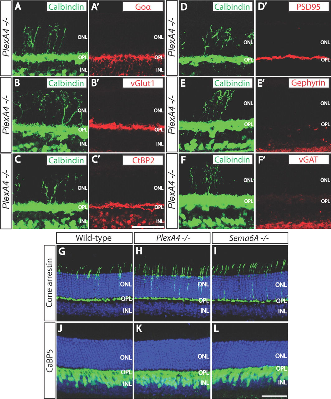
- Figure 4.
Aberrant horizontal cell neurites are not associated with ON bipolar cell dendrites, photoreceptor axon terminals, ribbon synapses, or excitatory or inhibitory synaptic markers in PlexA4−/− retinas. A–F′, PlexA4−/− adult retina sections were double immunostained with anti-calbindin (A–E, F), and anti-Goα (A′), anti-vGlut1 (B′), anti-C-terminal binding protein 2 (CtBP2, a ribbon synapse marker, C′), anti-PSD95 (D′), anti-Gephyrin (E′), or anti-vGAT (F′). Ectopic horizontal cell neurites located in the ONL of PlexA4−/− adult retinas are not accompanied by ON bipolar cell dendrites (A, A′) or photoreceptor axon terminals (B, B′), and therefore ribbon synapses are not formed on the aberrant horizontal cell neurites in the ONL (C, C′). Ectopic horizontal cell neurites located in the ONL of PlexA4−/− adult retinas are also not associated with excitatory postsynaptic regions (D, D′), inhibitory postsynaptic regions (E, E′), or inhibitory presynaptic regions (F, F′). G–L, WT (G, J), PlexA4−/− (H, K), and Sema6A−/− (I, L) adult retina sections were immunostained with anti-cone arrestin (G–I) or anti-calcium binding protein 5 (CaBP5, J–L). Cone photoreceptor axonal targeting to the OPL does not apparently differ among WT (G), PlexA4−/− (H), and Sema6A−/− (I) retinas. Rod bipolar cell as well as type 3 and type 5 cone bipolar cell dendrites do not exhibit aberrant neurite extension into the ONL of PlexA4−/− (K) and Sema6A−/− (L). Scale bars: C′ (for A–F′), L (for G–L), 50 μm.
- Figure 5.
PlexinA4 directs horizontal cell axon apposition within rod ribbon synapses, but PlexA4−/− retinas exhibit normal dark-adapted and light-adapted ERG responses. A–D, EM analysis of rod ribbon synapses reveals synaptic ultrastructure in WT (A, B) and PlexA4−/− (C, D) adult retinas. In WT retinas, rod ribbon synapses are in most cases surrounded by two horizontal cell neurites (A, B); however, in PlexA4−/− retinas, a significantly increased number of rod ribbon synapses lack one horizontal cell neurite (C, D). H, Horizontal cell neurite tips; B, bipolar cell dendritic tips. E, Quantification of ribbon synapses associated with one or no horizontal cell neurite (defined here as a “nonsurrounded ribbon”); 7.3 ± 1.7% for WT retinas and 26.2 ± 4.3% for PlexA4−/− retinas. The number of rod ribbon synapses quantified is 233 for WT and 234 for PlexA4−/− retinas (n = 3 retinas from three animals for each genotype). p = 0.015 by Student's t test. F–I, Representative dark-adapted ERGs (F) and light-adapted ERGs (H) obtained from PlexA4+/− and PlexA4−/− mice. Intensity response functions for the amplitude of the a-wave and b-wave of dark-adapted ERGs (G) and light-adapted ERGs (I) obtained from PlexA4+/− and PlexA4−/− mice. The amplitudes of the a-wave and the b-wave of dark-adapted ERGs (G) as well as light-adapted ERGs (I) are comparable between PlexA4+/− and PlexA4−/− mice. Data points indicate average ± SEM for four or more mice. Scale bar: (in D) A–D, 500 nm.
- Figure 6.
Sema6A and PlexinA4 are both localized in horizontal cell bodies and neurites. A–D″, WT retina sections from P14 (A–B″) and P5 (C–D″) mice were double immunostained with anti-calbindin (A′, B′, C′, D′, red) and either anti-Sema6A (A, C, green) or anti-PlexA4 (B, D, green). Both Sema6A and PlexA4 are localized to horizontal cell bodies (yellow arrows) and neurites within the OPL (A″, B″, C″, D″). Scale bar, 50 μm.
- Figure 7.
P5 PlexA4−/− horizontal cells exhibit reduced overall neurite coverage in the OPL, but show normal cell body spacing. A, B, P5 WT (A) and PlexA4−/− (B) whole-mount retinas were double immunostained with anti-calbindin (green) and anti-neurofilament (NF, red). In PlexA4−/− retinas, fewer horizontal cell neurites cover the surface of the OPL compared with WT retinas. C, Quantification of neurofilament+ horizontal cell neurite area (%) within the OPL of P5 WT and PlexA4−/− retinas (n = 3 retinas from three animals for each genotype). Average neurite area is 28.5 ± 0.9% for WT and 21.0 ± 1.8% for PlexA4−/− retinas. p = 0.0098 by Student's t test. D, E, P5 WT (D) and PlexA4−/− (E) whole-mount retinas were immunostained with anti-calbindin. Horizontal cell body spacing in WT and PlexA4−/− retinas is not apparently different. F, G, DRP analysis of WT (F) and PlexA4−/− (G) P5 retinas stained with anti-calbindin (n = 3 retinas from three animals for each genotype). Cell body spacing and cell number of horizontal cells measured by DRP analysis do not significantly differ between WT and PlexA4−/− retinas. Average horizontal cell densities in WT and PlexA4−/− retinas were 1019 and 1013 cells/mm2. Error bars indicate SEM. Scale bar: (in E) A, B, D, E, 50 μm.
- Figure 8.
Sema6A−/− retinas phenocopy the early postnatal horizontal cell neurite arborization defects observed in PlexA4−/− retinas. A, B, P5 WT (A) and Sema6A−/− (B) retina sections were immunostained with anti-neurofilament. Horizontal cells in Sema6A−/− retinas exhibit aberrant neurites within the INL (yellow arrows), as we observed in PlexA4−/− retinas during early postnatal retinal development. C, D, P5 WT (C) and Sema6A−/− (D) whole-mount retinas were double immunostained with anti-calbindin (green) and anti-neurofilament (NF, red). As observed in PlexA4−/− retinas, Sema6A−/− retinas exhibit fewer horizontal cell neurites covering the surface of the OPL compared with WT retinas. Scale bar, 50 μm.
- Figure 9.
PlexA4−/− horizontal cells exhibit reduced dendritic self-avoidance in vivo. A, B, Representative images of WT (A) and PlexA4−/− (B) adult horizontal cells filled with Alexa Fluor 555 fluorescence dye. A′, A″, B′, B″, Representative inverted images of WT (A′) and PlexA4−/− (B′) adult horizontal cells filled with Alexa Fluor 555 fluorescence dye. Horizontal cell neurites from these images were traced in A″ and B″. Red dots indicate sites where neurites cross. C, Quantification of average horizontal cell neurite length per neuron of WT and PlexA4−/− adult horizontal cells. The average horizontal cell neurite lengths per cell are 1238 ± 107 μm for WT (n = 7) and 1492 ± 115 μm for PlexA4−/− (n = 8) horizontal cells. p = 0.134, Student's t test. D, Quantification of the average number of self-neurite crossings in WT and PlexA4−/− adult horizontal cells. The average number of crossings per cell are 10.3 ± 1.6 for WT (n = 7) and 23.8 ± 2.5 for PlexA4−/− (n = 8) horizontal cells. p = 0.0007, Student's t test. Scale bar: (in A) for A–B″, 20 μm.


