

Article Figures & Data
Figures
- Figure 1.
Neuronal death induced by epileptic seizures is blocked in Lgals1 mutants. A, FJB staining showing degenerating neurons in different areas of the cerebral cortex and hippocampus, in wild-type mice injected with saline solution and in wild-type or Lgals1-mutant animals injected with pilocarpine. B, Quantification of the total number of FJB-positive neurons in the cortex and hippocampus of injected animals, per 30 μm section. Scale bar, 200 μm.
- Figure 2.
Lack of programmed cell death in Lgals1-mutant mice after pilocarpine-induced seizures. A, TUNEL staining in the parietal cortex, comparing wild-type and Lgals1-mutant mice 24 h after pilocarpine injection. Scale bar, 100 μm. B, Quantification of TUNEL-positive cells in the parietal cortex. C, Coimmunostaining for NeuN and active caspase-3 in the parietal cortex. Scale bar, 100 μm. D, Quantification of the NeuN-positive neuron proportion also positive for active caspase-3 staining. E, Colocalization of active caspase-3 and Gal-1 in the CA1 of the hippocampus following pilocarpine-induced seizures. Scale bar, 50 μm.
- Figure 3.
The number of Gal-1-expressing neurons decreases after epileptic seizures. A, Gal-1 immunoreactive cells are almost exclusively neurons. Coimmunostaining in the hippocampus and the cerebral cortex with antibodies against Gal-1 and NeuN. B, The proportion of Gal-1-positive cells identified as neurons is plotted as the percentage of Gal-1-immunoreactive cells also positive for NeuN immunostaining. C, The pattern of neuronal degeneration triggered by pilocarpine injection parallels the pattern of Gal-1 expression in basal conditions. D, In the parietal cortex, the number of neurons expressing Gal-1 decreases 24 and 72 h after pilocarpine-induced seizures. E, Quantification of the total number of Gal-1/NeuN double-positive neurons in the piriform cortex and hippocampus, per 30 μm section. F, p75NTR immunoreactivity in the hippocampus and piriform cortex 24 h after pilocarpine-induced seizures. Scale bars: A, C, F, 200 μm; D, 100 μm. n.s., not statistically significant.
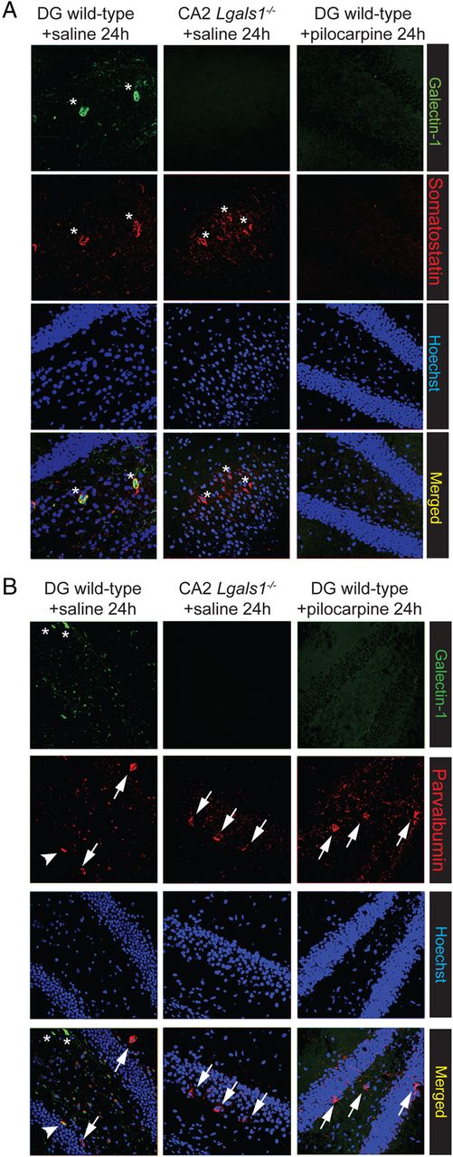
- Figure 4.
Gal-1-positive neurons are also somatostatin-positive. A, Colocalization of Gal-1-positive and somatostatin-positive neurons (asterisks) in the hippocampus of wild-type mice. Note that somatostatin neurons are still present in the hippocampus of Lgals1−/− mice. B, Most (arrows), but not all (arrowhead), paravalbumin-positive neurons do not express Gal-1 (asterisks).
- Figure 5.
Gal-1 expression increases in astrocytes 72 h after epileptic seizures. A, Coimmunostaining for Gal-1 and GFAP in the piriform cortex. B, Quantification of the number of Gal-1-positive cells also positive for GFAP in the cerebral cortex and the hippocampus. Scale bar, 100 μm.
- Figure 6.
Glial reactivity is blocked in Lgals1-mutant mice after pilocarpine injection. A, Astrocyte-specific GFAP immunostaining in the cerebral cortex and hippocampus, comparing wild-type and Lgals1-mutant mice, 24 and 72 h after injection. B, Microglia-specific CD11b immunostaining in the cerebral cortex and hippocampus in wild-type versus Lgals1-mutant mice 24 h after injection. Scale bar, 200 μm.
- Figure 7.
p75NTR is overexpressed in Lgals1 mutant mice after pilocarpine injection. A, p75NTR immunostaining in the parietal cortex and hippocampus, comparing wild-type and Lgals1-mutant mice, 24 h after injection. Scale bar, 200 μm. B, p75NTR is expressed in the Gal-1-positive neurons (asterisks) 24 h after pilocarpine injections in the hippocampus (arrows).
- Figure 8.
Gal-3 is expressed in microglial cells after epileptic seizures, but not in Lgals1-mutant mice. Coimmunostaining for Gal-3 and CD11b in the parietal cortex. Scale bar, 100 μm.
- Figure 9.
Pilocarpine-induced seizures trigger neuronal degeneration in Lgals3-mutant mice. A, Coimmunostaining for Gal-3 and CD11b in the parietal cortex 72 h after pilocarpine injection. B, Quantification of the total FJB-positive neurons in the cerebral cortex and hippocampus of injected animals. Scale bar, 100 μm. n.s., not statistically significant.


