

Article Figures & Data
Figures
- Figure 1.
Structure of the transgene and characterization of the transgenic mice. A, Partial structure of the PLP/Fv2E-PERK transgene. Transcription initiates within the PLP portion of the transgene and drives expression of the Fv2E-PERK open reading frame. B, Western blotting showed that the Fv2E-PERK protein was expressed in the brains of PLP/Fv2E-PERK mice (Tg), but not in littermate wild-type mice (WT). Moreover, Fv2E-PERK was not expressed in heart, lung, liver, kidney, or spleen in the transgenic mice. C, CC1 and FKBP12 double immunostaining showed that wild-type mice did not express Fv2E-PERK in the brain and that Fv2E-PERK was expressed specifically in oligodendrocytes in the brains of PLP/Fv2E-PERK mice. D, GFAP and FKBP12 double immunostaining showed that Fv2E-PERK was not expressed by astrocytes in the brains of PLP/Fv2E-PERK mice. E, NeuN and FKBP12 double immunostaining showed that Fv2E-PERK was not expressed by neurons in the brains of PLP/Fv2E-PERK mice. F, CD11b and FKBP12 double immunostaining showed that Fv2E-PERK was not expressed by microglia in the brains of PLP/Fv2E-PERK mice. G, PDGFαR and FKBP12 double immunostaining showed that Fv2E-PERK was not expressed by OPCs in the brains of PLP/Fv2E-PERK mice; N = 3 animals. Scale bar: C, 23 μm; D–G, 31 μm.
- Figure 2.
Persistent activation of PERK signaling specifically in oligodendrocytes was not detrimental to these cells or to myelin in adult animals. A, Real-time PCR analysis showed that AP20187-induced PERK signaling increased the expression of CHOP and GADD34 in the mixed glial cultures generated from neonatal PLP/Fv2E-PERK mice in a dose-dependent manner but did not significantly affect the expression of BIP. B, Real-time PCR analysis showed that AP20187 treatment increased the expression of CHOP and GADD34 in the spinal cords of PLP/Fv2E-PERK mice in a dose-dependent manner but did not significantly affect the expression of BIP. C, D, CC1 and p-eIF2α double immunostaining showed that p-eIF2α was undetectable in oligodendrocytes in the spinal cords of PLP/Fv2E-PERK mice treated with vehicle but became detectable in the oligodendrocytes of mice treated with AP20187. E, Cell counting revealed that the numbers of CC1-positive oligodendrocytes in the white matter of the spinal cords and the cerebellums of PLP/Fv2E-PERK mice treated with AP20187 were comparable with those of mice treated with vehicle. F, G, MBP immunostaining showed that AP20187 treatment did not alter myelin integrity in the spinal cords of adult PLP/Fv2E-PERK mice; N = 3 animals. Error bars indicate SD. ★p < 0.05. Scale bar: C, D, 12 μm; F, G, 200 μm.
- Figure 3.
Enhancing the PERK-mediated ISR specifically in oligodendrocytes attenuated EAE disease severity. A, Mean clinical score were recorded daily: 0 = healthy; 1 = flaccid tail; 2 = ataxia and/or paresis of hindlimbs; 3 = paralysis of hindlimbs and/or paresis of forelimbs; 4 = tetraparalysis; 5 = moribund or death. N = 12 animals. Error bars indicate SD. ★p < 0.05. B, Real-time PCR analysis showed that AP20187 treatment increased the expression of CHOP in the spinal cords of PLP/Fv2E-PERK mice at PID12; N = 4 animals. Error bars indicate SD. ★p < 0.05. C, D, CC1 and p-eIF2α double immunostaining showed that the level of p-eIF2α was markedly increased in oligodendrocytes in the lumbar spinal cords of PLP/Fv2E-PERK mice treated with AP20187 at PID12; N = 3 animals. Scale bar: C, D, 23 μm.
- Figure 4.
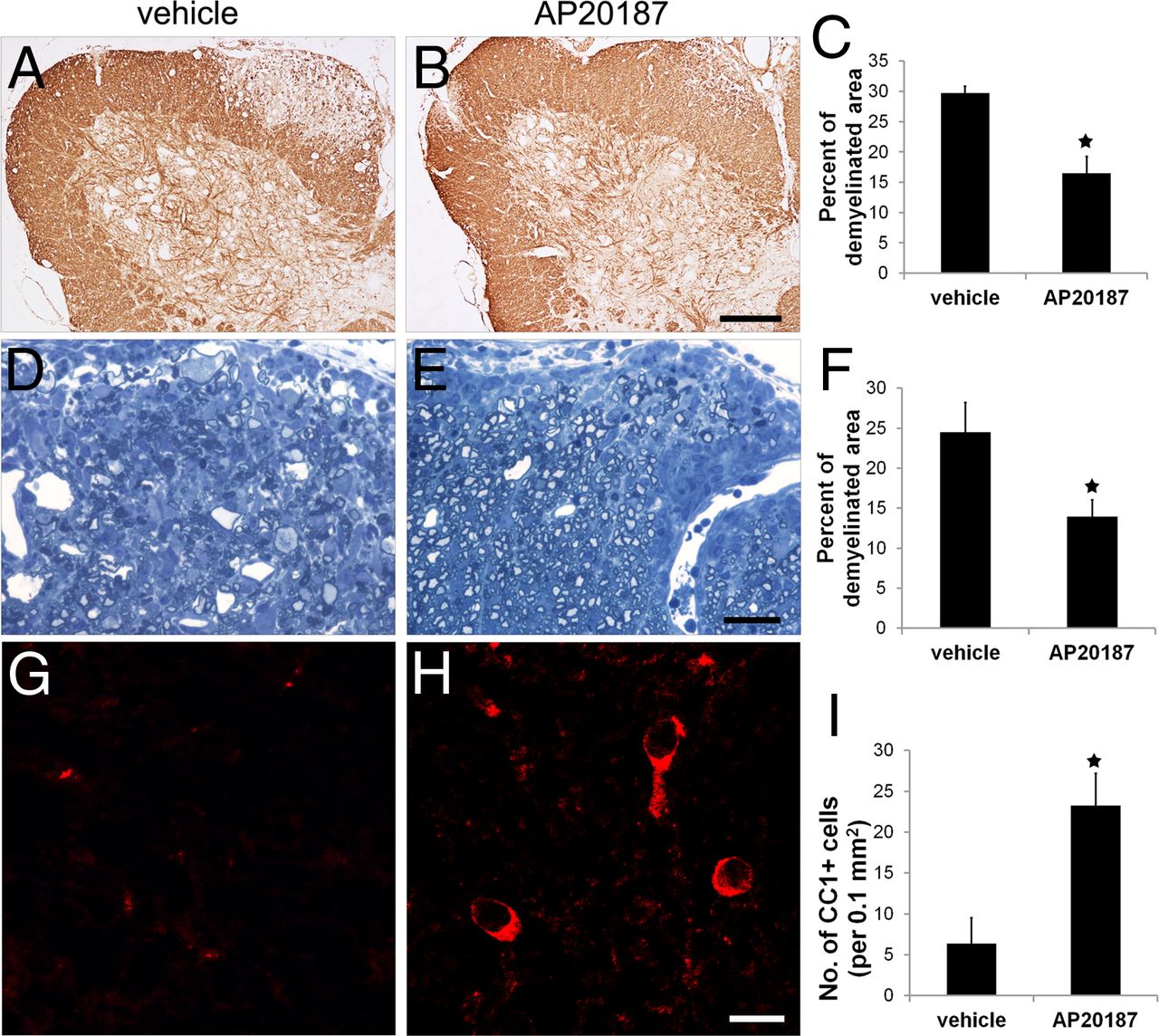
Enhancing the PERK-mediated ISR specifically in oligodendrocytes ameliorated EAE-induced demyelination and oligodendrocyte loss. A–C, At PID19, MBP immunostaining revealed large demyelinating lesions in the lumbar spinal cords of PLP/Fv2E-PERK mice treated with vehicle. Interestingly, AP20187 treatment significantly reduced the size of demyelinating lesions in PLP/Fv2E-PERK mice; 20 sections per mouse, N = 4 animals. D–F, Toluidine blue staining showed that AP20187 treatment significantly reduced the percentage of the white matter area of the lumbar spinal cord that was demyelinated in PLP/Fv2E-PERK mice at PID19; 5 serial sections per mouse, N = 4 animals. G–I, CC1 immunostaining showed that AP20187 treatment significantly attenuated the oligodendrocyte loss observed at PID19 in the demyelinating lesions in the lumbar spinal cord of PLP/Fv2E-PERK mice; 20 sections per mouse, N = 4 animals. Error bars indicate SD. ★p < 0.05. Scale bar: A, B, 200 μm; D, E, 30 μm; G, H, 15 μm.
- Figure 5.
Enhancing the PERK-mediated ISR specifically in oligodendrocytes ameliorated EAE-induced axonal degeneration. A, B, SMI-31 immunostaining of lumbar spinal cord longitudinal sections showed that AP20187 treatment markedly attenuated axon loss and axon degeneration in the demyelinating lesions in PLP/Fv2E-PERK mice at PID 19. C–F, EM analysis showed that AP20187 treatment significantly reduced the number of degenerating axons (*, arrow) and increased the number of total axons in the demyelinating lesions in PLP/Fv2E-PERK mice at PID19; N = 4 animals. Error bars indicate SD. ★p < 0.05. Scale bar: A, B, 10 μm; C, D, 2 μm.
- Figure 6.
AP20187 treatment did not directly act on T cells to influence their activity in PLP/Fv2E-PERK mice undergoing EAE. A, The BrdU cell proliferation assay showed that AP20187 treatment did not affect T-cell proliferation in response to MOG35–55 peptide. B, The MTT-cell viability assay showed that AP20187 treatment did not alter T-cell viability in response to MOG35–55 peptide. C, D, ELISA analyses showed that AP20187 treatment did not significantly affect the ability of T cells to produce the Th1 cytokine IFN-γ or the Th2 cytokine IL-4 in response to MOG35–55 peptide. N = 4 animals. Error bars indicate SD.
- Figure 7.
AP20187 treatment did not alter the degree of the inflammatory response in the CNS of PLP/Fv2E-PERK mice undergoing EAE. A, B, E, CD3 immunostaining showed that AP20187 treatment did not affect T-cell infiltration into the white matter of the lumbar spinal cord in PLP/Fv2E-PERK mice at PID12; 20 sections per mouse, N = 4 animals. C–E, CD11b immunostaining showed that AP20187 treatment did not change the number of microglia/macrophages present in the white matter of the lumbar spinal cord in PLP/Fv2E-PERK mice at PID12; 20 sections per mouse, N = 4 animals. F, Real-time PCR analysis showed that AP20187 treatment did not significantly alter the levels of the mRNAs encoding iNOS, TNF-α, IFN-γ, IL-4, IL-5, IL-12, IL-17, and IL-23 in the spinal cord of PLP/Fv2E-PERK mice at PID12; N = 4 animals. G, H, K, CD3 and MBP double immunostaining showed that AP20187 treatment did not change the number of T cells present in the white matter of the lumbar spinal cord in PLP/Fv2E-PERK mice at PID19; 20 sections per mouse, N = 4 animals. I–K, CD11b and MBP double immunostaining showed that AP20187 treatment did not change the number of microglia/macrophages present in the white matter of the lumbar spinal cord in PLP/Fv2E-PERK mice at PID19; 20 sections per mouse, N = 4 animals. L, Real-time PCR analysis showed that AP20187 treatment did not significantly alter the levels of the mRNAs encoding iNOS, TNF-α, IFN-γ, IL-4, IL-5, IL-12, IL-17, and IL-23 in the spinal cord of PLP/Fv2E-PERK mice at PID19; N = 4 animals. Error bars indicate SD. Scale bar: A, B, 20 μm; C, D, 60 μm; G, H, 25 μm; I, J, 25 μm.
- Figure 8.
Enhancing the PERK-mediated ISR specifically in oligodendrocytes protected these cells against immune attack. A–D, M, CC1, TUNEL, and DAPI triple labeling showed that there is no apoptotic oligodendrocytes in the lumbar spinal cords of 10-week-old naive PLP/Fv2E-PERK mice. E–H, M, At PID12, CC1, TUNEL, and DAPI triple labeling revealed a number of apoptotic oligodendrocytes (arrow) in the lumbar spinal cords of PLP/Fv2E-PERK mice treated with vehicle. Inset, Apoptotic oligodendrocytes were CC1- and TUNEL-positive with nuclear fragmentation. I–M. Nevertheless, the number of apoptotic oligodendrocytes was dramatically reduced in the lumbar spinal cords of PLP/Fv2E-PERK mice treated with AP20187; 20 sections per mouse, N = 4 animals. Error bars indicate SD. ★p < 0.05. Scale bar: A–L, 20 μm.
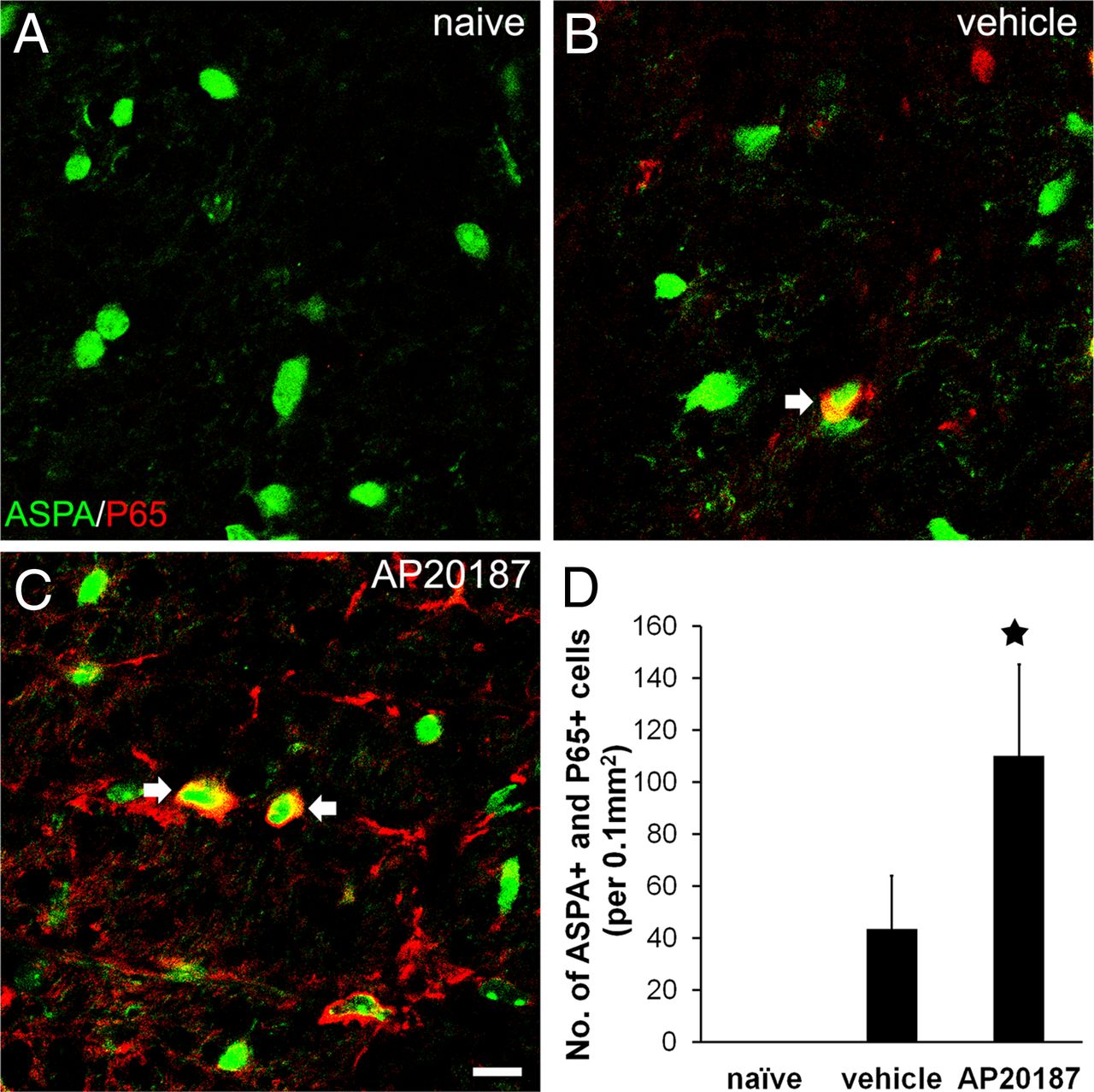
- Figure 9.
Enhancing the PERK-mediated ISR specifically in oligodendrocytes increased activation of the NF-κB pathway in the CNS of EAE mice. A, Double immunostaining for ASPA and the active form of NF-κB p65 showed that the active form of NF-κB p65 was undetectable in oligodendrocytes in the CNS of adult naive mice. B, However, the active form of NF-κB p65 was detectable at PID12 in a number of oligodendrocytes in the lumbar spinal cord white matter of PLP/Fv2E-PERK mice treated with vehicle. C, Interestingly, the number of oligodendrocytes positive for the active form of NF-κB p65 was further increased in the lumbar spinal cords of AP20187-treated mice. D, Quantitative analysis showed that the number of oligodendrocytes positive for the active form of NF-κB p65 in the lumbar spinal cord white matter of PLP/Fv2E-PERK mice treated with AP20187 was significantly increased compared with vehicle-treated mice; 20 sections per mouse, N = 4 animals. Error bars indicate SD. ★p < 0.05. Scale bar: A–C, 20 μm.
- Figure 10.
The PERK-mediated ISR activated the NF-κB pathway in oligodendrocytes. A, PLP and p-eIF2α double immunostaining showed that AP20187 treatment had no effect on eIF2α phosphorylation in differentiated oligodendrocytes cultured from wild-type mice. In contrast, AP20187 treatment resulted in eIF2α phosphorylation in the cells cultured from PLP/Fv2E-PERK mice. B, 2′,3′-Cyclic nucleotide phosphodiesterase and the active form of NF-κB p65 double immunostaining showed that AP20187 treatment had no effect on NF-κB p65 activation in differentiated oligodendrocytes cultured from wild-type mice. Interestingly, AP20187 treatment resulted in NF-κB p65 activation in the cells cultured from PLP/Fv2E-PERK mice. Scale bar: A, B, 20 μm.


