

Article Figures & Data
Figures
- Figure 1.
Retrogradely labeled neurons in LEC following viral injections into the olfactory bulb (GFP) or the aPCX (mCherry). Labeled neurons were distributed throughout the LEC, especially in layers II–V in the ipsilateral LEC, with the most robust labeling observed after aPCX infections. With these markers, no double-labeled cells were observed in any animal. A, Left, Section labeled with RV-GFP (olfactory bulb) and RV-mCherry (aPCX); right, annotated corresponding to the section (bregma 5.3) adapted from (Paxinos and Watson, 2009). Scale bar, 2 mm. B–E, Higher-magnification images of boxed regions in (A). B,D, High-magnification of lamina II–III. C, E, High-magnification of lamina V–VI, from adjacent sections for illustration. Scale bar, 100 μm. F, G, High-magnification images of the labeled neurons in lamina V–VI in contra lateral LEC. Scale bar, 100 μm. H, Schematic representation of the rabies viruses mediated retrograde tracing of the projection neurons from LEC to the olfactory bulb and aPCX. I, Quantification of mean cell counts across animals (n = 7) in the different lamina ipsilateral and contralateral to the OB and aPCX infections. Here and in all subsequent figures, data are presented as mean ± SEM.
- Figure 2.
Spontaneous activity recorded in aPCX of urethane anesthetized rats was enhanced by muscimol infusion into the ipsilateral LEC. A, Representative example of increased aPCX single-unit firing rate within a few minutes of muscimol infusion into the ipsilateral LEC. B, Of the 10 cells tested, nine showed enhanced activity after LEC infusion. C, There was no spontaneous activity change after control (saline or no infusion) manipulations. D, Silencing of the LEC with muscimol significantly enhanced ipsilateral aPCX single-unit (n = 10) spontaneous activity (asterisk represents significant difference from controls, p < 0.01). E, Representative example of raw LFP recording in aPCX and pseudocolor sonograph of the same data before (E) and after (F) muscimol infusion into the ipsilateral LEC. Pseudocolor show higher power in yellows and reds. Resp, Respiration from chestwall movement. Time scale (2 s) and voltage scale are shown. G, Representative quantitative example of change in LFP spontaneous activity in the aPCX and LEC (percentage change in power from pre-infusion) following muscimol infusion into the LEC. Note the relative depression of LEC activity and relative enhancement of aPCX activity. H, Mean-fold change in aPCX odor-evoked LFP activity following (10–30 min) LEC muscimol infusion (n = 4). Note particular enhancement in the beta and low gamma range in these anesthetized animals. Asterisks represent significant change from baseline. Asterisks represent significant change, p < 0.05.
- Figure 3.
aPCX single-unit entrainment to aPCX LFP beta oscillations is reduced by LEC muscimol in anesthetized animals. A, Representative example of aPCX single-unit entrainment to LFP beta oscillation recorded in aPCX before and during ipsilateral LEC muscimol infusion. Average beta cycle shown at top. B, Polar plot of activity from the cell shown in A relative to beta oscillation cycle before and during LEC muscimol. Number in the top-left quadrant signifies maximal circle radius. Note the increase in firing rate after LEC muscimol and reduction in beta entrainment in this cell. C, Vector describing activity from the cell shown in A relative to beta oscillation cycle before and during LEC muscimol. Note the dramatic reduction in vector length during LEC suppression, from a statistically significant 0.45 (possible range 0–1) during baseline to 0.13, 30 min after LEC muscimol. D, Mean aPCX single-unit activity (n = 9 cells) relative to beta oscillation phase before and after ipsilateral LEC muscimol infusion. Note the increase in firing rate after LEC muscimol and reduction in beta entrainment.
- Figure 4.
A, Animals were trained in a go-left, go-right, two-alternative forced-choice odor discrimination task. B, Comparison of discrimination task difficulty as determined by initial rate of acquisition from 4 representative animals in each task. The 10c versus 10-1 discrimination required significantly more trials to reach criterion than the 10c versus 10cR1 task as previously reported (Barnes et al., 2008; Chapuis and Wilson, 2012). C, An example of LFP beta-band activity (signals digitally filtered 15–35 Hz) recorded in aPCX of a rat performing in the two-alternative forced-choice task under control conditions and during unilateral infusion of muscimol (0.5 mg) or saline into the LEC. The treatments occurred over the course of three consecutive days in the order shown. “Odor sample” marks the time spent in the odor sampling port on a given trial, whereas “left water” marks the time spent in the left water delivery port. For simplicity no choices to the right are shown. LEC silencing enhanced aPCX oscillations (beta activity during odor sampling shown) in behaving rats, which recovered to normal the following day under saline infusion. Top, Time × frequency pseudocolor plots of power aligned to the LFP recordings and behavioral markers immediately below. Total power within the beta frequency band during odor sampling are plotted for the three conditions at bottom.
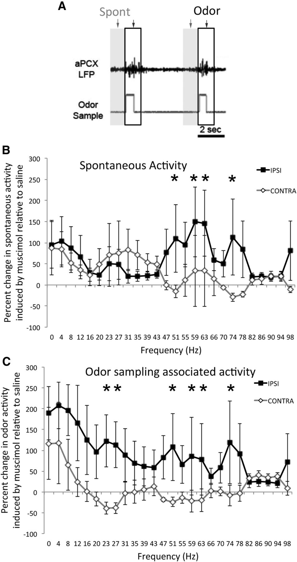
- Figure 5.
Unilateral muscimol infusions into the LEC enhanced LFP oscillations during odor sampling in the ipsilateral aPCX of rats performing in an odor discrimination task. A, FFT analyses were performed during the 1 s immediate pre-odor sampling period (“spontaneous”, gray shaded region) or during 1 s beginning at the start of odor sampling (outlined region), across all trials in a session. B, Change in LFP power during LEC muscimol infusions relative to control (saline) infusions for both the aPCX ipsilateral to the infusion and contralateral. LEC silencing induced a significant enhancement in spontaneous gamma frequency oscillations in the ipsilateral aPCX compared with the contralateral aPCX (within animal comparisons, n = 6). C, During odor sampling both beta and gamma frequency oscillations were enhanced ipsilateral to the silenced LEC. Asterisks signify significant (p < 0.05) post hoc comparisons between ipsilateral and contralateral change.
- Figure 6.
Bilateral muscimol infusion into the LEC of rats performing an two-alternative forced-choice task impaired fine odor discrimination but did not impact gross odor discrimination. A, Bilateral infusion of 0.5 mg of muscimol into the LEC had no effect on a simple odor discrimination (10c vs 10R1). Each manipulation in this animal was performed on different days in the order shown. Infusion of larger volumes (1 mg) did impair discrimination but also impaired response rats and general behavior. B, Bilateral infusion of 0.5 mg of muscimol in the LEC impaired discrimination of two highly similar odorant mixtures, without any reduction in general performance (trials initiated). C, Bilateral LEC silencing impaired performance on a well learned but difficult discrimination task (n = 8), but had no effect on a well learned simpler task (n = 4). Asterisk signifies significant reduction (p < 0.05) in percentage correct in the muscimol condition for the 10c versus 10c-1 discrimination compared with control conditions.
- Figure 7.
Bilateral infusion of muscimol into the aPCX impaired gross odor discrimination (10c vs 10cR1). A, Although LEC muscimol infusions had no effect on this discrimination task (Fig. 5), mucimol infusion into the aPCX was sufficient to impair discrimination in all animals (n = 5) compared with control conditions. B, Performance during muscimol infusion was significantly impaired compared with control conditions. Asterisk represents significant difference from controls, p < 0.05.
- Figure 8.
Impairment the discrimination task in LEC infused rats was not due to general behavioral performance deficits. A, Bilateral LEC muscimol infusions significantly increased error rate in the difficult task comparably between left and right choices. Asterisks signify significant difference between difficult muscimol condition from all other conditions (p < 0.05). B, Odor sampling time (duration in the odor sampling port) was not significantly modified by bilateral LEC muscimol infusion. There was a trend toward extended sampling times in the difficult task (10c vs 10c-1; n = 8 rats) compared with the easy task (10c vs 10R1, n = 4 rats), though this difference was not significant and was not significantly affected by LEC muscimol. C, D, Latency to move from the odor sampling port to the water reward ports was not changed in either task by LEC muscimol. Mean distribution of response latencies are shown for both tasks and infusion conditions.


