

Article Figures & Data
Figures
- Figure 1.
Calpain activity is required for synaptic NMDAR-dependent neuroprotection against starvation and oxidative stress. A, Trophic deprivation for 3 d induced cultured cortical neuron death. Incubation with 20 μm Bic and 100 μm 4-AP during starvation period (Bic + 4-AP) or before starvation (Bic + 4-AP, then MK801 + TTX) reduced neuronal death. Coapplication of 10 μm CI-III with Bic and 4-AP blocked the neuroprotective effect of Bic and 4-AP. Incubation of neurons under either normal condition or starvation with 10 μm CI-III alone for 3 d did not cause cell death. Neuronal death was observed and quantified by Hoechst staining; 300–500 cells were counted for each group in each independent experiment. *p < 0.05; ns, not significantly different; one-way ANOVA followed by Bonferroni test. n = 3. Error bar indicates SEM. B, Similar experimental design as in A except that neuronal death was induced by a 24 h-H2O2 insult (20 μm). In all cases, results are means ± SEM of three independent experiments with 300–500 cells for each group in each independent experiment. *p < 0.05; ns, not significantly different; one-way ANOVA followed by Bonferroni test. C, Representative photos of Hoechst staining. Hoechst brightly stains condensed and/or fragmented nuclei of apoptotic neurons but only dimly stains normal nuclei of healthy neurons. Scale bar, 10 μm.
- Figure 2.
Synaptic NMDAR-dependent calpain activation degrades PHLPP1 and activates Akt and ERK1/2 pathway. A, Treatment of rat brain P2 fraction aliquots with purified μ-calpain (4 μg/ml) in the presence of 200 μm Ca2+ or purified m-calpain (4 μg/ml) in the presence of 2 mm Ca2+ for 1 h reduced the levels of full-length PHLPP1α and PHLPP1β, as compared with treatment with μ-calpain or m-calpain alone. B, Western blots for rat acute hippocampal slices treated with 20 μm Bic and 100 μm 4-AP for 30 min. In another group, 10 μm CI-III was applied 20 min before Bic and 4-AP treatment. PHLPP1α, PHLPP1β, phospho-Akt S473 (pAkt), Akt, phospho-ERK 1/2 (pERK 1/2), and ERK1/2 were detected by Western blot. C, Quantitative analysis of Western blots similar to those shown in B. The ratios of PHLPP1 to Akt (loading control), p-Akt to Akt, and p-ERK 1/2 to ERK 1/2 were compared. All ratios were normalized to control values before analysis. Bic and 4-AP treatment decreased PHLPP1/Akt and increased p-Akt/ Akt and p-ERK/ ERK. Preapplication of CI-III blocked those changes. *p < 0.05; ns, not significantly different; one-way ANOVA followed by Bonferroni test; n = 3. D, Cultured cortical neurons (DIV12) were treated with 20 μm Bic and 100 μm 4-AP for 30 min. In another group, 10 μm CI-III was applied 10 min before Bic and 4-AP treatment. The levels of PHLPP1, p-Akt, Akt, p-ERK1/2, ERK1/2, and PTEN were detected by Western blot. E, Quantitative analysis of Western blots similar to those shown in D. Bic and 4-AP treatment decreased PHLPP1/Akt and increased p-Akt/Akt and p-ERK/ERK, effects which were blocked by CI-III. PTEN levels were not affected by treatments. *p < 0.05; ns, not significantly different; one-way ANOVA followed by Bonferroni test; n = 3. Error bars indicate SEM.
- Figure 3.
Lentiviral shRNA knockdown of PHLPP1 mimics the neuroprotective effect of synaptic NMDAR activation and eliminates the effect of calpain inhibition on neuroprotection. A, Cultured cortical neurons were infected by PHLPP1 shRNA lentivirus or scrambled shRNA lentivirus. The levels of indicated proteins in infected neurons were assessed by Western blot. B, shRNA lentivirus-infected neurons were treated with Bic and 4-AP or with CI-III, Bic, and 4-AP for 30 min. The levels of indicated proteins were assessed by Western blot. C, Quantitative analysis of Western blots similar to those shown in A and B. Bic and 4-AP treatment reduced PHLPP1α and PHLPP1β levels and increased p-Akt/Akt levels in scrambled RNAi-infected neurons but not in PHLPP1 RNAi-infected neurons. CI-III blocked Bic and 4-AP-induced changes in PHLPP1, p-Akt/Akt, and p-ERK/ERK levels in scrambled RNAi-infected neurons but not in PHLPP1 RNAi-infected neurons. *p < 0.05; ns, not significantly different; one-way ANOVA followed by Bonferroni test; n = 3. Error bars indicate SEM. D, shRNA lentivirus-infected neurons were subjected to starvation for 3 d. PHLPP1 RNAi-infected neurons showed decreased neuronal death compared with scrambled RNAi-infected neurons. Bic and 4-AP incubation along with starvation decreased neuronal death in scrambled RNAi-infected neurons but not in PHLPP1 RNAi-infected neurons. CI-III blocked Bic and 4-AP induced reduction of neuronal death in scrambled RNAi-infected neurons but not in PHLPP1 RNAi-infected neurons. Approximately 300–500 cells were counted for each group in each independent experiment. *p < 0.05; ns, not significantly different; one-way ANOVA followed by Bonferroni test. n = 3. Error bar indicates SEM.
- Figure 4.
Extrasynaptic NMDAR-induced calpain activation degrades STEP but not PHLPP1 and mediates neurotoxicity. A, Cortical neurons were treated with 40 μm MK801 and 10 μm Bic for 2 min, washed three times, then treated with 100 μm NMDA and 10 μm glycine for 1 h to induce extrasynaptic NMDAR activation. In another group, 10 μm CI-III was applied 10 min before addition of NMDA and glycine. The levels of indicated proteins were assessed by Western blot. B, Quantitative analysis of Western blots similar to those shown in A. Extrasynaptic NMDAR activation decreased STEP61 levels and increased STEP33 levels. CI-III blocked extrasynaptic activation-induced STEP cleavage. PHLPP1, pAkt/Akt, and pERK/ERK levels were not affected by extrasynaptic NMDAR activation. *p < 0.05; ns, not significantly different; one-way ANOVA followed by Bonferroni test; n = 3. Error bars indicate SEM. C, Extrasynaptic NMDAR activation for 2 h induced neuronal death. Addition of 10 μm CI-III along with extrasynaptic activation reduced neuronal death. At least 300–500 Hoechst-stained cells were counted for each group in each independent experiment. *p < 0.05; one-way ANOVA followed by Bonferroni test. n = 3. Error bar indicates SEM.
- Figure 5.
An m-calpain-specific inhibitor blocks extrasynaptic NMDAR-induced neurotoxicity but not synaptic NMDAR-induced neuroprotection. A, Cortical neurons were treated with 20 μm Bic and 100 μm 4-AP for 30 min and lysed for Western blot. In another group, 200 nm mCalp-I was applied 10 min before Bic and 4-AP treatment. The levels of indicated proteins were assessed by Western blot. B, Quantitative analysis of Western blots similar to those shown in A. mCalp-I (200 nm) did not affect Bic- and 4-AP-induced changes in PHLPP1, pAkt/Akt, and pERK/ERK levels. *p < 0.05; ns, not significantly different; one-way ANOVA followed by Bonferroni test; n = 3. Error bars indicate SEM. C, mCalp-I at 5 μm but not 200 nm blocked Bic- and 4-AP-induced neuroprotection against starvation in cortical neurons. At least 300–500 Hoechst-stained cells were counted for each group in each independent experiment. *p < 0.05; ns, not significantly different; one-way ANOVA followed by Bonferroni test. n = 3–6. Error bar indicates SEM. D, 5 μm but not 200 nm mCalp-I blocked Bic- and 4-A-induced neuroprotection against H2O2 insult. n = 3. E, Cortical neurons were treated with the protocol of extrasynaptic NMDAR activation and then lysed for Western blot. In another group, 200 nm mCalp-I was applied 10 min before extrasynaptic activation. F, Quantitative analysis of Western blots similar to those shown in E. mCalp-I (200 nm) reduced the cleavage of STEP induced by extrasynaptic NMDAR activation. *p < 0.05; one-way ANOVA followed by Bonferroni test; n = 3–5. Error bars indicate SEM. G, Pretreatment with 200 nm or 5 μm mCalp-I reduced neuronal death induced by extrasynaptic NMDAR activation. Approximately 300–500 Hoechst-stained cells were counted for each group in each independent experiment. *p < 0.05; one-way ANOVA followed by Bonferroni test. n = 4. Error bar indicates SEM.
- Figure 6.
A μ-calpain-specific inhibitor blocks synaptic NMDAR-induced neuroprotection but not extrasynaptic NMDAR-induced neurotoxicity. A, Cortical neurons were treated with 20 μm Bic and 100 μm 4-AP for 30 min and lysed for Western blot. In another group, 2 μm μCalp-I was applied 10 min before Bic and 4-AP treatment. The levels of indicated proteins were assessed by Western blot. B, Quantitative analysis of Western blots similar to those shown in A. μCalp-I (2 μm) blocked Bic- and 4-AP-induced changes in PHLPP1, pAkt/Akt, and pERK/ERK levels. *p < 0.05; one-way ANOVA followed by Bonferroni test; n = 3. Error bars indicate SEM. C, μCalp-I (2 μm) blocked Bic- and 4-AP-induced neuroprotection against starvation in cortical neurons. Incubation of neurons with 2 μm μCalp-I alone for 3 d did not cause cell death. At least 300–500 Hoechst-stained cells were counted for each group in each independent experiment. *p < 0.05; ns, not significantly different; one-way ANOVA followed by Bonferroni test. n = 3–5. Error bar indicates SEM. D, μCalp-I (2 μm) blocked Bic- and 4-AP-induced neuroprotection against H2O2. n = 3. E, Cortical neurons were treated with the protocol for extrasynaptic NMDAR activation and then lysed for Western blot. In another group, 2 μm μCalp-I was applied 10 min before extrasynaptic NMDAR activation. F, Quantitative analysis of Western blots similar to those shown in E. μCalp-I (2 μm) did not affect STEP degradation induced by extrasynaptic NMDAR activation (ns, not significantly different; two-tailed t test; n = 3). Error bars indicate SEM. G, μCalp-I (2 μm) did not affect neuronal death induced by extrasynaptic NMDAR activation. Approximately 300–500 Hoechst-stained cells were counted for each group in each independent experiment. ns, not significantly different; two-tailed t test; n = 4–5. Error bar indicates SEM.
- Figure 7.
μ-calpain but not m-calpain knockdown blocks PHLPP1 degradation and Akt and ERK activation induced by synaptic NMDAR activation. A, Cultured cortical neurons at DIV 7 were infected with scrambled siRNA, μ-calpain siRNA, or m-calpain siRNA separately. Two days after infection, the levels of indicated proteins were assessed by Western blot. B, Quantitative analysis of Western blots similar to those shown in A. μ-calpain and m-calpain siRNA significantly reduced μ-calpain and m-calpain levels, respectively. *p < 0.05; one-way ANOVA followed by Bonferroni test. n = 3. C, siRNA-infected neurons were treated with Bic and 4-AP for 30 min. The levels of indicated proteins in infected neurons before or after the treatment were assessed by Western blot. D, Quantitative analysis of Western blots similar to those shown in C. The levels of PHLPP1α and PHLPP1β were significantly higher and the ratios of pAkt/Akt and pERK/ERK were significantly lower in μ-calpain siRNA-infected neurons than that in scrambled siRNA-infected neurons. *p < 0.05; ns, not significantly different; one-way ANOVA followed by Bonferroni test; n = 3.
- Figure 8.
μ-calpain but not m-calpain is recruited to synaptic NMDAR complex following synaptic NMDAR activation. A, NR2A coprecipitated μ-calpain,NR2B, and PSD95 but not m-calpain from lysates of cultured cortical neurons. Treatment of neurons with 20 μm Bic, 100 μm 4-AP, and 10 μm CI-III for 1 h increased the association of μ-calpain with NR2A. IP with nonimmune rabbit IgG was included as a negative control. B, Statistical analysis of three independent co-IP experiments. The ratios of μ-calpain or NR2B to NR2A in IP fraction were compared. All ratios were normalized to the average ratio of basal group before t test (*p < 0.05; ns, no significant difference; two-tailed t test; n = 3). C, PHLPP1 coprecipitated μ-calpain and NR2A but not m-calpain from lysates of cultured cortical neurons. Treatment of neurons with Bic, 4-AP, and CI-III for 1 h increased the association of μ-calpain with PHLPP1. D, Statistical analysis of three independent co-IP experiments. The ratios of μ-calpain or NR2A to PHLPP1 in IP fraction were compared (*p < 0.05; ns, no significant difference; two-tailed t test; n = 3). E, F, Representative images of μ-calpain/m-calpain and PSD95 costaining in dendrites of cultured cortical neurons before and after 1 h of Bic (20 μm) and 4-AP (100 μm) treatment. Scale bar, 2 μm. G, Quantitative analysis of cluster density in images similar to those shown in E and F. Bic and 4-AP treatment did not affect cluster densities of μ-calpain and m-calpain. H, Quantitative analysis of percentage of clusters immunopositive for μ-calpain and m-calpain and PSD95 in images similar to those shown in E and F. Bic and 4-AP treatment increased the colocalization of PSD95 with μ-calpain but not with m-calpain. Approximately 800 clusters of each protein from three separate experiments were quantified; *p < 0.05; ns, not significantly different; two-tailed t test. Error bars indicate SEM.
- Figure 9.
m-calpain inhibition decreases while μ-calpain knockdown increases NMDA-induced LDH release in acute hippocampal slices. A, Acute hippocampal slices from wild-type mice on postnatal day 15 (P15) were treated with NMDA (100 μm) ± CI-III (10 μm) or mCalp-I (200 nm) for 1 h. The levels of indicated proteins were detected by Western blot. B, Quantitative analysis of Western blots similar to those shown in A. NMDA treatment significantly increased the levels of STEP33 (calpain-mediated STEP breakdown product) and p-ERK and decreased the level of full-length PHLPP1β. CI-III blocked all those changes. mCalp-I blocked the increase of STEP33 but not the decrease in PHLPP1β. *p < 0.05; one-way ANOVA followed by Bonferroni test. n = 3–5. C, Levels of μ-calpain and m-calpain in cortical and hippocampal homogenates from wild-type and μ-calpain KO mice. D, Acute hippocampal slices from P15 μ-calpain KO mice were treated with NMDA (100 μm) ± CI-III (10 μm) for 1 h. The levels of indicated proteins were detected by Western blot. E, Quantitative analysis of Western blots similar to those shown in D. NMDA treatment of slices from μ-calpain KO mice significantly increased STEP33 but did not affect PHLPP1 or p-ERK. *p < 0.05; one-way ANOVA followed by Bonferroni test. n = 3. F, NMDA-induced LDH release in acute hippocampal slices from P15 wild-type and μ-calpain KO mice. Slices were treated with NMDA (100 μm) in the absence or presence of CI-III (10 μm) for 3 h. LDH release was normalized to protein concentration and results are shown as fold of the average value of the control group. *p < 0.05; one-way ANOVA followed by Bonferroni test. n = 4 (16–24 slices each treatment from 4 independent experiments). Error bar indicates SEM. G, NMDA-induced LDH release from slices of P15 wild-type and μ-calpain KO mice was expressed as fold of the respective control and compared. *p < 0.05; two-tailed t test. n = 4. Error bars indicate SEM.
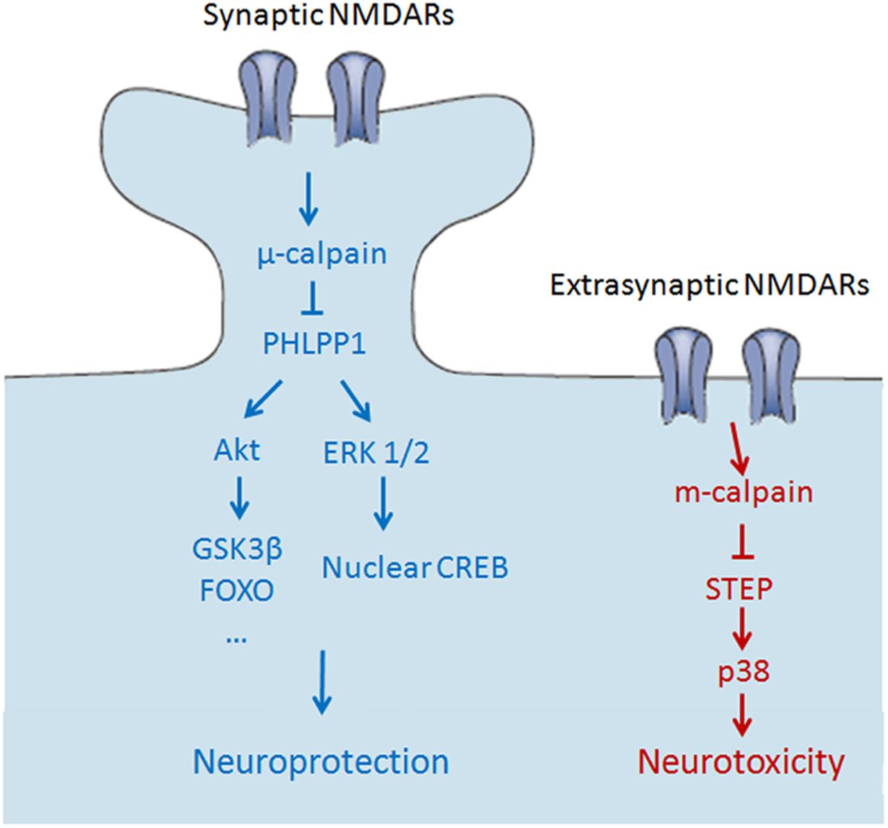
- Figure 10.
Schematic illustration of the separate pathways downstream of synaptic and extrasynaptic NMDARs. Left, Synaptic NMDAR activation stimulates μ-calpain, resulting in PHLPP1degradation and in the activation of Akt and ERK1/2 and their downstream prosurvival pathways. Right, Extrasynaptic NMDAR activation induces m-calpain activation and the resulting STEP degradation and in the activation of p38 prodeath pathway, leading to neurotoxicity. There are no cross-talks between those two pathways.


