

Article Figures & Data
Figures
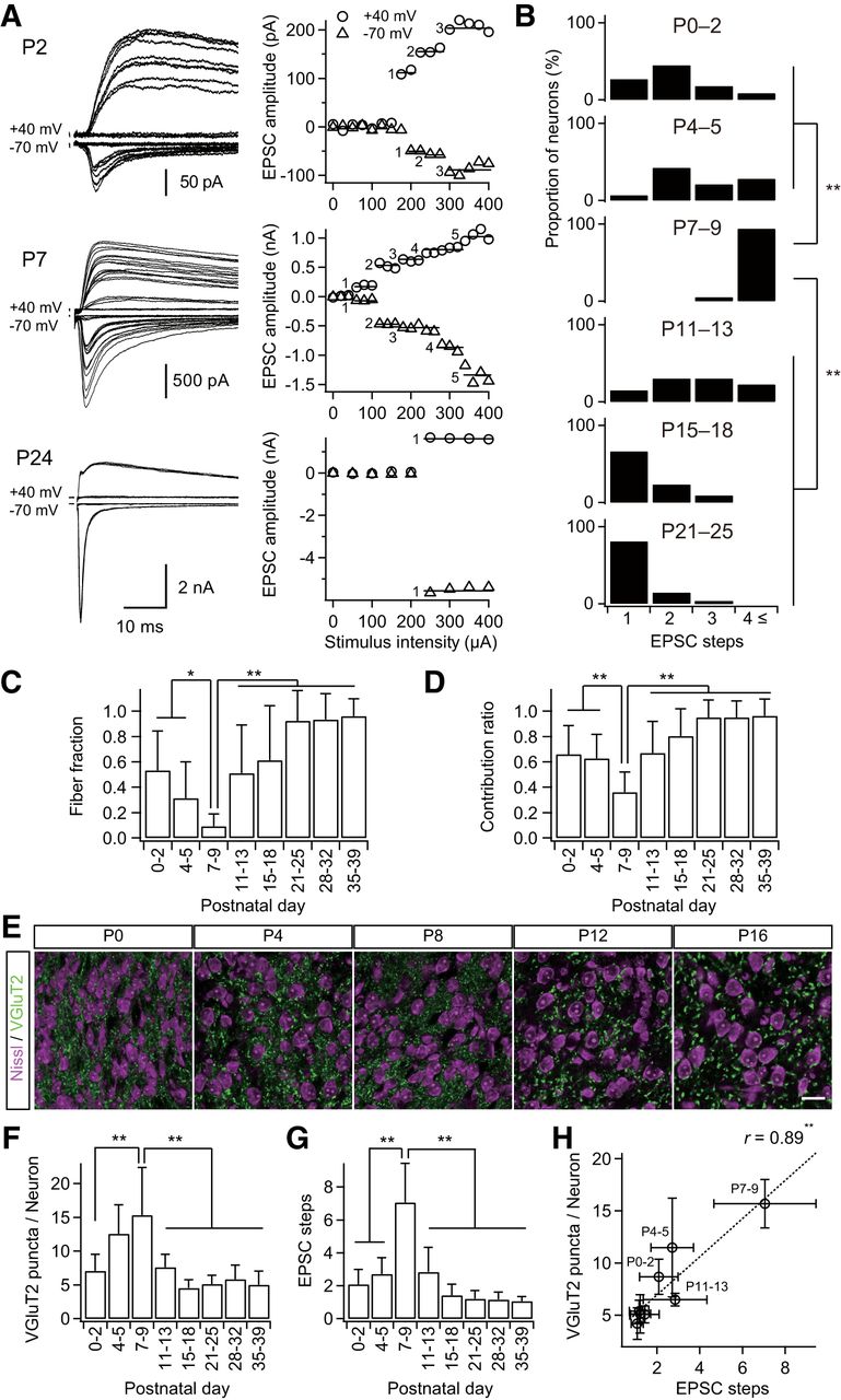
- Figure 1.
Formation and elimination of lemniscal synapses during postnatal development. A, Developmental increase and decrease in number of innervating lemniscal fibers on a neuron in the whisker-related somatosensory thalamus. The number of innervating fibers was estimated by counting steps of lemniscal fiber-mediated EPSCs (lemniscal EPSCs) in response to increasing stimulus intensity applied to the lemniscal fiber bundle. Holding potentials were at +40 and −70 mV. Left, Several raw traces with different stimulus intensities are superimposed at each holding potential. Right, Search of EPSC steps. Peak amplitudes of lemniscal EPSCs are plotted against stimulus intensities. EPSC steps are indicated by horizontal bars and integers. B, Distributions of thalamic neurons with different numbers of lemniscal inputs throughout postnatal development. C, Developmental change in the fiber fraction, the first single fiber-meditated EPSC divided by the saturated currents of the same thalamic neuron. D, Developmental change in the contribution ratio, the ratio of the largest single-fiber-mediated EPSC to the saturated currents of the same thalamic neuron. E, Density of thalamic neurons decreased during the first postnatal week, whereas that of VGluT2 puncta decreased during the second but not first postnatal week in the V2 VPM. Scale bar, 20 μm. F, VGluT2 puncta per thalamic neuron within 1 μm optical slice first increased and then decreased during postnatal development. G, Electrophysiological evidence for the change in the number of lemniscal fiber inputs on a thalamic neuron. H, Correlation between VGluT2 puncta per neuron and EPSC steps, structural and functional data on synapse formation and elimination. r, Pearson's correlation coefficient. Broken line is the regression line. Values are represented as mean + SD. n = 13–42 thalamic neurons for each developmental bin for electrophysiological data; n = 12–40 images from 6–8 mice for each developmental bin for structural data. Statistical significance was tested by multiple t test with Bonferroni correction after one-way ANOVA for B, C, D, F, and G, and with the t test for noncorrelation for H. *p < 0.05; **p < 0.01; two-tailed test.
- Figure 2.
Genetic visualization of PrV2-origin lemniscal fibers. A, Strategy for genetic visualization of PrV2-origin (maxillary Pr5-origin) lemniscal fibers. Top, Alleles of Krox20-Cre and Ai14 mice. These lines were crossed. Bottom, Schematic drawing of selective labeling of PrV2-origin lemniscal fibers with tdTomato. Selective expression of Cre recombinase in the V2 subregion of the Pr5 drives tdTomato in the double heterozygote (Krox20-Ai14 mice). B, Krox20-Ai14 mice have strong tdTomato expression in almost all neurons in the PrV2 that project to the maxillary (V2) VPM. The layer-six neurons in the somatosensory cortex of Krox20-Ai14 mice that project to the V2 VPM do not colocalize with tdTomato expression. CTB was injected in the V2 VPM on P28. Scale bars: Left, 400 μm; Right, 50 μm. C, Krox20-Ai14 mice have strong tdTomato expression in >85% of neurons in the PrV2, but not in the mandibular Pr5 (PrV3). Such expression was observed as early as P2. Scale bars: Left, 200 μm; Right, 50 μm. D, tdTomato-labeled lemniscal fibers from PrV2 to the VPM in the P28 Krox20-Ai14 mouse. Top, tdTomato signals throughout lemniscal fibers. Scale bar, 200 μm. Bottom: Terminals of tdTomato-labeled lemniscal in the V2 VPM. Terminals were clustered in barreloid hollows surrounded ring-shaped alignment of thalamic neurons. Broken lines indicate barreloid units. Scale bar, 20 μm. E, Serial thalamic sections of P28 Krox20-Ai14 mice were subjected to CO staining, tdTomato immunostaining, and acetylcholine esterase (AChE) staining. Cutting plane was perpendicular to the longest axis of barreloid units. tdTomato-immunoreactive puncta were confined to the most caudal region of the dorsolateral part of the VPM (V2 VPM). Scale bar, 200 μm. VPL, ventral posterolateral thalamic nucleus. F, Time course of barreloid establishment in the Krox20-Ai14 mice. Scale bar, 200 μm. Top and Middle: Adjacent CO and fluorescent Nissl stained sections are shown. A–E, Barreloid rows; 1-3, barreloid arcs. Bottom, Eight-bit signals in the tdTomato channel of confocal images shown in middle. R, rostral direction; VM, ventromedial direction.
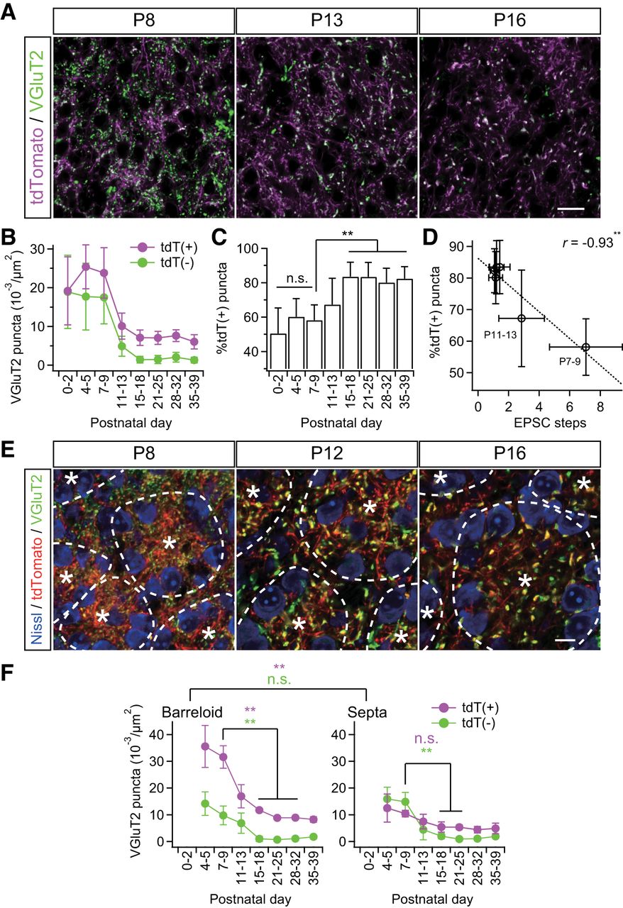
- Figure 3.
Somatotopic information is refined via fiber-origin-dependent synapse elimination. A, VGluT2-immunostained thalamic sections of Krox20-Ai14 mice. PrV2-origin lemniscal boutons are characterized as tdTomato-positive VGluT2 puncta in white, whereas non-PrV2-origin afferent boutons as tdTomato-negative VGluT2 puncta in green. Scale bar, 20 μm. B, tdT(+) = tdTomato-positive VGluT2 puncta; tdT(−) = tdTomato-negative VGluT2 puncta. Densities of both types of puncta decreased during the synapse elimination phase, but density of tdT(−) decreased more evidently. C, Proportion of tdT-positive puncta significantly increased during the synapse elimination phase. D, Proportion of tdT-positive puncta significantly correlated with EPSC steps after the onset of synapse elimination. r, Pearson's correlation coefficient. Broken line is the regression line. E, Cross-sectional images of barreloid units surrounded by interbarreloid septa. *Center of the barreloid unit characterized by ring-shaped alignment of thalamic neurons and clustered lemniscal fibers within it. Broken lines indicate border of barreloid units and septal region. Inside of a barreloid unit is the barreloid hollow, whereas outside is the septal region. Scale bar, 10 μm. F, Developmental changes in density of VGluT2 puncta in barreloid hollow and at interbarreloid septa. Values are represented as mean ± SD. n = 12–40 images from 6–9 mice for A–D and six images from three mice for E–F for each developmental bin. Statistical significance was tested with a t test for noncorrelation for D. Differences between barreloid and septa were tested as a factor of two-way repeated ANOVA for F. Differences between particular ages were tested by post hoc multiple t test with Bonferroni correction for C and F. *p < 0.05; **p < 0.01; n.s., not significant, two-tailed test.
- Figure 4.
Somatotopic information is refined in neuropil rather than on the soma. A, Analysis of somatic and neuropil puncta on a particular thalamic neuron. Somatic puncta were counted directly as shown. The number of neuropil puncta was calculated by subtracting somatic puncta on the neuron from the average number of puncta per neuron in the image. *Analyzed neuron; arrows, somatic puncta; arrowheads, neuropil puncta (quarter). Scale bar, 5 μm. B, The number of neuropil puncta per neuron changed dramatically during development, whereas that of somatic puncta did not. C, Proportion of tdT-positive neuropil puncta increased significantly during development. D, Neuropil puncta per neuron correlated well with steps of lemniscal EPSCs. r, Pearson's correlation coefficient. Broken line is the regression line. E, Proportion of tdT-positive neuropil puncta correlated well with steps of lemniscal EPSCs after the onset of the synapse elimination phase. F, Projection images of biocytin-labeled thalamic neurons after acute patch-clamp recordings. Scale bar, 100 μm. G, Sholl-analysis of dendrites of thalamic neurons using a Neurolucida system. Dendritic complexity increased significantly during the synapse formation and elimination phases. Bin was 10 μm. Values are represented as mean + SD. n = 30 thalamic neurons in six images from three mice for A–E and five thalamic neurons for F–G for each developmental bin. Statistical significance was tested by Bonferroni correction after one-way ANOVA for B, one-way ANOVA for C, t test for noncorrelation for D and E, and multiple t test with Bonferroni correction after two-way repeated ANOVA for G. *p < 0.05; **p < 0.01; n.s., not significant, two-tailed test.
- Figure 5.
VGluT2 immunoreactivity colocalized with PSD95 and GluA3 immunoreactivities. Representative images of whisker sensory thalamic sections double immunostained for VGluT2 and PSD95 (A) or GluA3 (B). VGluT2-immunoreactive puncta colocalized well with intense signals of PSD95 and GluA3. Scale bars, 5 μm.
- Figure 6.
Strengthening of PrV2-origin lemniscal synapses during postnatal development. A, VGluT2-immunoreactive puncta in the V2 VPM before and after synapse elimination. Scale bar, 5 μm. B, Distributions and cumulative probabilities of tdT(+) and tdT(−) VGluT2 puncta before and after synapse elimination. Data from six and eight mice were pooled for P7–P9 and P15–P18, respectively. C, Miniature lemniscal EPSCs before and after synapse elimination. Miniature lemniscal EPSCs were evoked in the modified ACSF with Sr2+ ions instead of Ca2+ ions, whereas lemniscal fibers were stimulated electrically. D, Averaged miniature lemniscal EPSCs before and after synapse elimination. The numbers of averaged events were 489 and 763 for P7–P9 and P15–P18, respectively. E, The mean amplitude of miniature lemniscal EPSCs of a neuron significantly increased during the synapse elimination phase. Analysis was conducted on 512–814 events at 6–10 neurons from three mice for each developmental bin. F, The amplitude of miniature lemniscal EPSCs correlated well with the size of VGluT2 puncta during development. r, Pearson's correlation coefficient. Broken line is the regression line. G, Proportion of tdT-positive puncta correlated well with the size of tdT-positive puncta during development. Values are represented as mean ± SD. Statistical significance was tested by multiple t test with Bonferroni correction after one-way ANOVA for developmental changes in E and t test for noncorrelation for F and G. **p < 0.01; n.s., not significant, two-tailed test.
- Figure 7.
Sensory-experience-dependent somatotopic refinement via synapse elimination. A, Experimental schedule and schematic view of whisker deprivation. B, Acute slice patch-clamp recordings from Krox20-Ai14 mice. Clouds of tdT-positive terminals were observed in the dorsal part of the VPM via a confocal laser scanning unit and the whole-cell recordings were then made from thalamic neurons under the infrared-differential interference contrast (IR-DIC) viewing. After the recordings, slices were fixed and we confirmed that the recorded thalamic neurons had been in the clouds of tdTomato-positive lemniscal fiber terminals. The arrow indicates the recorded thalamic neuron. Scale bars, 200 μm. ml, Medial lemniscus; Rt, reticular nucleus of thalamus. C, Representative traces of lemniscal EPSCs from intact and deprived mice under voltage-clamp conditions at +40 and −70 mV. Several raw traces with different stimulus intensities are superimposed at each holding potential. EPSCs from an intact mouse showed all-or-none responses, whereas those from a deprived mouse showed stepwise increments in amplitude. D, Distributions of neurons with different numbers of steps of lemniscal EPSCs. n = 46 and 47 neurons of seven and nine mice for intact and deprived groups, respectively. E, VGluT2-immunostained whisker-sensory thalamic sections of intact and deprived Krox20-Ai14 mice. Scale bars, 20 and 5 μm for left and right, respectively. F, G, Density and size of tdT(+)- and tdT(−)-VGluT2 puncta of intact and deprived Krox20-Ai14 mice. Forty images from 7–8 mice were analyzed for each group. H, Proportion of tdT-positive puncta plotted against EPSC steps. Whisker deprivation at P12–P13 consistently disrupted synapse elimination and somatotopic refinement. r, Pearson's correlation coefficient. The broken line is the regression line. Values are represented as mean ± SD. Statistical significance was tested by Kolmogorov–Smirnov two-sample test for D and Student's t test for F and G. *p < 0.05; **p < 0.01; n.s., not significant, two-tailed test.
- Figure 8.
Developmental change in origin of afferent fibers innervating the whisker sensory thalamus. A CTB solution was injected to the right V2 VPM to retrogradely label the origin of nuclei of afferent fibers. Survival time was 2 d. Arrows indicate ectopic labeling. Three to six mice were examined for each group. Cu, Cuneate nucleus; Gr, gracile nucleus; SpC, caudal subnucleus of spinal trigeminal nuclei. Scale bars, 400 μm.
- Figure 9.
Schema of postnatal development of the whisker sensory thalamus of mice. During P0–P5, the afferent fibers and VPM neurons forms the whisker pattern. Just after the map formation, VPM relay neurons (neurons in cyan) receive intermingled innervations of PrV2-origin lemniscal fibers (magenta fibers with white terminals) and non-PrV2-origin afferent fibers (black fibers with green terminals). During P6–P9, many afferent fibers newly innervate VPM neurons with rough somatotopy. After P9, redundant, non-PrV2-origin afferent fibers are selectively and experience,dependently eliminated. By P21, the fine somatotopy with mono-innervations of PrV2-origin lemniscal fibers are established in the V2 VPM. Left, A–E, Barreloid rows; 1–7, barreloid arcs; α–δ, barreloid units representing the four posterior whiskers.


