

Article Figures & Data
Figures
- Figure 1.
Dual immunofluorescence localization of PSD-95 and pCaMKII T286/287 was used to map colocalization across 42 sample zones in rostral hippocampus. A, Representative images show immunolabeling for pCaMKII alone (left, red), and for pCaMKII plus PSD-95 (right, red and green, respectively) in stratum radiatum (SR) of field CA1b. Scale bar, 2 μm. Note, both antigens are exclusively and densely localized to small puncta, some of which were double labeled (arrows, 2 examples of colocalization). B, Plot shows the immunofluorescence intensity frequency distribution for pCaMKII+ labeling colocalized with PSD-95 (mean values from 3 representative cases). The vertical line marks the intensity value above which labeling was considered intense; elements with this pCaMKII immunolabeling intensity, or higher, were counted as being double labeled in calculations of the percentage PSD-95+ synapses enriched in pCaMKII. C, PSD sizes were distributed according to a Poisson curve (R2 = 0.84). The intensity of pCaMKII labeling colocalized with PSDs did not correlate with PSD size (R2 = 0.0018, p = 0.74). D, Photomicrograph of a rostral hippocampal section processed for the Timm's stain for heavy metals (brown to black) and Nissl staining (violet) to illuminate major lamina and cellular layers, respectively. Sampling zones used for automated counting of pCaMKII+ and PSD-95+ elements are indicated with dotted lines. Each of the major hippocampal subdivisions (CA1, CA3ab, CA3c, DG) were divided into 4–6 zones as illustrated for CA1 SR: these numbered zones (1–5) extended across the three lamina in fields CA1 and CA3. Numbering within CA3 and the DG molecular layer began with CA3a and the lateral aspect of the upper leaf, respectively. LM, Lacunosum-moleculare; SO, stratum oriens; UL, upper leaf of the DG molecular layer; LL, lower leaf of the DG molecular layer.
- Figure 2.
Behavioral analyses demonstrate differences in exploratory behavior between experimental groups. One group (contingency) explored freely for 5 min, after which entry into one compartment (Room 1) triggered a flashing light and a buzzer over the remaining 25 min; the second (unsupervised “exploration”) group was allowed explore both rooms with no contingencies for 30 min. A, B, Heat maps show the time spent at different locations over 30 min in the two compartments by representative rats from each group (red > yellow > green > blue). C, Quantification of time spent in Room 1 for each group, in 5 min time segments over the 30 min session (n = 8/group). Note the steep drop after minute 5 for the contingency group. D, Distance traveled, in both compartments, by unsupervised “exploration” and response “contingency” groups demonstrates similar habituation curves. E, Comparison of distance traveled per 5 min bin in a separate group (n = 8) of unsupervised exploration rats tested on 2 consecutive days. The between-day difference in these habituation curves was highly significant, as was the total distance traveled over 30 min (p < 0.0001, 2-way ANOVA and t test respectively). F, Same curves as in E but for contingency rats (n = 8; separate set from those in C and D); there were no detectable differences between days 1 and 2.
- Figure 3.
Induction of LTP is accompanied by long-lasting phosphorylation of CaMKII in a subpopulation of hippocampal synapses. A, Plot summarizes the mean field EPSP slopes for a group of slices receiving TBS and recorded for the following 60 min. Arrow indicates the delivery of TBS. B, Double-immunofluorescence localization of total (phosphorylated and unphosphorylated) CaMKII-positive (red) and PSD-95-positive (green) elements in CA1 stratum radiatum of an adult hippocampal slice. The higher resolution inset describes a densely labeled contact (arrow). Scale bar, 10 μm; inset scale bar, 1 μm. C, Blind automated counting shows that TBS increased the number of pCaMKII+ synapses (mean ± SEM) in slices collected at 7 or 60–90 min after stimulation (**p < 0.01, t test vs control slices (con)).
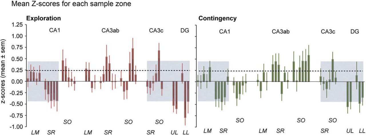
- Figure 4.
Mean Z-score values for doubled-labeled synapses across 42 sampling zones demonstrate broad regions with values that are correlated between unsupervised exploration and contingency groups. Values (Z-score means ± SEM) were calculated from all sections collected from eight rats per group. The dashed line indicates the mean of positive values for both experimental groups; the height of the gray boxes equals 1 SD above that mean. The gray boxes highlight areas (21 zones) for which the scores in the two groups were strongly correlated. Patterns in the remaining areas were not correlated and were statistically different (p = 0.005). See Figure 1 for abbreviations identifying the lamina.
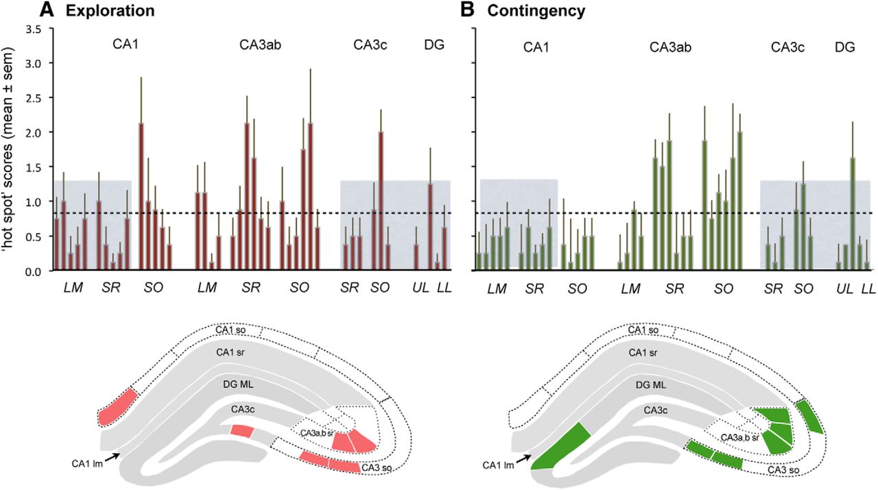
- Figure 5.
Distribution of sample zones containing a high percentage of PSDs associated with dense concentrations of pCaMKII T286/287. Z-scores for each section were assigned to one of three numerical categories (0, 1, 2) and then values for each sampling zone were summed across sections from individual rats, thereby generating a “hot spot” value for each animal. Plots show means (±SEM) for the Exploration (A) and Contingency (B) groups (n = 8 each). In each plot, the dashed line represents the mean value across all sampling zones for both groups; the height of the gray boxes is 1 SD above that mean. The z-score distributions were correlated between the experimental groups for two collections of contiguous zones (gray boxes) but not for the intervening zone. The overall distributions were statistically distinct between groups with the major differences being in regions without the gray highlight (Fig. 1, abbreviations). Line drawings of hippocampal cross sections at the bottom of each panel illustrate, with color fill, the distribution of sample zones with hot spot scores >1 SD from the overall mean score; the gray fill indicates zones with correlated labeling patterns between the two experimental groups.
- Figure 6.
”Hot spot” scores for unsupervised Exploration and Contingency groups following subtraction of mean values from home-cage animals. As previously, the dotted line is the mean of positive values for the two groups and the height of the gray boxes is 1 SD above that mean. Subtracting home-cage scores reduced several strongly positive values in the exploration animals, mainly in stratum oriens (SO) of CA3a,b, thereby increasing the relative sizes of the remaining hot spots. The same subtraction procedure eliminated all values >1 SD above the mean in the contingency group; despite this, the 21 sampling fields within the gray boxes were highly correlated with the corresponding sites in the unsupervised exploration group. The overall patterns were statistically different between the groups, particularly in the noncorrelated region between the gray boxes. The x's indicate three regions that were robustly different (p < 0.001) than ≥2 of the remaining sampling zones in the same Exploration group.
- Figure 7.
Schematic summarizing effects of learning on numbers of pCaMKII+ PSDs in the different hippocampal subfields. As in schematics within Figure 4, zones with gray shading exhibited numbers of pCaMKII+ PSDs that were correlated between the unsupervised exploration and contingency groups (i.e., same regions plotted over gray backdrop in Figs. 3–5). For areas highlighted in rose color, numbers of densely pCaMKII+ PSDs were, in free-exploration rats only, both >1 SD from the mean value for the two learning groups and significantly >1 SD from the mean in ≥2 other sample zones in the same group. Hot spots meeting these same criteria were not present in the contingency group. Abbreviations as in Figure 1; DG ML, DG molecular layer.


