

Article Figures & Data
Figures
- Figure 1.
HBCs possess primary cilia. Immunofluorescence staining was performed on tissue from the olfactory epithelium of 3- to 6-week-old wild-type, Arl13b-EGFPtg, and EGFP-CETN2 mice. A, p63-labeled HBCs possess ARL13B-labeled cilia (inset, magnified image). Scale bars, 10 μm. B, Cilia extend from γ-tubulin-labeled basal bodies (arrows). Low-magnification view (left) and high-magnification view (right) of ciliated HBC. Scale bars, 5 μm. C, In Arl13b-EGFPtg mice, K5-labeled HBCs possess GFP+ cilia (inset, magnified image). Scale bars, 10 μm. D, HBC cilia labeled with canonical cilia markers AC3 and ARL13B (arrowheads). E, In Arl13b-EGFPtg mice, AC3 labels GFP+ cilia (arrowheads). F, p63-labled HBCs possess AC3-labeled cilia. G, In EGFP-CETN2 mice, GFP-expressing basal bodies possess AC3+ cilia (arrowheads). H, In Arl13b-EGFPtg mice, GFP+ cilia (arrows) project from p63-labeled HBCs into the interstitial space between HBCs and K18-labeled SUS cell end feet (inset, magnified image). Scale bars, 5 μm. I, In wild-type mice intranasally infected with adenovirus containing GFP, K5-labeled HBCs possess ARL13B-labeled cilia (see arrow) that project into the interstitial space between an HBC and GFP+ end foot of a SUS cell (see inset for higher magnified image). Scale bar, 10 μm. N = 10 total mice for all immunostaining. Dashed lines, Basement membrane.
- Figure 2.
HBCs are the predominant ciliated olfactory basal stem cell. Immunofluorescence staining was performed in the olfactory epithelium of wild-type mice. A, The canonical GBC marker MASH1 colocalizes with a subset of SEC8+ GBCs, while, LSD1 colocalizes with a larger subset of SEC8+ GBCs. B, Few SEC8-labeled GBCs possess ARL13B-labeled cilia (see arrows) compared with K5-labeled HBCs (inset, magnified image). Scale bars, 10 μm. Dashed line, Basement membrane. †Occasional migrating GBC. C, Quantified data of SEC8+ cells that are either MASH1+ (N = 4) or LSD1+ (N = 4). D, The percentage of HBCs (N = 6) and GBCs (N = 6) that possess cilia. ****p < 0.0001 by Student's t test. Data are shown as the mean ± SEM.
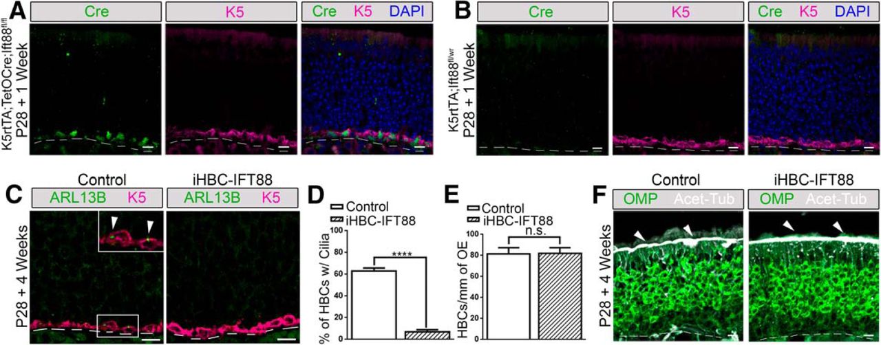
- Figure 3.
Cell-specific deletion of Ift88 in HBCs results in the loss of HBC cilia with no effects on OSN cilia. Control and iHBC-IFT88 mutant mice (referred to as iHBC-IFT88) were administered a dox-containing diet at P28 for 1 or 4 weeks, and immunofluorescence staining of the olfactory epithelium was performed. A, After 1 week of the dox-containing diet, Cre is present in K5-labeled HBCs of mice with K5rtTA and TetOCre alleles. B, After 1 week of the dox-containing diet, Cre is absent in mice lacking the TetOCre allele. C, After 4 weeks of the dox-containing diet, in control mice but not in iHBC-IFT88 mice, K5-labeled HBCs possess Arl13b-labeled cilia. D, E, Quantified data show that the percentage of HBCs that are ciliated in control mice is significantly reduced by ∼88.5% in iHBC-IFT88 mice, with no significant difference in the number of HBCs per millimeter of OE in control and iHBC-IFT88 mice after 4 weeks of the dox-containing diet. F, Acet-Tub-labeled cilia are still present in OMP-labeled mature OSNs (see short arrows). Scale bars, 10 μm. Dashed lines, Basement membrane. N = 5 for both groups. ****p < 0.0001. n.s., No significance by Student's t test. Data are shown as the mean ± SEM.
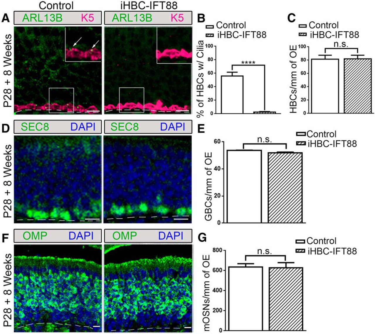
- Figure 4.
Loss of HBC cilia has no homeostatic effects on the cell composition of the OE. Control and iHBC-IFT88 mice were administered a doxycycline-containing diet at P28 for 8 weeks. A, In control mice, but not in iHBC-IFT88 mice, K5-labeled HBCs possess ARL13B-labeled cilia (see arrows; inset, magnified image). B, C, Quantified data show a significant loss of ciliated HBCs in iHBC-IFT88 mice, with no change in the number of HBCs per millimeter of OE. D, SEC8-labeled GBCs in the OE of control and iHBC-IFT88 mice. E, Quantified data show no significant difference in the number of GBCs per millimeter of OE in both groups. F, OMP-labeled mature OSNs in control and iHBC-IFT88 mice. G, Quantified data show no significant difference in the number of mature OSNs per millimeter of OE between both groups. Scale bars, 10 μm. Dashed lines, Basement membrane. N = 2 for control mice, N = 4 for iHBC-IFT88 mice. ****p < 0.0001. n.s., No significance by Student's t test. Data are shown as the mean ± SEM.
- Figure 5.
Loss of HBC cilia results in the improper regeneration of the OE and loss of TH expression in the OB. Control and iHBC-IFT88 mice were administered a dox-containing diet at P28 for 4 weeks and then were given an intraperitoneal injection of 75 mg/kg methimazole to ablate the OE. Following 8 weeks of recovery, immunofluorescence staining was performed. A, In the OE of control mice, but not of iHBC-IFT88 mice, K5-labeled HBCs possess ARL13B-labeled cilia. B, SEC8-labeled GBCs in the OE of control and iHBC-IFT88 mice. C, D, Quantified data show that the percentage of HBCs that are ciliated in control mice is significantly reduced in iHBC-IFT88 mice with no difference in the number of HBCs per millimeter of OE between both groups. E, Quantified data show a significant reduction in the number of GBCs per millimeter of OE. F, OMP-labeled mature OSNs and Acet-Tub-labeled cilia in the OE of control and iHBC-IFT88 mice. G, Quantified data show a significant reduction in the number of mature OSNs per millimeter of OE. H, A significantly thinner OE in iHBC-IFT88 mice. I, J, No difference in the number of cleaved caspase-3-labeled apoptotic cells was observed in the OE of control and iHBC-IFT88 mice. K, TH expression within glomeruli (dotted circles) in the OBs of control and iHBC-IFT88 mice. L, Quantified data show that the intensity of TH measured in arbitrary units is significantly reduced in the OBs of iHBC-IFT88 mice. Scale bars, 10 μm. Dashed lines, Basement membrane. N = 4 for both groups. *p < 0.05, ****p < 0.0001. n.s., No significance by Student's t test. Data are shown as the mean ± SEM.
- Figure 6.
Loss of Arl13b in HBCs results in the improper regeneration of the OE. Control and iHBC-ARL13B mice were administered a doxycycline-containing diet at P28 for 4 weeks to induce the deletion of Arl13b. Mice were then given an intraperitoneal injection of 75 mg/kg methimazole to ablate the OE and were allowed 8 weeks of recovery. A, K5-labeled HBCs in the OE of iHBC-ARL13B mice do not possess ARL13B-labeled cilia. B, The number of HBCs per millimeter of OE in both control and iHBC-ARL13B mice. C, SEC8-labeled GBCs in the OE of control and iHBC-ARL13B mice. D, Quantified data show a significant reduction in the number of GBCs per millimeter of OE. E, OMP-labeled mature OSNs and Acet-Tub-labeled cilia in the OE of control and iHBC-ARL13B mice. F, Quantified data show a significant reduction in the number of mature OSNs per millimeter of OE. Scale bars, 10 μm. Dashed lines, Basement membrane. N = 3 for both groups. *p < 0.05, ***p < 0.001. n.s., No significance by Student's t test. Data are shown as the mean ± SEM. ARL13B, ADP-ribosylation factor-like protein 13b; mOSN, mature OSN.
- Figure 7.
Loss of HBC cilia results in the impaired development of regions in the OE. Control and iHBC-IFT88 mice were treated with a doxycycline-containing diet at E16 for 4.5–5 weeks and were analyzed at P28 with immunofluorescence staining of the OE. A–F, OMP-labeled mature OSNs in anterior–posterior sections of control and iHBC-IFT88 mice. Scale bars, 200 μm. G, Illustration depicting the location of sections A–F in a sagittal view of the mouse olfactory organ. H, Magnified view of the boxed region in D with OMP-labeled mature OSNs and Acet-Tub-labeled cilia in the OE of control and iHBC-IFT88 mice. Scale bars, 50 μm. I, Quantified data show that the number of mature OSNs per millimeter of OE in iHBC-IFT88 mice is significantly reduced specifically in dorsal-lateral and medial regions of the OE, but not in ventral regions. N = 5 for both groups. *p < 0.05 by Student's t test. Data are shown as the mean ± SEM. DL, Dorsal-lateral; DM, dorsal-medial; VL, ventral-lateral; VM, ventral-medial.


