

Using Tension to Connect Neurons
Margaret H. Magdesian, G. Monserratt Lopez-Ayon, Megumi Mori, Dominic Boudreau, Alexis Goulet-Hanssens, et al.
(see pages 979–987)
Tension is a fundamental driver of neurite growth. During initial neurite extension, receptors located in growth cones seek extracellular binding partners, while their intracellular domains interact with the actin cytoskeleton. When the receptors bind to fixed ligands, they anchor actin filaments, which are pulled continually toward the neurite shaft by myosin motors. When actin is anchored, the force exerted by myosin causes tension to build up along the filaments. This tension causes microtubules to advance into the growth cone and promotes consolidation of a new neurite segment. Tension is also thought to drive neurite growth after synaptogenesis; in this case, the tension results from growth of the surrounding tissue, which stretches axons. Neurite growth can also be induced by experimentally applied tension (Suter and Miller, 2011, Prog Neurobiol 94:91).
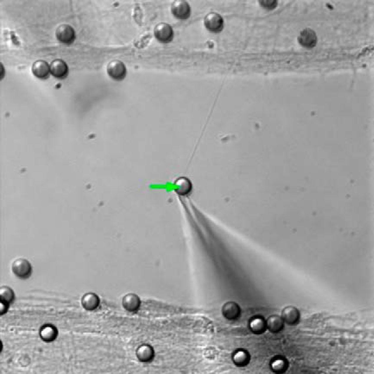
Applying force to a bead (arrow) can be used to drive axon growth and connect neuronal populations. For details and to watch the video, see the article by Magdesian et al.
Magdesian et al. now show that tension-stimulated neurite growth can be used to connect neuronal populations. Rat hippocampal neurons were grown for 2–3 weeks on glass coverslips in microfluidic devices that prevented contact between populations. The microfluidic devices were then removed and beads coated with polylysine—an adhesive molecule—were placed on neurites. Atomic force microscopy probes or micromanipulators were used to move an attached bead toward a second neuronal population, where the bead was set down. As the bead was moved, it pulled out new neurites, and within 30 min of contact with the second neuronal population, a stable connection formed between the newly formed neurites and the target neurons. Importantly, newly formed neurites appeared to form functional synaptic connections with the second population, as indicated by the occurrence of more EPSCs in the target population 100 ms after a presynaptic spike than in the 100 ms preceding spikes. Such a pattern was not detected in unconnected populations.
These results demonstrate that mechanical tension can be used to promote formation of functional connections between neuronal populations. This technique should allow researchers to establish defined neural networks in culture to test hypotheses about circuit function and dynamics. In the more distant future, similar techniques may even allow surgeons to help severed spinal axons grow across lesions to reestablish connections and restore function after injury.
Transcriptional Repression by Apolipoprotein E
Veena Theendakara, Clare A. Peters-Libeu, Patricia Spilman, Karen S. Poksay, Dale E. Bredesen, et al.
(see pages 685–700)
Most Alzheimer's disease (AD) cases are late-onset sporadic forms, the causes of which remain unknown. By far the strongest genetic risk factor for sporadic AD is possession of the ε4 allele of apolipoprotein E (ApoE4), a protein involved in lipid transport. People with two copies of this allele are ∼8 times more likely to develop AD than people who have only ε2 or ε3 alleles. The ε4 and ε3 alleles differ by a single amino acid, but this is sufficient to change several functional properties. ApoE4 is less efficient at transporting cholesterol—necessary for maintaining cell shape and creating lipid rafts for signaling molecules—than ApoE3, and ApoE4 promotes, while ApoE3 suppresses, inflammation, which is thought to contribute to AD. In addition, ApoE4 promotes generation of toxic amyloid-β from amyloid precursor protein (APP), whereas ApoE3 promotes generation of the growth-promoting cleavage product soluble APPα (sAPPα). Impaired glucose metabolism and mitochondrial dysfunction have also been proposed to contribute to the increased risk of AD in ε4 carriers (Yu et al., 2014, Ann Rev Neurosci 37:79).
Theendakara et al. have now discovered a new mechanism by which ApoE4 may influence AD risk. The authors' previous work showed that ApoE4, but not ApoE3, reduced expression of Sirtuin 1 (SirT1), a protein that activates transcription of a secretase that generates sAPPα from APP. They now report that transfected ApoE entered the nucleus of human neuroblastoma cells, where it bound to the SirT1 promoter. Genome-wide studies using ApoE chromatin precipitation followed by DNA sequencing revealed that >3000 genomic sequences interacted with ApoE4, and approximately half of these did not bind ApoE3. Notably, several of these genes have previously been linked to AD. In addition to SirT1, these include genes involved in axon guidance, neuronal survival, cell death, energy metabolism, and inflammation. Further experiments revealed that transfected ApoE4 reduced transcription of at least two genes besides SirT1 in glioblastoma cells; both of these genes are involved in inflammatory processes.
These results indicate that in addition to transporting lipids, ApoE can act as a transcriptional repressor. By investigating genes differentially regulated by ApoE4 and ApoE3, researchers may learn more about how this allele increases AD risk and how to reduce that risk.
Footnotes
This Week in The Journal is written by Teresa Esch, Ph.D.


