

Article Figures & Data
Figures
- Figure 1.
Attentive motion-discrimination task: design and overall cortical activation pattern during active task performance. A, Stimulus sequence and configuration. Subjects initiated a trial by foveating the central fixation spot (FP), surrounded by eight saccade targets (ST). After a 500 ms delay, a cue appeared (shown is the spatial cue of task variants 1 and 2, extending from the fixation spot, pointing either left or right), indicating to which side attention had to be deployed. Inset shows the two cueing procedures: in task variants 1 and 2 (top), a spatial cue was used (bar to the left for attend left; bar to the right for attend right), in variant 3 (bottom), the target was cued symbolically by the color of the fixation point (monkey Q: red for left; green for right; monkey M: green for left; red for right). After 500 ms, two RDSs appeared to the left and the right of the fixation spot at equidistant positions. B, Event sequence of one of the RDSs. While both RDSs were changing their motion direction every 60 ms, the subject had to track the target stimulus for 20–60 direction changes until the translation direction ceased changing for 500 ms (PME) in monkey Q and 800 ms in monkey M, respectively, followed again by rapid direction changes. Monkeys were required to respond to the target surface PME by a saccade to the corresponding ST. C, Temporal sequence of behavioral conditions (attentive motion discrimination and passive fixation) during scanning. Monkeys alternated between paying attention to the left (L, top), passive fixation (F, center), and paying attention to the right (R, bottom) in sequence LFRFLFR. During the L and R blocks, subjects had to complete seven trials successfully (hits). Thus, block duration was variable, lasting on average ∼30 s. Passive fixation blocks lasting 5 s each separated the two attention conditions. D, Statistical parametric maps of contrast task versus fixation overlaid on the inflated (top) and flattened (bottom) right hemispheres of monkey Q (left) and monkey M (right). Yellow and red colored regions were significantly more activated by performance of the attentive motion-discrimination task than by passive fixation at p < 0.005, corrected for multiple comparisons, while cyan-blue regions were significantly less active during attention task performance. Activity in the somatosensory and primary motor cortex was reduced during active task performance. Scale bar, 1 cm. LS, Lunate sulcus; IPS, intraparietal sulcus; CS, central sulcus; AS, arcuate sulcus. Bottom, Same parametric maps overlaid on flattened posterior hemispheres. Dashed and solid black lines mark vertical and horizontal meridians, respectively. Motion-sensitive areas (black outlines) in the STS, MT, MST, and FST areas, and in the intraparietal sulcus, area VIP, and area LIP, mapped with a motion localizer (see Materials and Methods).
- Figure 2.
Spatial attention modulates activity in specific cortical areas of the occipital, temporal, parietal, and frontal lobes. A, Schematic of the two stimulus conditions contrasted in Figure 2: attention to the contralateral RDS (attend contra) versus attention to the ipsilateral RDS (attend ipsi). Attention location is indicated by a colored ring over the cued surface. Significant response enhancements for the attend contralateral versus the attend ipsilateral condition are shown in yellow and red; significant response enhancements to the opposite condition are in blue. B, Statistical parametric maps for the contrast attend contra vs attend ipsi; conventions as in Figure 1D, top. C, Same parametric maps overlaid on flattened posterior hemispheres; conventions as in Figure 1D, bottom; thresholds at p < 0.05, corrected. Left, Numbers point to regions of significant activation shown on coronal slices in D: 1, area V1 lower hemifield; 2, foveal representation; 3, area V1 upper hemifield; 4, area V2; 5, area V3; 6, posterior parietal area LIP; 7, area V4t; 8, area MT; 9, area PITd. D, Parametric maps on coronal slices of high-resolution anatomy, left hemisphere on the right. Cyan and blue indicate higher activity for contrast attend left > attend right; yellow and red indicate higher activity for contrast attend right > attend left.
- Figure 3.
Spatial attention modulates activity in cortical ROIs in the occipital, temporal, parietal, and frontal lobes. A, β Values of the GLM for monkey Q (BOLD, top) and monkey M (Sinerem, bottom) across ROIs (left to right: V1, V2, V3a, V3, V4d, V4t, MT, MST, FST, PITd, 7a, VIP, LIP, and FEF) in the attentive discrimination task with central bar cue (Fig. 1A, var1). Responses during the attend contralateral (Attend contra) condition are shown in gray; responses during the attend ipsilateral (Attend ipsi) condition in black. Significant activation differences in the two conditions: *p < 0.01 (ANOVA, multiple-comparison corrected); ***, most significant differences; **, differences with at least half the F value of the most significant difference. B, β Values for the motion-discrimination task with color cue (Fig. 1A, var3; averaged across monkeys Q and M, Sinerem both). Responses to the passive fixation task condition (neutral) are shown in white. Significant activation differences between the two attention conditions (Attend contra and Attend ipsi) are indicated by asterisks following the same convention as in A. FEF modulation was marginally significant (p = 0.03). C, Left, Scatter plot of activation differences (Attend contra vs Attend ipsi, vertical axis) for each ROI versus mean activation during the two attention conditions (horizontal axis) averaged across attentive motion-discrimination tasks variant 1 (vars1) and variant 3 (vars3; A and B). Areas MST, FST, VIP, and 7a show weak and insignificant activation and attention modulation across tasks and animals. The dotted line indicates conditions with response enhancement during the Attend contra condition that is enhanced by 67% (corresponding to an AI of 0.25) relative to the response during the Attend ipsi condition. Right, Bar graph of AIs of ROIs with both visual activation and attentional modulation. Areas V1, V2, V3a, and V3 show similar and lower levels of attentional modulation; motion-selective areas V4t and MT show more; and area PITd, area LIP, and the FEF show the strongest. The AI for the FEF, which typically exhibited little mean activation, was the largest of these three areas, and was cutoff at the top for display purposes.
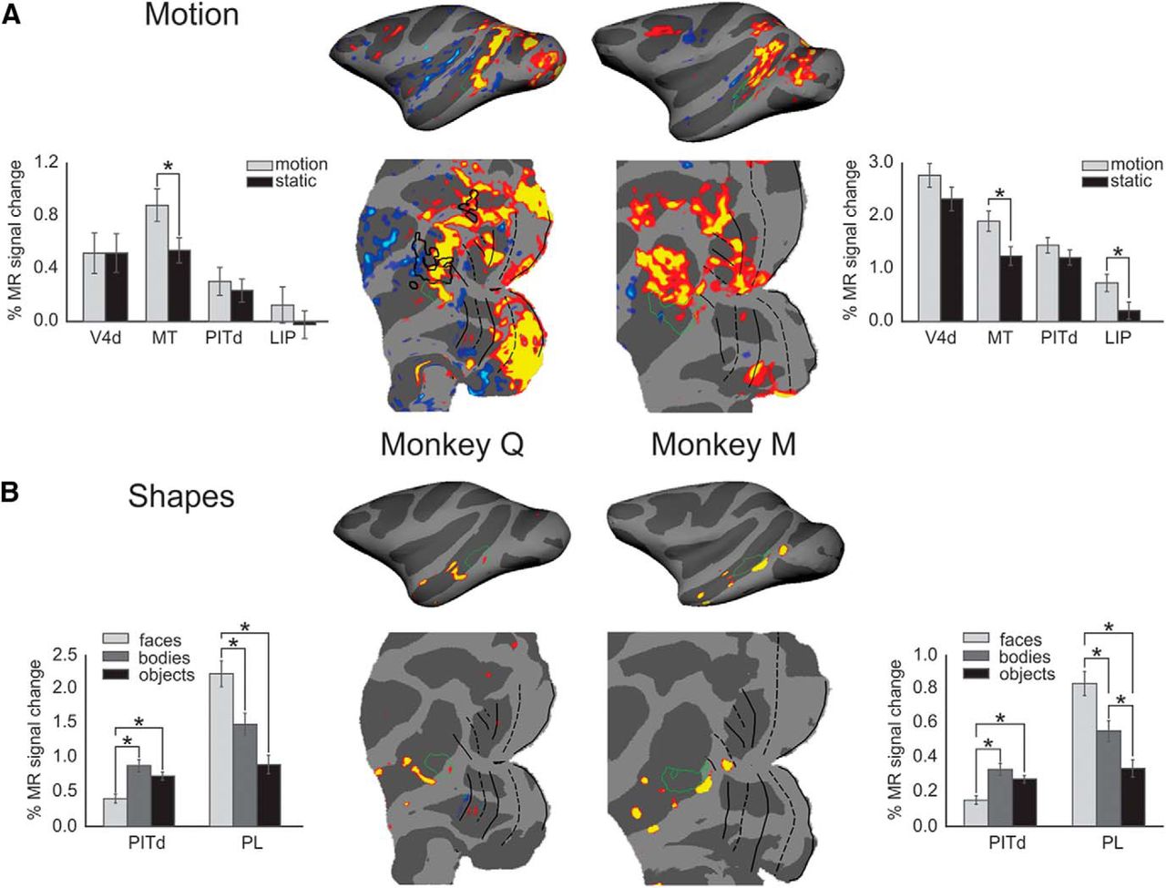
- Figure 4.
Attentionally modulated parts of area PITd are located adjacent to motion-selective and face-selective areas. A, Areas activated by motion stimuli are shown in red/yellow (p < 0.05) overlaid onto inflated and flatmap representations of the right hemispheres for both monkeys. Green outlines depict the location of attentionally modulated area PITd. There is some overlap in one of monkey Q's hemispheres between areas involved in motion processing and area PITd. Asterisks in bar graphs mark significant differences (Wilcoxon signed-rank test at p < 0.01). There is little overlap between area PITd and motion-selective areas. B, Face-selective areas shown in red/yellow (p < 0.05). There is little if any overlap between area PITd and the face-selective cortex. In fact area PITd responds less to faces than to other objects. Asterisks in bar graphs mark significant differences (Wilcoxon signed-rank test at p < 0.01).
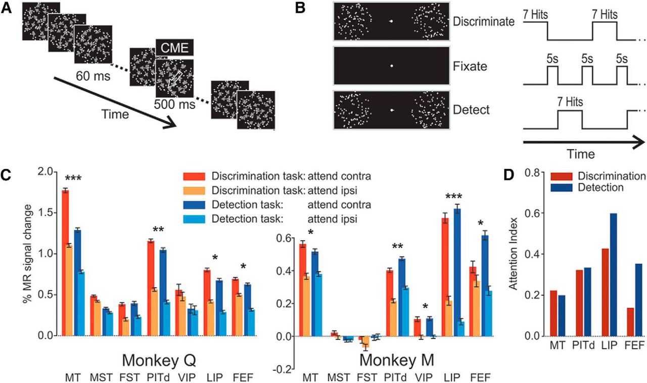
- Figure 5.
Design of attentive motion-detection task and comparison of activation patterns during attentive discrimination and detection tasks. A, Event sequence of one of the RDSs. The sequence of events was identical to that in the motion-discrimination task (Fig. 1B), but motion during the brief 60 ms periods was incoherent, while motion during the task-relevant prolonged motion event was 10% coherent (CME). The duration of the CME was ≤500 ms for monkey Q and ≤800 ms for monkey M. Monkeys were required to respond to the target surface CME by a saccade onto the target surface, regardless of motion direction. B, Temporal sequence of behavioral conditions (attentive motion discrimination, passive fixation, and attentive motion detection) during scanning. The attentive motion-detection task (E) employed a spatial cue, rapid serial visual stimulus presentation, and alternated with the attentive motion-discrimination task (I) and passive fixation periods (F) in a sequence IFEFIFE. During attentive discrimination and attentive detection, monkeys had to complete seven trials successfully (hits). Thus, duration of these blocks was variable, on average ∼30 s. Passive fixation blocks separated the two attention paradigms and lasted 5 s. C, β Values for four attention conditions are shown as bar graphs. Significant activation differences between the two conditions attend contralateral (attend contra) and attend ipsilateral (attend ipsi) are indicated, when they occurred in both subjects, as follows: *p < 0.01 (ANOVA, multiple-comparison corrected); ***, most significant differences; **, differences with at least half the F value of the most significant difference. Because the occipital lobe was not covered by slices in these scans, early cortical areas could not be included in the analysis. D, AIs for the four ROIs with significant activation and attention modulation across animals and conditions. Areas MT and PITd show the same degree of modulation in both attention tasks, while area LIP and the FEF show stronger attentional modulation during motion-detection tasks than during motion-discrimination tasks. AIs in area PITd are very similar to those in Figure 3.


