

Article Figures & Data
Figures
- Figure 1.
COLXV-B deposition during primary myogenesis. A–I, Immunostaining of embryos at 8–10 somites (A–D) and 18 hpf (E–I) with COLXV-B antibodies (red) and anti-β-catenin (green). Dorsal views of anterior and posterior part of the same embryonic trunk show morphological changes of adaxial cells at 8–10 somite stage: elongated adaxial cells (A), cuboid adaxial cells (D, arrow), and interleafing stage (D, asterisk). A, COLXV-B is deposited by elongated adaxial cells. Arrowheads indicate somite boundaries. B, C, Orthogonal views of areas 1 and 2 as indicated in A. Dorsal views (E, G), cross sections (F, H), and lateral view (I) of anterior and posterior trunk of 18 hpf embryos. E, Arrowheads indicate vertical myosepta. COLX-B is deposited in the motor path and vertical myosepta. J–M, Immunostaining of embryos at 18 hpf (J, K) and 24 hpf (L, M) with COLXV-B antibodies (red) and F59 (green). Cross section at 24 hpf (M) shows the persistence of COLXV-B staining in the motor path when F59-positive slow fibers (green) have migrated to the myotome periphery. M, Arrowhead indicates COLXV-B staining in contact with MPs. N, Production, characterization of polyclonal antibodies against zebrafish COLXV-B. Left, Schematic representation of the domain structure of the zebrafish COLXV-B α1 chain and location of the NC10 (residues 255–270) epitope. Collagenous domains (green) are numbered from the C terminus. Black represents noncollagenous linker domains. NC10 domain contains a TSPN (thrombospondin N-terminal like domain) domain and the NC1 the restin domain. Right, Western blotting with anti-COLXV-B antibodies of conditioned HEK293 media transfected with zebrafish col15a1b full-length cDNA incubated with (+) or without (−) collagenase. Lane 1: no treatment; lanes 2, 3: incubated at 37°C without collagenase (lane 2) and with collagenase (lane 3). The COLXV-B α1 chain is detected as an immunoreactive band migrating >200 kDa in undigested samples incubated (lane 2) or not (lane 1) at 37°C. After collagenase digestion, the band corresponding to COLXV-B α1 chain disappears and a collagenase digestion product is observed at the expected size for NC10 domain, attesting to the specificity of the anti-COLXV-B antibodies. O–Q, COLXV-B coincides with chondroitin sulfate signal in the motor path (P, Q, arrowheads). Single confocal plane of immunofluorescence staining of 24 hpf embryo cross section with antibodies to COLXV-B (O, red) and C-6-S (P, green) and merge image (Q). Hoechst solution was used to reveal nuclei. SC, Spinal cord (F, H, M, dotted line); N, notochord; ant, anterior trunk; post, posterior trunk. For lateral and dorsal views, anterior is left.
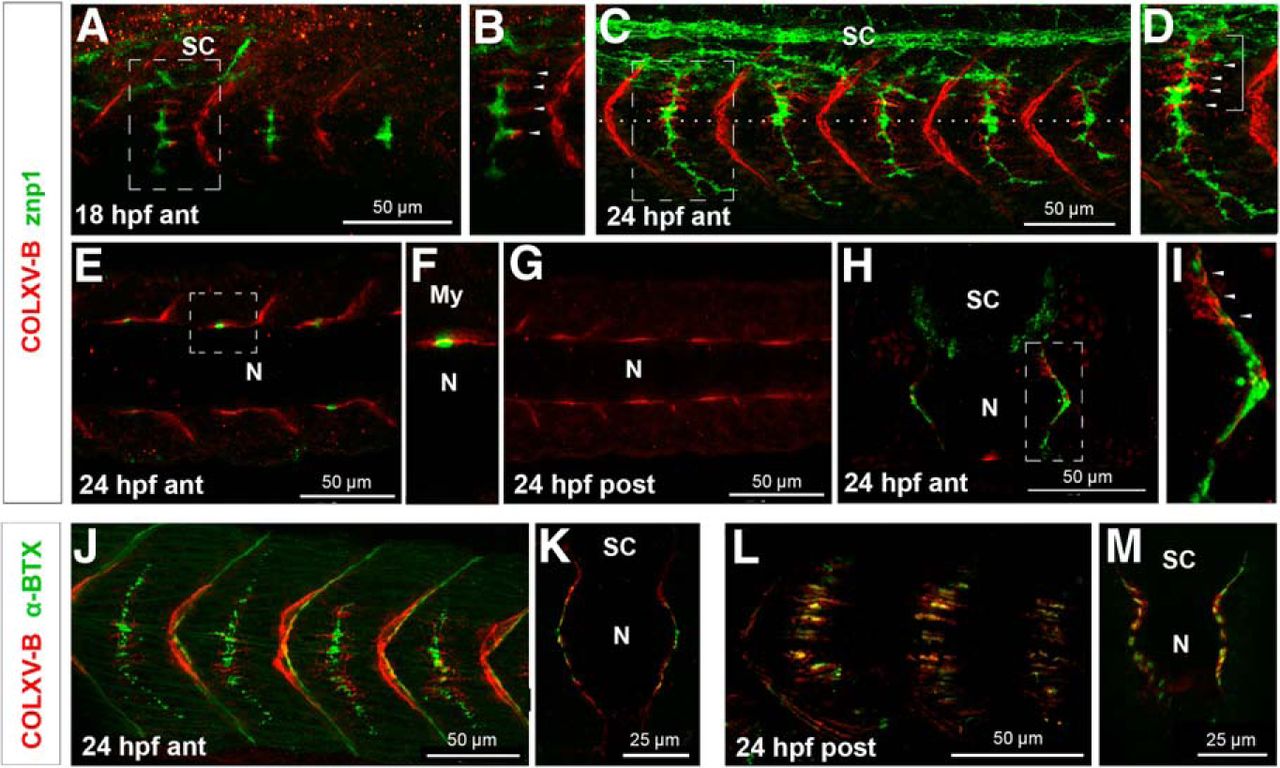
- Figure 2.
COLXV-B is present in the pathway of motor axons and coincides with aneural AChR clusters. Double staining of embryos at 18 hpf (A, B) and 24 hpf (C–M) with anti-COLXV-B antibodies (red) and znp1 (green) to label primary motoneurons (A–I) or conjugated α-bungarotoxin (α-BTX, green) to stain AChR (J–M). A–D, Lateral views of anterior mid-trunk (boxed areas in A, C indicate higher magnification shown in B and D, respectively). B, D, Arrowheads point to the ladder-type structural organization of COLXV-B staining. D, Bracket represents COLXV-B signal enrichment along the common path. C, Dotted line indicates the position of the horizontal myoseptum. E–G, Dorsal views of anterior (E, F) and posterior (G) trunk. F, Higher magnification of the boxed area in E showing that COLXV-B deposit embraces CaP axon. H, I, Strong COLXV-B signal is observed along the common path (arrowheads) wrapping around motoneuron axon. H, Cross section. I, Higher magnification of boxed area in H. J–M, Lateral views (J, L) and cross sections (K, M) at the level of neural (anterior mid-trunk) and aneural clusters (posterior trunk). COLXV-B is present within the neural clusters of AChR in the anterior trunk (J, K) and colocalizes with the aneural clusters of AChR in the posterior trunk (L, M). N, Notochord; SC, spinal cord; My, myotome; ant, anterior mid-trunk; post, posterior trunk. For lateral and dorsal views, anterior is to the left.
- Figure 3.
col15a1b expression in the myotome depends on Hh/Gli signaling. A, Costaining with anti-COLXV-B (red) and F59 (green) of 24 hpf embryos treated with cyclopamine to inhibit HH signaling or DMSO as control. Lateral views with anterior to the left. B, Costaining with anti-COLXV-B (red) and F59 (green) of 24 hpf double homozygous ptch1−/−/ptch2−/− mutants and wild-type/heterozygous clutch mates ptch1+/+/ptch2+/− or embryos injected with dnPKA or Shh mRNA and uninjected (uninj.) control embryos; Hoechst solution was used to reveal nuclei. Cross sections with dorsal up. C, In situ hybridizations with col15a1b probe of 18 hpf dnPKA or Shh mRNA-injected embryos and uninjected (uninj.) control embryos. Dashed lines indicate the whole somite. Arrowheads indicate adaxial cells. N, Notochord; SC, spinal cord. Lateral views with anterior to the left.
- Figure 4.
Col15a1b is expressed in slow muscle as a direct target gene of Hh signaling effectors. A, Schematic representation of col15a1b:GFP reporter constructs driving GFP expression under the control of 2.5 kb col15a1b zebrafish proximal promoter encompassing the wild-type (GGGTGGTC, col15a1b:GFP) or mutated (GGCTCCTC; col15a1bMut:GFP) putative Gli binding site (GBS, blue box) at position −1726. The Icse I restriction site is indicated. GFP-transgenic embryos at 24 hpf were selected, and GFP-positive slow muscle fibers/embryo were quantified. Data are expressed as mean percentage of embryos ± SEM for three independent experiments (n = number of injected embryos). *p < 0.05 (Student's t test). ***p < 0.001 (Student's t test). B, Single confocal plane at the trunk level (lateral view anterior left) of embryos injected with col15a1b:GFP and stained with F59 (red). Blue represents nuclei. C, D, GFP expression in embryos injected with col15a1b:GFP construct alone (C) or in combination with Shh mRNA to induce ectopic activation of Hedhehog signaling (D).
- Figure 5.
unp/MuSK signaling organizes COLXV-B deposition in the motor path. A, Lateral views of 18 hpf wild-type (unp+/+) and homozygous unplugged (unp−/−) clutch mate embryos in HB9:GFP background (green) that are stained with COLXV-B antibodies (red). Arrows point to motor path. Asterisks indicate vertical myosepta. Right panels, Boxed regions at higher magnification. B, In situ hybridizations with col15a1b probe of 18hpf unp+/+ and unp−/− embryos. Embryos are anterior to the left.
- Figure 6.
col15a1b morpholino-knockdown efficiency. A, Analysis of col15a1b transcripts by RT-PCR in 24 hpf uninjected and injected embryos with splice-blocking morpholino (sMO15b) alone or in combination with p53MO. The shift in size of the PCR product corresponds to exon 2 skipping (as verified by sequencing). B, COLXV-B staining of 24 hpf uninjected embryos (uninj.) and embryos injected with standard morpholino (ctl-MO), morpholino targeted to the ATG site (tMO15b), or to exon 2 splicing (sMO15b). No COLXV-B immunoreactivity is observed in MO15b-injected embryos. Pictures are acquired with identical confocal settings and are equally post-treated with ImageJ to respect differences in signal intensity. C, Bright-field images of 2 dpf uninjected embryos (uninj.) or embryos injected with standard morpholino (ctl-MO), morpholino targeted to the ATG site (tMO15b), or splice-blocking morpholino (sMO15b) in combination with p53MO (sMO15b) showing no morphological defects or developmental delay. Lateral views with anterior to the left.
- Figure 7.
COLXV-B influences MP fate. A, Immunofluorescence of 24 hpf uninjected (uninj.) and MO15b-injected (MO15b) embryos with F59 (green) and collagen XII antibodies that stain vertical myosepta (COLXII, red). Orthogonal views at the level of the white line in A, B (right panels) show positions of migrated SSFs that were not affected in morphants (green). B, In situ hybridization of 18 hpf uninjected and MO15b-injected embryos with the MP marker engrailed2a (eng2a) probe. C, Immunostaining of 24 hpf uninjected and MO15b-injected embryos with the slow muscle marker Prox1 (red) and 4D9 (engrailed, green) antibodies that mark SSFs (red), MPs (yellow), and medial fast fibers (green) nuclei. D, Quantification of MP number (left) and total number of slow muscle cells (right, MPs + SSFs) per somite hemisegment of 24 hpf tMO15b-injected (n hemisegments = 56) and uninjected embryos (n hemisegments = 59) confirms an increase in the number of MPs in morphants. Data are mean ± SEM. NS, Nonsignificant. ***p < 0.001 (Student's t test). E, In situ hybridization of 18 hpf uninjected and MO15b-injected embryos with the patch2 receptor (ptch2) probe, a direct target of Hh signaling. Lateral views with anterior to the left. Cross sections were prepared at the position indicated by the dashed line in the corresponding lateral view.
- Figure 8.
Loss and gain of col15a1b expression provokes pathfinding errors in primary motor axons. A, B, col15a1b knockdown. A, Immunofluorescence of 27 hpf embryos injected with ctl-MO or M015b with znp1 antibody to reveal primary motoneuron axons. Arrows point to axon navigation defects in MO15b-injected embryos. CaP axons stall at choice point, branch at this level, or do not exit from spinal cord. Dashed line in embryo injected with ctl-MO indicates the level of horizontal myoseptum. All embryos are lateral views. B, Quantification. C–E, col15a1b overexpression. C, Embryos injected with smyhc1:col15a1b construct, with smyhc1:GFP construct, or uninjected as controls stained at 27 hpf with znp1 antibody. znp1-positive CaP axons of transgenic embryos show aberrant branches at the choice point (white arrowheads), early branching (arrow), and presence of an additional axon (yellow arrowhead). D, Lateral views and cross sections of 24 hpf uninjected embryos or injected with smyhc1:col15a1b stained with anti-COLXV-B. E, Quantification. B, E, For quantification, 10 hemisgments were analyzed in each embryo. F–I, col15a1b mutants. DNA sequencing chromatograms (F) and immunofluorescence with COLXV-B antibodies (G, single confocal planes are shown) or znp1 (H) of 27 hpf wild-type (col15a1b+/+), heterozygote (col15a1b+/−), and homozygote (col15a1b−/−) fish derived from incross of col15a1bsa12573 heterozygotes. A single base change (G/T) results in the introduction of a premature stop codon in the col15a1b ORF that is predicted to lead to truncation of COLXV-B at position 822 (F, asterisk). I, Quantification. Eighteen hemisegments were analyzed in each embryo. For all quantification, error bars are the SEM. J, K, Double staining of 24 hpf ctl-MO and MO15b-injected embryos with α-bungarotoxin (red) and znp1 (green) imaged at the anterior (J; ant) and posterior (K; post) trunk levels showing that lack of COLXV-B does not affect AChR prepatterning and neuromuscular junction formation. Asterisks indicate absence of AChR clusters at the level of truncated CaP axons. Lateral views with anterior to the left. *p < 0.05, **p < 0.01, ***p < 0.001 (Student's t test). NS, Nonsignificant.
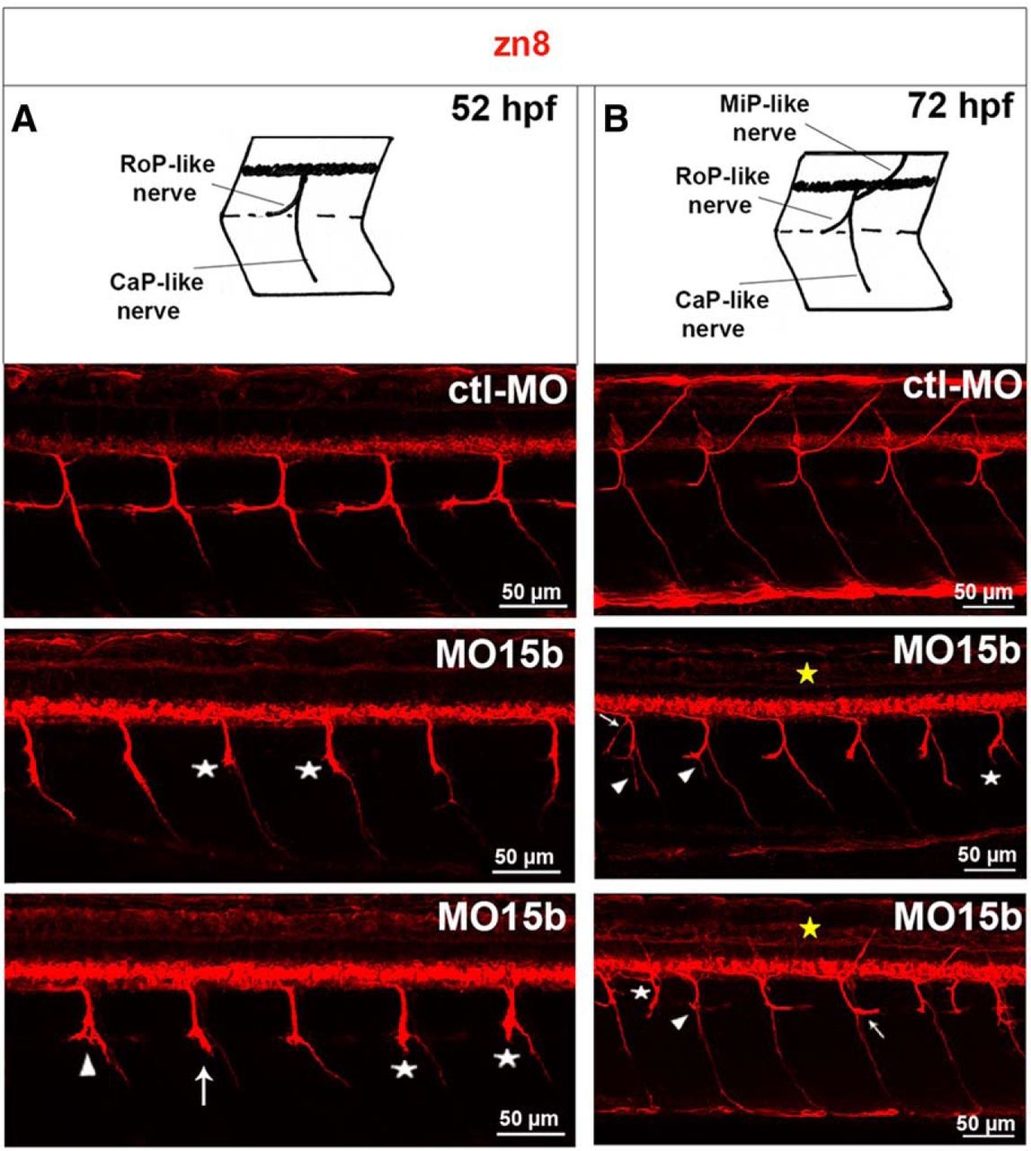
- Figure 9.
Lack of COLXV-B compromises secondary motor axon navigation. Immunostaining with zn8 antibody of 52 hpf (A) and 72 hpf (B) embryos injected with ctl-MO or MO15b. Diagrams depicting regular trajectories of RoP-, MiP-, and CaP-like nerves at 52 and 72 hpf. A, *Abnormal extension of RoP-like nerves. Arrowheads indicate axon failure to separate properly at the choice point; arrow points to absence of CaP-like nerves. B, MO15b, Top, Asterisk and arrowheads indicate respective abnormal ventral projection or additional ventral branching of RoP-like nerves; arrow indicates branching of common nerves. MO15b, Bottom, *Ectopic nerve exit from the spinal cord. Arrow and arrowhead indicate caudal projections and absence of RoP-like nerves, respectively. Yellow asterisks indicate absence of MiP-like nerves. All lateral views, anterior to the left.
- Figure 10.
Knockdown of col15a1b results in skeletal muscle atrophy. A, Triple fluorescence staining with conjugated phalloidin to visualize actin (red), β-catenin antibody to label cell membrane (green), and Hoechst solution to stain nuclei (blue) of 3 dpf uninjected (uninj.) and tMO15b-injected embryos (tMO15b). Trunk cross sections represent the center (left) and the periphery (right) of the myotome. tMO15b-injected embryos show drastic reduction of myofibril content (asterisks compared with control), a significant decrease in fast fiber (FF) size, and an abnormal shape of SSFs. B, Cross sections of whole-mount in situ hybridization of 3 dpf uninjected and tMO15b-injected embryo probed with the slow fiber marker smyhc1 confirming the broader shape of slow fibers in morphants. C, Quantification of cross-sectional areas of individual fast fibers from ¼ of the total myotome surface in uninjected and tMO15b-injected embryos (n = number of cells from 5 separate embryos). ***p < 0.001 (Mann–Whitney's test). Cross sections at the level of the yolk extension; dorsal to the top.
- Figure 11.
Col15a1b knockdown impairs embryo motility. A, Single frame from videos showing escape response upon tactile stimulus of 2 dpf controls (uninjected or ctl-MO-injected embryos), morphants (embryos injected with tMO15b or sMO15b morpholinos), and rescued embryos (embryos injected with tMO15b and 50 pg of smyhc1:col15a1b construct). B, Quantification of the escape response expressed as the time it takes for the embryo to reach out of the larger circle. C, Quantification of the escape response is expressed as the mean of escape time ± SEM. Partial rescue of the motility phenotype is observed in 2 dpf embryos coinjected with tMO15b and smyhc1:col15a1b. NS, Nonsignificant. *p < 0.05 (Student's t test). ***p < 0.001 (Student's t test).
- Figure 12.
Model of the matrix COLXV-B fingerprint laid down by committed adaxial cells for motor axon navigation. A, Expression of col15a1b in adaxial cells depends on Hh/Gli signaling. B, COLXV-B protein is deposited and organized in the motor path upon unplugged/MuSK signaling. C, COLXV-B is present in the motor path at the time of axon growth to provide a positional cue that guides axons toward intermediate targets.


Tables
52 hpf 72 hpf Total RoP-like nerve Total RoP-like nerve MiP-like nerve CaP-like nerve Common nerve embryos Absence embryos Absence Ventral projection Branching Absence Absence Branching Uninjected 49 19.59 ± 4.04 44 15.45 ± 2.92 0.45 ± 0.32 0.23 ± 0.23 0.68 ± 0.38 0 0.23 ± 0.23 Ctl-MO 50 12.85 ± 2.31 (NS) 47 6.80 ± 1.58 (NS) 0.43 ± 0.29 (NS) 0.43 ± 0.30 (NS) 1.48 ± 0.6 (NS) 0.21 ± 0.21 (NS) 3.40 ± 1.15** tMO15b 51 85.88 ± 3.79*** 52 27.5 ± 3.73* 9.03 ± 2.0*** 11.35 ± 2.18*** 59.04 ± 6.08*** 5.19 ± 2.10* 6.35 ± 1.40*** sMO15b 47 68.94 ± 5.07*** 44 29.32 ± 4.12** 2.05 ± 0.62* 2.95 ± 0.90*** 39.09 ± 5.90*** 3.18 ± 1.48* 5.23 ± 1.36*** ↵aQuantitative analysis of secondary motor axon/nerve defects per embryo under the different conditions. Results are expressed as mean frequencies of affected hemisegments per embryo ± SEM; 10 hemisegments per embryo were examined. NS, Nonsignificant.
↵*p < 0.05;
↵**p < 0.01;
↵***p < 0.001; Student's t test.


