

Article Figures & Data
Figures
- Figure 1.
ChR2-eYFP expression in the ATN and in their axon terminals in the presubiculum. A, Mouse brain injected with AAV5-ChR2-eYFP (green) in the ATN. The 320-μm-thick horizontal section with DAPI staining (blue). B, ChR2-eYFP expression at the injection site. AD, Anterodorsal nucleus; AV, anteroventral nucleus; LD, laterodorsal nucleus; sm, stria medullaris; V3, third ventricle; LHb, lateral habenula. C, D, ChR2-eYFP-expressing axon terminals in the presubiculum at different dorsoventral levels, bregma −2 and −3 mm. Presubicular layers 1–6 are indicated. More details about the presubicular anatomy, ChR2-eYFP expression patterns, and recording locations can be found Figure 1-1 and Figure 1-2. E, Firing of a thalamic cell induced by a twofold rheobase depolarizing current. Hyperpolarization induced by a negative current pulse is followed by a rebound burst. F, Top, Blue light stimulation (0.1 mW, 300 ms) evoked spikes and depolarization block in the thalamic cell. Bottom, ChR2-mediated photocurrent recorded in voltage-clamp in response to the same photostimulation. Blue bar represents the light pulse. Recordings made in the presence of CNQX and d-AP5.
- Figure 2.
ATN axons contact MEC-projecting neurons. A, Schematic showing unilateral injection sites for AAV5-ChR2 in ATN (green) and retrobeads in the MEC (red). B, Left, Horizontal section of the parahippocampal formation with retrobeads injected in the MEC (orange). Blue represents DAPI staining. Thalamic axons expressing ChR2-eYFP target the presubiculum. DG, Dentate gyrus; PrS, presubiculum; Sub, subiculum; PaS, parasubiculum. Right, Magnification of the presubiculum (B, inset) stained with DAPI (blue), ChR2-eYFP-expressing thalamic axons (green), retrobeads (red), and a merged image (right). C, Confocal image of the soma of a biocytin-filled neuron in superficial layer 3. The soma (gray) contains red retrobeads and is surrounded by thalamic axons (green). D, Light-evoked EPSCs in the same neuron. More details about the illumination area and calibration for the blue LED light can be found in Figure 2-1 and Figure 2-2. E, Average EPSC latencies (left) and amplitudes (right) from 11 bead-labeled neurons. Horizontal and vertical lines indicate mean ± SEM. F, Light stimuli repeated at 30 Hz initiated AP firing (top, black; same neuron as in C). TTX (1 μm) and 4-AP (100 μm) abolished spikes, whereas direct EPSPs persisted (bottom, red). G, Light-evoked currents at holding potentials of 40 and at −70 mV revealed glutamatergic EPSCs with a NMDA and AMPA receptor-mediated component. Recordings in the presence of 10 μm gabazine and 10 μm CGP 52432. EPSCs were entirely abolished by 100 μm APV and 10 μm NBQX.
- Figure 3.
ATN-driven sequences of excitation-inhibition versus unbalanced excitation or inhibition. A, B, ATN-evoked currents recorded from presubicular pyramidal neurons at 0 mV holding potential, in the presence of APV. The current was biphasic, with an initial inward current followed by an outward current. A, The outward current, mediated by GABAA receptors, was entirely blocked by gabazine. B, NBQX abolished both the inward current and the disynaptic GABAergic component. C, D, Disynaptic IPSCs induced by ATN stimuli have longer-onset latencies and more jitter than the ATN-driven EPSCs. Wilcoxon matched-pairs signed rank test: *p < 0.05; ****p < 0.001. Data are mean ± SEM. E, Different possibilities of afferent connectivity: a pyramidal neuron may receive (Ea) excitation and feedforward inhibition, (Eb) excitation only, or (Ec) inhibition only. F, Schematic of the experiment showing the presubiculum (PrS) with the recording pipette and the PC soma (red), and the grid of laser light stimulation sites (black circles, spacing 40 μm). PaS, Parasubiculum; Sub, subiculum. Ga, Sequences of excitatory-inhibitory responses. Gb, Excitatory responses only. Gc, Inhibitory responses only. Recordings from the same pyramidal neuron for different laser light stimulation sites across the presubiculum. H, Probability to elicit excitatory-inhibitory responses (E-I, blue), excitatory responses only (E, red), or disynaptic inhibitory responses only (I, green). Gray represents no response.
- Figure 4.
Relative AP timing in pairs of pyramidal neurons and interneurons. A, Left, Current-clamp recording of a PV interneuron (green), a PC (black), and SST interneuron (purple), with two different light intensities for stimulation of ATN axon terminals (0.2 and 1.1 mW). Right, Summary data. Higher stimulus intensities led to shorter AP latencies in the same neuron. B, APs initiated by light activation of thalamic fibers in a simultaneously recorded PC-PV pair or (C) in a PC-SST pair. D, E, AP latencies from 7 PC-PV pairs, and from 6 PC-SST pairs. Each dot represents 1 neuron. AP latencies in PC cells were longer than in PV interneurons but shorter than in SST interneurons. Wilcoxon matched-pairs signed rank test: *p < 0.05; **p < 0.01.
- Figure 5.
Long-range excitatory inputs from ATN to pairs of PV or SST interneurons and pyramidal neurons in layer 3. A, D, Presubicular slice with two biocytin-filled neurons. A, B, Pyramidal neuron and PV interneuron. D, E, Pyramidal neuron and SST interneuron. Green represents ATN axons in the superficial layers. Scale bars, 100 μm. B, E, Reconstructed cell pairs. Blue represents PC dendrites. Yellow represents PC axons. Green represents interneuron dendrites. Red represents interneuron axons. C, Dual records of PV interneuron (green) and pyramidal neuron (black) or (F), SST interneuron (purple) and pyramidal neuron (black) respectively. Left, Firing pattern in response to a twofold rheobase current injection. Right, Light-evoked EPSCs from the illustrated pair of simultaneously recorded pyramidal neuron and interneuron. Red represents the presence of TTX (1 μm) and 4-AP (100 μm) in the bath solution. Amplitudes (G) and latencies (H) from double-recorded principal neuron and interneuron EPSCs. Wilcoxon matched-pairs signed rank test: **p < 0.01; ***p < 0.001. I, Light stimulation reliably generated APs in a PC. EPSCs in a simultaneously recorded SST interneuron had a 30% failure rate (30 trials). J, Bar plots of success rate for synaptic events in different cell types: calculated from 30 trials for each neuron tested; 13 PV interneurons (green), 13 PC (black), and 11 SST interneurons (purple). ****p < 0.0001 (Kruskal–Wallis and Dunn's multiple-comparison post hoc test). Data are mean ± SEM.
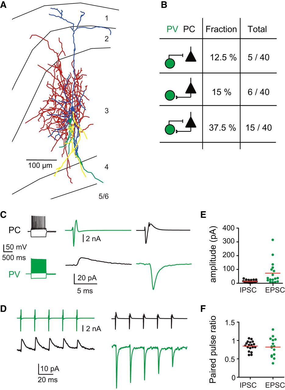
- Figure 6.
Presubicular layer 3 PV interneurons and PCs are highly interconnected. A, Anatomical reconstruction of a pair of reciprocally connected PV interneuron and pyramidal neuron in presubicular layer 3. Blue represents PC dendrites. Yellow represents PC axons. Green represents PV dendrites. Red represents PV axons. Left, Subiculum. Right, Parasubiculum. B, Summary of connectivity between PV and PC cells. C, Paired recording of PV-PC pair shown above. Left, Firing patterns in response to a negative and positive (twofold rheobase) current injection. An evoked action current in the PV cell (green, −70 mV, voltage clamp) initiated a short-latency IPSC in the PC (black, −50 mV, voltage clamp). Conversely, an evoked action current in the PC elicited an EPSC in the PV cell. Traces are averaged from 10 trials. D, Unitary IPSCs (black) and EPSCs (green) from the same cell pair induced by 5 action currents at 50 Hz. Absolute amplitudes (E) and PPR (F) of IPSCs in PC neurons (black) and EPSCs in PV interneurons (green). Each dot represents averaged recordings from one neuron.
- Figure 7.
PV interneurons mediate ATN-driven feedforward inhibition in the presubiculum. A, Blue (470 nm) and yellow (585 nm) illumination via the same light path was used to activate ChR2 alone, or in combination with eNpHR. B, A depolarizing current step produced high-frequency firing in an eNpHR-expressing PV cell. Yellow light activated eNpHR and strongly hyperpolarized this interneuron. C, Action currents recorded in cell-attached mode from an eNpHR-expressing PV cell in response to photoactivation of ATN inputs with blue light, before, during, and after illumination with yellow light. Yellow light was triggered 5 ms before the onset of the 0.5 ms blue light pulse and remained on for 20 ms. D, Synaptic currents induced in a layer 3 pyramidal neuron by photostimulation (blue) of ATN fibers in the absence, in the presence, and again in the absence of yellow light. Yellow light stimuli (middle) reversibly inhibited PV interneurons and suppressed evoked IPSCs recorded as outward currents at 0 mV holding potential. E, IPSCs were suppressed when PV interneurons were silenced. **p < 0.01 (Kruskal–Wallis and Dunn's multiple-comparison post hoc test). F, EPSCs recorded in PC cells at −60 mV holding potential were not affected by yellow light stimuli.
- Figure 8.
Synaptic dynamics of responses to photostimulation of ATN inputs in layer 3 pyramidal neurons and interneurons. A, D, G, EPSCs evoked by 10 successive light stimuli repeated at 10 Hz, and (B, E, H) at 30 Hz in PV interneurons (light green, dark green), PC cells (gray, black), and SST interneurons (purple, violet). C, F, I, EPSC amplitudes to stimuli at 10 Hz and 30 Hz for the three cell types, respectively. Box plots represent minimum, maximum, lower and upper quartiles, median, and mean values. Anderson–Darling test and Hochberg–Benjamini multiple-comparison test: *p < 0.05; **p < 0.01; ***p < 0.001.
- Figure 9.
Cell type-specific spiking probabilities following optical stimulation of ChR2-expressing ATN fibers. A, Variation of spiking probability as a function of light intensity. A spiking probability of 1 corresponds to 10 spikes for a train of 10 stimuli. At higher intensities, a single pulse sometimes induced multiple spikes in PV interneurons (green). Black represents PC neurons. Purple represents SST interneurons. Data are mean ± SEM. Examples of responses of a PV interneuron (B), pyramidal neuron (C), and SST interneuron (D) to 10 Hz and 30 Hz stimulation at resting potential or near AP threshold. D, Red arrow indicates the absence of a spike for the first pulse. E–G, AP spiking probabilities for each cell type for 10 Hz and 30 Hz represented by box plots showing the lower and upper quartiles, the median, and the mean. Anderson–Darling test and Hochberg–Benjamini multiple-comparison test, to compare the spiking probability of each pulse to the first pulse: *p < 0.05; **p < 0.01; ***p < 0.001; ****p < 0.0001.
- Figure 10.
Schematic illustration of connectivity of ATN fibers targeting presubicular layer 3 neurons. Presubicular PCs projecting to MEC receive direct excitation from thalamic fibers. PV-expressing interneurons also receive direct excitation from thalamic fibers, and they provide transient feedforward inhibition onto PCs. The activation of single thalamic fibers may either lead to (a) PC excitation followed by feedforward inhibition, or (b) only excitation, or (c) only rapid disynaptic inhibition of PCs (compare Fig. 3). SST interneurons (d) are recruited in a feedback manner following AP discharge of local PCs (Fig. 4) at high frequency (Fig. 9). SST-PC connectivity is high (compare Simonnet et al., 2017) and provides activity-dependent feedback or lateral inhibition.


Tables
PV SST PC PC beads+ Mean SEM N Mean SEM N Mean SEM N Mean SEM N Membrane potential (mV) −67 1.10 13 −54 2 15 −72 3 17 −74 1.1 15 Time constant (ms) 19 3 10 38 8 10 28 6 8 27 4 11 Sag ratio 1.10 0.01 12 1.20 0.02 15 1.04 0.01 16 1.05 0.01 16 Input resistance (mΩ) 147 12 13 438 58 14 362 68 15 359 34 14 Threshold current (pA) 183 18 13 59 19 15 63 18 13 70 11 16 I-O initial gain (Hz/pA) 1.06 0.09 13 0.90 0.07 15 0.36 0.06 16 0.27 0.02 15 Firing frequency at 2× rheobase 191 12 8 40 6.20 14 28 3.30 14 27 1.40 16 AP threshold (mV) −39 1.20 13 −34 0.90 15 −38 0.80 17 −30 0.60 16 AP amplitude (mV) 77 1.10 13 76 1.80 15 81 1.03 17 80 1.40 16 AP half-duration (ms) 0.20 0.01 13 0.30 0.01 15 0.60 0.01 17 0.65 0.02 16 AP maximum depolarization (V/s) 624 22 13 507 23 15 478 17 17 452 22 16 AP maximum repolarization (V/s) −464 22 13 −272 14 15 −127 4 17 −121 4 16 - Table 2.
EPSC and IPSC properties from synaptically connected PV interneuron-pyramidal neuron pairs
EPSCs IPSCs Mean SEM N Mean SEM N Rise time (ms) 0.31 0.02 17 0.52 0.07 19 Decay time (ms) 0.91 0.07 17 2.90 0.20 19 Latency onset (ms) 0.74 0.03 17 0.68 0.03 19 Transfer rate 0.79 0.06 17 0.71 0.07 19
Figure 1-1
Anatomy of horizontal slices containing the presubiculum, including expression patterns of Channelrhodopsin and locations of biocytin filled recorded neurons (Extended data supporting Figure 1). A-D. The shape of presubiculum becomes increasingly broader from ventral (A) to dorsal (D) levels. Left column, horizontal DAPI (blue) stained sections, for dorso-ventral planes as indicated. A corresponds to the ventral presubiculum (close to DV -4 mm from Bregma), D corresponds to the dorsal part of the presubiculum (close to DV -2.8 mm from Bregma). Second from left, corresponding expression pattern of eYFP tagged Channelrhodopsin (green), for dorso-ventral levels A to D. Examples for individual slices containing biocytin filled presubicular neurons (white), matched to the dorso-central levels A to D. Two examples of recorded and biocytin filled neurons (MN903c1c2 and MNo09c1c2) have been reconstructed and are part of Figure 5. *indicates same slices within a row. DG, Dentate Gyrus; Sub, subiculum; PaS, parasubiculum; MEC, medial entorhinal cortex; ChR2, eYFP-tagged channelrhodopsin. Download Figure 1-1, TIF file
Figure 1-2
Expression pattern of eYFP tagged channelrhodopsin. (Extended data supporting Figure 1). (A) Fluorescence profile across presubicular layers 1 to 6. (B) Measurement of fluorescence intensity (normalized). Download Figure 1-2, EPS file
Figure 2-1
60x whole field illumination area estimated by bleaching of eYFP fluoresence. (Extended data supporting Figure 2). (A). The location of the center of the 60x field of view is indicated by a red square. The pictures were taken with a 4x objective. (B-C). Following several minutes of illumination with blue LED light through the 60x objective, photobleaching occurred in a circular zone of diameter 200 µm, corresponding to the most intensely illuminated area under the 60x objective. Download Figure 2-1, EPS file
Figure 2-2
Calibration of the LED light duration and intensity (Extended data supporting Figure 2). (A) LED stimulation of thalamic fibers recorded in a pyramidal neuron at -60 mV, same intensity (0.3 mW) and 1 ms (black trace) or 10 ms duration (pink trace). (B) Calculated area of the light-evoked current plotted as a function of the duration for 6 pyramidal neurons. (C) Recordings from a presubicular layer 3 pyramidal neuron for LED stimulation at 0 (black), 0.7 (green) or 2 (blue) mW. (D) Calculated area of the light evoked currents as a function of intensity for 11 pyramidal neurons. Download Figure 2-2, EPS file


