

Article Figures & Data
Figures
- Figure 1.
The expression of pC/EBPβ in the SCDH in the peripheral gp120 neuropathic pain model. A, Rats received peripheral gp120 application onto the left sciatic nerve, and the sham group received RSA as vehicle. Time course of mechanical withdrawal threshold was measured using von Frey filaments to ipsilateral hindpaws and contralateral hindpaws. Peripheral gp120 induced a persistent lowered mechanical withdrawal threshold of the ipsilateral hindpaw as measured by von Frey filaments compared with sham rats; F(12,96), interaction = 2.238, p < 0.05; F(4,96), main effect time = 2.611, p < 0.05; F(3,24), main effect treatment = 11.05, p < 0.0001, two-way repeated-measures ANOVA; n = 7. We also observed that there was a significant lowered mechanical threshold in ipsilateral (gp120-ipsi) compared with that contralateral hindpaws (gp120-contra) in gp120 rats at days 7, 10, and 14 (**p < 0.01, ***p < 0.001 vs gp120-contralateral paw; two-way ANOVA, Bonferroni post-tests, n = 7. Mechanical threshold in ipsilateral hindpaws in gp120 rats (gp120-ipsi) was significantly lower than that in ipsilateral hindpaws in sham rats (sham-ipsi) at days 7, 10, and 14 (##p < 0.01, ###p < 0.001 vs sham-ipsilateral paw; two-way ANOVA, Bonferroni post-tests; n = 7. There was no significant difference in mechanical threshold between ipsilateral hindpaw (sham-ipsi) and contralateral paws (sham-contra) in sham rats. B, Western blots showed no difference in the expression of pC/EBPβ in sham rats between ipsilateral (sham-I) and contralateral (sham-C) SCDH in the SCDH 2 weeks post-gp120. C, pC/EBPβ in the ipsilateral SCDH in gp120 rats (gp120-I) was significantly higher than that in the contralateral SCDH (gp120-C); **p < 0.01, t test, n = 6. D, No difference in the expression of C/EBPβ in sham rats between ipsilateral (sham-I) and contralateral SCDH (sham-C) at 2 weeks post vehicle. E, C/EBPβ in the ipsilateral SCDH in gp120 rats (gp120-I) was significantly higher than that in the contralateral SCDH (gp120-C); **p < 0.01, t test, n = 6. F, RT-PCR showed that gp120 increased the expression of mRNA of C/EBPβ; ***p < 0.001 versus sham, t test; n = 5. G, Western blots assay showed that gp120 evoked overexpression of pC/EBPβ in the ipsilateral SCDH compared with either naive or sham groups; #p < 0.05 versus sham **p < 0.01 versus naïve; one-way ANOVA, post hoc PLSD test; n = 4. H, HIV gp120 induced the overexpression of C/EBPβ in the ipsilateral SCDH; ***p < 0.001 versus naïve; ###p < 0.001 versus sham; one-way ANOVA, post hoc PLSD test; n = 4. I, Low-magnification image showed pC/EBPβ immunostaining in the L4/5 lamina I–V of the SCDH. Scale bar, 100 μm. J–L, Double-immunostaining showed that pC/EBP-beta-positive cells were colocalized with NeuN (a neuron marker; J), but few of pC/EBP-beta-positive cells overlaid with GFAP (astrocytes marker; K) or with OX42 (a microglia marker; L) in the SCDH in rats with gp120 application at 2 weeks. Scale bar, 50 μm. M, Twelve days after gp120 application, intrathecal administration of C/EBPβ siRNA or mmRNA was given once a day for 2 d. Intrathecal C/EBPβ siRNA significantly increased mechanical threshold (F(5,50), interaction = 8.59, p < 0.0001; F(5,50), main effect time = 9.52, p < 0.0001; F(1,10), main effect treatment = 7.96, p < 0.05; two-way repeated-measures ANOVA). Mechanical withdrawal threshold in the C/EBPβ siRNA group was higher than that in mmRNA at day 1 and 2; ***p < 0.001 versus mmRNA; two-way ANOVA, Bonferroni tests; n = 6. N, Western blots displayed that C/EBPβ siRNA reversed the upregulated pC/EBPβ in the SCDH. **p < 0.01 versus sham+mmRNA; ##p < 0.01 versus gp120+mmRNA; one-way ANOVA, post hoc PLSD test; n = 4–5.
- Figure 2.
The increased expression of TNFα, TNFRI, and mtO2·− in the SCDH in the gp120 model. A–C, The expression of TNFα between ipsilateral and contralateral SCDH in both sham and gp120 rats at 2 weeks after gp120, using Western blots. A, No significant difference in TNFα between ipsilateral (sham-I) and contralateral (sham-C) SCDH in sham rats. B, TNFα in the ipsilateral SCDH of gp120 rats (gp120-I) was higher than that in the contralateral SCDH (gp120-C); *p < 0.05 versus gp120-C; t test, n = 6. C, At 2 weeks after gp120, peripheral gp120 increased spinal TNFα at ipsilateral SCDH ***p < 0.001 versus Naive; ###p < 0.001 versus sham-I, one-way ANOVA, posthoc PLSD test. n = 4. D, No significant difference in TNFRI between ipsilateral and contralateral SCDH in sham animals. E, TNFRI in the ipsilateral SCDH of gp120 rats (gp120-I) was higher than that in the contralateral SCDH (gp120-C) at 2 weeks after gp120; ***p < 0.001, t test; n = 6. F, HIV gp120 increased spinal TNFRI at ipsilateral SCDH (gp120-I) ***p < 0.001 versus Naive; ###p < 0.001 versus sham-I, one-way ANOVA, posthoc PLSD test; n = 4. G–I, Double-immunostaining showed that TNFRI was colocalized with NeuN. Scale bar, 50 μm. J, K, Representative images of MitoSox-positive cells in the SCDH in rats with gp120 and sham surgery at 2 weeks. Scale bar, 50 μm. L, HIV gp120 application increased MitoSox-positive cells in the lamina I–II, III–V, and total dorsal horn (laminar I–V) in the ipsilateral SCDH; **p < 0.01, t test; n = 5. M, The number of MitoSox-positive cells between ipsilateral and contralateral SCDH in sham rats, and in gp120 rats; ***p < 0.001, t test; n = 4–5.
- Figure 3.
The expression of pCREB and its binding location at the C/EBPβ gene promoter regions in the peripheral gp120 neuropathic pain model. The expression of pCREB and CREB between ipsilateral and contralateral SCDH in both sham and gp120 rats at 2 weeks after gp120. No significant difference in pCREB (A) and CREB (B) between ipsilateral (sham-I) and contralateral SCDH (sham-C) in sham animals. C, Expression of pCREB in the ipsilateral SCDH of gp120 rats (gp120-I) was higher than that in the contralateral SCDH (gp120-C); ***p < 0.001 versus gp120-C, t test; n = 6. D, No significant change of CREB in the ipsilateral and contralateral SCDH of gp120 rats. E, Western bolts showed that gp120 increased the expression of pCREB in the ipsilateral SCDH compared with naive or sham group; **p < 0.01 versus naïve; ##p < 0.01 versus sham-I; one-way ANOVA, post hoc PLSD test; n = 4. F, There was no significant difference between sham and gp120 group in CREB in the ipsilateral SCDH. G, Low-magnification image showed pCREB immunostaining in the L4/5 lamina I–V of the SCDH. Scale bar, 100 μm. H, Double-immunostaining showed that almost all of pCREB was colocalized with NeuN. Scale bar, 50 μm. I, Alignment of rat C/EBPβ gene promoter regions, and pCREB binding location. We marked the CRE and ChIP-qPCR primer areas at the location before the TSS of the C/EBPβ gene at rat chromosome 3 (ACCESSION #NC_005102.4). J, Application of gp120 increased the enrichment of pCREB at the C/EBPβ gene promoter regions in the ipsilateral SCDH compared with sham ipsilateral side; *p < 0.05 versus sham-I, t test; n = 5.
- Figure 4.
The effect of TNFα signal blockage with intrathecal rTNFSR on the expression of MitoSox cells, pCREB, and pC/EBPβ in the SCDH. A–C, Representative MitoSox image in sham+saline (A), gp120+saline (B), and gp120+rTNFSR (C) was shown. Scale bar, 50 μm. D, The number of MitoSox-positive cells in the ipsilateral SCDH was displayed; **p < 0.01 versus the sham+saline group; ##p < 0.01 versus gp120+saline group; one-way ANOVA, post hoc PLSD test; n = 5–6. E, Western blots showed that the effect of intrathecal rTNFSR on pCREB in rats with gp120; **p < 0.01 versus sham+saline; #p < 0.05 versus gp120+saline; one-way ANOVA, n = 4. F, There was no significant change in the expression of CREB among groups of sham+saline, gp120+saline, and gp120+rTNFSR. G, Effect of rTNFSR on pC/EBPβ in rats with gp120 using Western blots; *p < 0.05 versus sham+saline; ##p < 0.01 versus gp120+saline; one-way ANOVA; n = 4–5. H, Effect of rTNFSR on C/EBPβ in rats with gp120 using Western blots; **p < 0.01 versus sham+saline; ##p < 0.01 versus gp120+saline; one-way ANOVA, n = 4–5. I, The effect of rTNFSR on TNFa in rats with gp120 using Western blots; **p < 0.01 versus sham+saline; #p < 0.05 versus gp120+saline; one-way ANOVA, n = 4–5.
- Figure 5.
The effect of TNFRI knockdown on the expression of mtO2·−, pCREB, and pC/EBPβ in the ipsilateral SCDH in the gp120 model. A, Ten days after gp120, AS-TNFRI or mmODN was intrathecally administered once daily (20 μg) for 5 d. Intrathecal AS-TNFRI increased mechanical threshold; F(6,60), interaction = 4.44, p < 0.001; F(6,60), main effect time = 4.54, p < 0.001; F(1,10), main effect treatment = 16.09, p < 0.01; two-way repeated-measures ANOVA; n = 6. Mechanical threshold in the AS-TNFRI group was higher than that in mismatch ODN at days 2–5; *p < 0.05; ***p < 0.001 versus mmODN; two-way ANOVA, Bonferroni tests. B, HIV gp120 increased TNFRI expression compared with sham+mmODN; **p < 0.01; one-way ANOVA, PLSD test, n = 4. TNFRI in gp120+AS-TNFRI was lower than that in gp120+mmODN; ##p < 0.01, one-way ANOVA, post hoc PLSD test; n = 4. C, MitoSox-positive images in groups of sham+mmODN, gp120+mmODN, or gp120+AS-TNFRI are shown. D, The number of MitoSox-positive cells in lamina I–V of ipsilateral SCDH in the sham+mmODN, gp120+mmODN, and gp120+AS-TNFRI groups; ***p < 0.001 versus sham+mmODN; ###p < 0.001 versus gp120+mmODN; one-way ANOVA, post hoc PLSD test; n = 4–5. E, Effect of knockdown of TNFRI on pCREB in the SCDH; ***p < 0.001 versus sham+mmODN; ###p < 0.001 versus gp120+mmODN; one-way ANOVA, post hoc PLSD test; n = 4. F, Effect of knockdown of TNFRI on CREB in the SCDH. G, AS-TNFRI suppressed the increased expression of pC/EBP; **p < 0.01 versus sham+mmODN; ##p < 0.01 versus gp120+mmODN; one-way ANOVA, post hoc PLSD test; n = 4. H, AS-TNFRI suppressed the increased expression of C/EBPβ; **p < 0.01 versus sham+mmODN; ##p < 0.01 versus gp120+mmODN; one-way ANOVA, post hoc PLSD test; n = 4.
- Figure 6.
The effect of inhibition of mtO2·− signal and the knockdown of CREB on the gp120 neuropathic pain. A, HIV gp120 neuropathic rats received intrathecal Mito-Tempol (Mito-T, 50 μg) or saline (vehicle). Mito-T significantly increased mechanical threshold compared with vehicle; F(5,55), interaction = 8.32, p < 0.0001; F(5,50), main effect time = 7.80, p < 0.0001; F(1,11), main effect treatment = 9.71, p < 0.01; two-way repeated-measures ANOVA. Mechanical withdrawal threshold in the Mito-T group was higher than that in vehicle at 30–90 min; **p < 0.01, ***p < 0.001, two-way repeated-measures ANOVA with Bonferroni test; n = 6–7. B, The effect of Mito-Tempol on pCREB in the ipsilateral SCDH using Western blots; *p < 0.05 versus sham+saline group; ##p < 0.01 versus gp120+saline group; one-way ANOVA, post hoc PLSD test; n = 4. C, The effect of Mito-Tempol on pC/EBPβ in the gp120 model using Western blots; **p < 0.01 versus sham+saline group; ##p < 0.01 versus gp120+saline group; one-way ANOVA, post hoc PLSD test; n = 4. D, The effect of the knockdown of CREB using antisense ODN against CREB on mechanical threshold in the gp120 model. Intrathecal AS-CREB significantly increased mechanical threshold; F(8,88), interaction = 8.86, p < 0.0001; F(8,88), main effect time = 7.00, p < 0.0001; F(1,11), main effect treatment = 30.04, p < 0.001; two-way repeated-measures ANOVA; n = 6–7. Mechanical withdrawal threshold in the antisense ODN of CREB group was higher than that in mismatch ODN at days 3–6; ***p < 0.001, two-way repeated-measures ANOVA with Bonferroni test; n = 6–7. E, The effect of AS-CREB on the expression of pCREB in the ipsilateral SCDH; **p < 0.01 versus sham+mmODN group; ##p < 0.01 versus gp120+mmODN group; one-way ANOVA, post hoc PLSD test; n = 4. F–H, Knockdown of spinal CREB on the C/EBPβ mRNA, C/EBPβ, or pC/EBPβ in the ipsilateral SCDH of gp120 neuropathic rats. Intrathecal AS-CREB (once a day for 5 d) reversed the upregulation of C/EBPβ mRNA using RT-PCR (F) and C/EBPβ protein using Western blots (G); **p < 0.01 versus sham+mmODN group; #p < 0.05, ##p < 0.01 versus gp120+mmODN group; one-way ANOVA, n = 4. Similarly, intrathecal AS-CREB reversed the upregulation of pC/EBPβ using Western blots (H); **p < 0.01 versus sham+mmODN group; ##p < 0.01, versus gp120+mmODN group; one-way ANOVA, n = 4.
- Figure 7.
The effect of intrathecal MitoSox on pCREB, pC/EBPβ or C/EBPβ in naive rats, and the effect of the knockdown of CREB and C/EBPβ on mtO2·− in the gp120 neuropathic pain rats. A–D, Seventy minutes after rats received intrathecal MitoSox or vehicle injection, the SCDH was harvested. Western blots showed that there was no significant difference in the expression of pCREB (A), CREB (B), pC/EBPβ (C), or C/EBPβ (D) in the SCDH 70 min after MitoSox compared with vehicle. E, Effect of knockdown of spinal CREB on mitochondrial superoxide in the gp120 pain model. Ten days after gp120, AS-CREB or mmODN was intrathecally administered once daily (20 μg) for 5 d. One hour after the last ODN, MitoSox was intrathecally injected. Animals were perfused 70 min after MitoSox, and the spinal cord was harvested for MitoSox imagine. There was a significant increase in MitoSox-positive cells in gp120+mmODN group compared with that in sham+mmODN. ***p < 0.001, one-way ANOVA; n = 5. MitoSox-positive cells in gp120+AS-CREB group was lower than that in gp120+mmODN. ###p < 0.001, one-way ANOVA; n = 5. F, Effect of knockdown of spinal C/EBPβ on mitochondrial superoxide in the gp120 pain model. Ten days after gp120, C/EBPβ siRNA or mmRNA was intrathecally administered once daily for 2 d. One hour after the last C/EBPβ siRNA or mmRNA, MitoSox was intrathecally injected. Animals were perfused 70 min after MitoSox, and the spinal cord was harvested for MitoSox imagine. There was a significant increase in MitoSox-positive cells in gp120+mmRNA group compared with that in sham+mmRNA. ***p < 0.001, one-way ANOVA; n = 5. MitoSox-positive cells in gp120+C/EBPβ siRNA group was lower than that in gp120+mmRNA. ###p < 0.001, one-way ANOVA; n = 5. G, The proposed signaling pathways that TNFα–mtO2·−–pCREB signaling pathway triggered the pC/EBPβ in gp120-related neuropathic pain in the spinal cord dorsal horn. HIV gp120 induced glia activity to release spinal TNFα (Zheng et al., 2011). TNFα binds its receptor TNFRI on the neurons. TNFRI signal increases mitochondrial ROS through unknown factors. ROS can lead to the activation of various other cell signaling pathway components, for example, transcriptional factor pCREB. pCREB may induce C/EBP expression (for review, see Alberini, 2009). C/EBPβ promotes recycling of NMDA receptors from endosomes to the plasma membrane (Wang et al., 2013). NMDA receptors increase Ca2+ influx (Ohno et al., 2002; Valnegri et al., 2015). Mitochondria are the major sites of ROS production in neurons, and participate in the regulation of cellular Ca2+ homeostasis, which is implicated in mitochondrial ROS generation (Camello-Almaraz et al., 2006; Kann and Kovacs, 2007; Douda et al., 2015). Thus, based on our results and previous evidence, we think that it is possible that pathway of TNFα/TNFRI signal triggering ROS–pCREB–pC/EBP through unknown pathways, is involved in the gp120 neuropathic pain state.
- Figure 8.
Recombinant TNFα induced mitochondrial superoxide, pCREB, and pC/EBPβ in the cultured B35 neuronal cell line. A, Representative image of MitoSox for mtO2·− in the cultured neuronal cells. Scale bar, 50 μm. B, The treatment with rTNFα significantly increased MitoSox signals in the B35 neuronal cells compared with vehicle treatment. **p < 0.01, t test; n = 3. C, The treatment with rTNFα increased pCREB compared with that with vehicle (**p < 0.01, t test; n = 3–4), but not total CREB protein (D). E, ChIP assay showed that treatment with rTNFα increased the binding of pCREB on the C/EBPβ promoter region; **p < 0.01 versus vehicle; t test, n = 3. F, Treatment with rTNFα increased the expression of C/EBPβ mRNA in the cultured B35 cells; **p < 0.01 versus vehicle; t test, n = 3. G, The treatment with rTNFα in the B35 cells increased pC/EBPβ protein expression compared with vehicle. **p < 0.01, t test. H, C/EBPβ protein was increased by rTNFα in the cultured neurons. *p < 0.05, t test; n = 3.
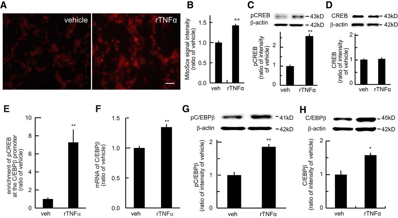
- Figure 9.
The relationship among mtO2·−, pCREB, and pC/EBPβ in the cultured B35 neuronal cells treated with rTNFα. A, Representative imaging of MitoSox-positive cells in vehicle with vehicle (Aa), rTNFα with vehicle (Ab), and rTNFα with Mito-T (Ac). Scale bar, 50 μm. B, The effect of Mito-T on the MitoSox signals in the cultured B35 cells. MitoSox signals in the groups of vehicle+vehicle, rTNFα+vehicle, and rTNFα+Mito-T; ***p < 0.001 versus vehicle+vehicle; ###p < 0.001 versus rTNFα+vehicle; one-way ANOVA, post hoc PLSD test; n = 3. C, The effect of Mito-T on pCREB in the cultured B35 cells treated with rTNFα; ***p < 0.001 versus vehicle+vehicle; ###p < 0.001 versus rTNFα+vehicle; one-way ANOVA, post hoc PLSD test; n = 4. D, The effect of Mito-T on pC/EBPβ in the B35 cells treated with rTNFα; **p < 0.01 versus vehicle+vehicle; ##p < 0.01 versus rTNFα+vehicle; one-way ANOVA; n = 4. E, The effect of Mito-T on C/EBPβ in the B35 cells treated with rTNFα; ***p < 0.001 versus vehicle+vehicle; ##p < 0.01 versus rTNFα+vehicle; one-way ANOVA; n = 4. F, The effect of AS-CREB on pCREB in the B35 cells treated with rat rTNFα or vehicle; **p < 0.01 versus mmODN+vehicle; ##p < 0.01 versus mmODN+rTNFα; one-way ANOVA; n = 3. G, The effect of AS-CREB on pC/EBPβ in the B35 cells; **p < 0.01 versus mmODN+vehicle; ##p < 0.01 versus mmODN+rTNFα; one-way ANOVA; n = 3. H, AS-CREB on total C/EBPβ in the B35 cells; **p < 0.01 versus mmODN+vehicle; ###p < 0.001 versus mmODN+rTNFα; one-way ANOVA, n = 3. I, The possible pathway in the rat neuronal B35 cells treated with rTNFα. TNFα triggered overproduction of mitochondrial superoxide. The oxidative stress induced pCREB. Furthermore, pCREB caused pC/EBPβ in the neuronal B35 cells.
- Figure 10.
The effect of intrathecal rTNFα on mechanical threshold, MitoSox image, pCREB, and pC/EBPβ in naive rats. Intrathecal rTNFα (0.3 ng, twice a day for 2 d) or saline (vehicle) was administered. At day 3 (16 h post the last rTNFα), mechanical threshold was measured and the SCDH harvested. A, Intrathecal rTNFα decreased mechanical threshold at day 3; **p < 0.01 versus vehicle, t test; n = 5. B, Representative imaging of MitoSox with vehicle or rTNFα. C, Intrathecal rTNFα increased the number of MitoSox-positive cells in the SCDH lamina I–II and III–V (***p < 0.001 vs vehicle, t test; n = 5). D, Intrathecal rTNFα increased pCREB (*p < 0.05 vs vehicle, t test; n = 5), but not total CREB E, F, ChIP-qPCR assay showed that intrathecal rTNFα increased the enrichment of pCREB on the C/EBPβ gene promoter regions compared with vehicle (**p < 0.01 vs vehicle, t test; n = 4–5). G, RT-PCR displayed that mRNA of C/EBPβ was increased by intrathecal rTNFα compared with vehicle (***p < 0.01 vs vehicle, t test; n = 5). Western blots also showed an increase in pC/EBPβ (H) and C/EBPβ (I) induced by intrathecal rTNFα (**p < 0.01 vs vehicle, t test; n = 5).
- Figure 11.
The relationship among mtO2·−, pCREB, and pC/EBPβ in the SCDH in rats treated with intrathecal rTNFα. A, Naive rats received intrathecal injection of saline (vehicle) or rTNFα (twice a day for 2 d; arrowhead). At day 3, 16 h post the last rTNFα injection, rats received intrathecal mitochondrial superoxide scavenger Mito-T or vehicle, and mechanical threshold was measured. The SCDH was harvested at 2 h after intrathecal compounds. Arrows showed vehicle or Mito-T injection. Intrathecal Mito-T significantly reversed mechanical threshold lowered by rTNFα; F(10,70), interaction = 9.54, p < 0.0001; F(5,70), main effect time = 15.06, p < 0.0001; F(2,14), main effect treatment = 13.81, p < 0.001; two-way repeated-measures ANOVA; n = 5–7. Mechanical withdrawal threshold in the rTNFα+Mito-T group was higher than that in rTNFα+vehicle at 30–120 min; *p < 0.05; ***p < 0.001 versus rTNFα+vehicle; two-way repeated-measures ANOVA with Bonferroni post hoc test; n = 6–7. B, The effect of intrathecal Mito-T on pCREB in rats treated with rTNFα; *p < 0.05 versus vehicle+vehicle group; ##p < 0.01 versus rTNFα+vehicle group; one-way ANOVA, post hoc PLSD test; n = 4–5. C, The effect of Mito-T on pC/EBPβ in rats treated with rTNFα; *p < 0.05 versus vehicle+vehicle group; #p < 0.05 versus rTNFα+vehicle; one-way ANOVA; n = 4–5. D, The effect of intrathecal AS-CREB on mechanical threshold in rats with rTNFα. Arrows show the ODN injections. AS-CREB significantly reversed the lowered mechanical threshold; F(3,30), interaction = 3.57, p < 0.05; F(3,30), main effect time = 9.93, p < 0.001; F(1,11), main effect treatment = 7.43, p < 0.05; two-way repeated-measures ANOVA; n = 6. Mechanical withdrawal threshold in the rTNFα+AS-CREB group was higher than that in rTNFα+mmODN at 16–18 h after the last rTNFα; *p < 0.05, two-way repeated-measures ANOVA with Bonferroni test; n = 6. E, The effect of intrathecal AS-CREB on spinal pCREB in rat treated with rTNFα; **p < 0.01 versus vehicle+mmODN; #p < 0.05 versus rTNFα+mmODN; one-way ANOVA; n = 4–5. F, The effect of intrathecal AS-CREB on pC/EBPβ in rat treated with intrathecal rTNFα; **p < 0.01 versus vehicle+mmODN; ##p < 0.01 versus rTNFα+mmODN; one-way ANOVA; n = 4–5. G, The effect of intrathecal AS-CREB on total C/EBPβ in rat treated with intrathecal rTNFα; ***p < 0.001 versus vehicle+mmODN; ###p < 0.001 versus rTNFα+mmODN; one-way ANOVA; n = 4–5. H, The effect of intrathecal C/EBPβ siRNA or mmRNA on mechanical threshold in rats with intrathecal rTNFα. Arrows show the siRNA injections. Intrathecal C/EBPβ siRNA significantly increased mechanical threshold compared with mmRNA; F(3,24), interaction = 3.19, p < 0.05; F(3,24), main effect time = 18.31, p < 0.0001; F(1,8), main effect treatment = 14.36, p < 0.01; two-way repeated-measures ANOVA; n = 5. Mechanical withdrawal threshold in the rTNFα+siRNA group was higher than that in rTNFα+mmRNA at 16–18 h after the last rTNFα; *p < 0.05, **p < 0.01; two-way repeated-measures ANOVA with Bonferroni test; n = 6–7. I, The possible pathway in rats treated with intrathecal rTNFα in naive rats. TNFα triggered mitochondrial oxidative stress inducing the phosphorylation of CREB, which furthermore induced pC/EBPβ. Phospho-C/EBPβ plays important role in the neuropathic pain state.


