

Article Figures & Data
Figures
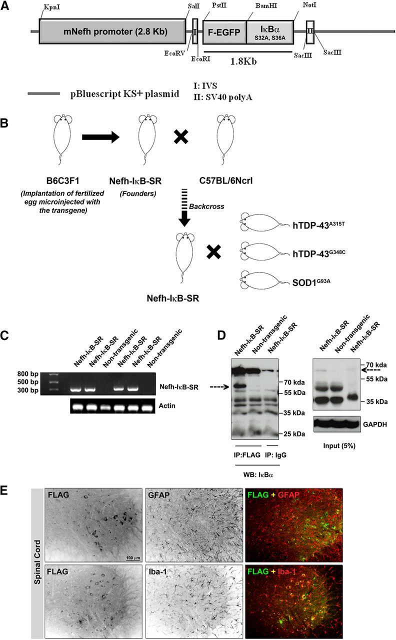
- Figure 1.
Generation and characterization of the IκB super-repressor mice. The NFH promoter was amplified from mouse genomic DNA and subcloned into the pBluescript KS+ backbone. The Flag-EGFp and IκB (SS32, 36AA) fragments were cloned from pEGFP-N3 and PCMV4-3HA/IκBα (SS32, 36AA) plasmids, respectively, and inserted into the pBSKS-Nefh promoter plasmid (A). Oocytes that were microinjected with this plasmid were implanted into B6C3F1 mouse to generate the Nefh-IκBSS32,36AA mouse (also known as the IκB super-repressor [SR] mouse). The founders were backcrossed with C57BL/6Ncrl mice before cross-breeding with ALS mouse models (viz TDP-43A315T, TDP-43G348C, and SOD1G93A) (B). Genotyping of IκB-SR mice was performed to check a 329 bp fragment from the EGFP gene, which was absent in nontransgenic littermates (C). Spinal cord lysates from littermate IκB-SR and WT mice were immunoprecipitated with Flag-M2 antibody and probed with IκBα antibody. Data are a representative image of three separate experiments. Results clearly showed a prominent band at ∼63 kDa corresponding to the combined molecular weight of FLAG-EGFP and IκBα (dotted arrow), which was absent in the WT. This band could not be detected when the Flag-M2 antibody was replaced with normal mouse IgG (D). To determine that the transgene was not expressed in other non-neuronal cell types, spinal cord sections were costained for Flag and Iba-1 (microglia marker) or Flag and GFAP (astrocyte marker). Flag expression could not be detected from either Iba-1-positive or GFAP-positive cells (E); Extended data Figure 1-1.
- Figure 2.
Phospho-P65-NF-κB expression in spinal cord neurons and NF-κB-TDP-43 interaction. To assess the level of inhibition of p65-NF-κB activation, achieved due to the super-repression, and compare them with that in ALS mice models, spinal cord sections were stained with neuron-specific NeuN antibody and phopho-p65 NF-κB antibody. Nuclei were stained with DAPI. Single-channel images from IκB-SR;TDP-43A315T (Ai-Aiii), TDP-43A315T (Bi-Biii), IκB-SR;TDP-43G348C (Ci-Ciii), TDP-43G348C (Di-Diii), IκB-SR (Ei-Eiii), and WT (Fi-Fiii) mice were analyzed separately using Fiji in a three step process. First, three corresponding single-channel images (NeuN, p65-NF-κB, DAPI) of every field were opened and stacked together. At the second step, neuronal boundaries were drawn (based on NeuN staining) using the free-hand selection tool, and the area was measured. Only neurons with an area > 250 µm2 were considered for this study. The nuclear boundary was also marked similarly (based on DAPI). Finally, the p65-NF-κB signal in the area marked as nuclear was measured and corrected for background fluorescence. For representational purposes, all channels in F are showing original boundary markings. Neurons from at least six sections per mouse with 3 mice in each group were considered. Original magnification ×20. The highlighted sections in each single channel images were cropped using Adobe Photoshop CS5, enlarged 300-fold, merged, and displayed in actual colors (Aiv, Biv, Civ, Div, Eiv, Fiv). Scale bar, 100 µm. Data were tabulated and the mean ± SEM plotted. ANOVA was performed using Kruskal-Wallis test followed by comparison between groups using Dunn's post-test. G, Adjusted p values. The neuronal nuclear phospho-p65 NF-κB expression was found to be significantly elevated in both the ALS mice model compared with IκB-SR and WT. Neurons in the spinal cord of double-transgenic mice (IκB-SR;TDP-43 mutants) showed marked reduction in presence of phospho-p65 NF-κB compared with respective TDP-43 mutants only. To assess whether the inhibition of p65-NF-κB release from the inhibitory κB complex affected its binding with TDP-43, spinal cord lysates from double-transgenic mice (IκB-SR;TDP-43 mutant) and its littermate TDP-43 mutant and IκB-SR were immunoprecipitated with anti-P65-NF-κB antibody and probed with anti-hTDP-43 antibody. Results showed a clear reduction in binding between the two proteins due to IκBα super-repression (dotted arrow). No interaction was expectedly observed in IκB-SR mice, which lacked the human TDP-43 expression. H, Representative of three independent experiments; Extended data Figure 2-2.
- Figure 3.
TDP-43 expression in spinal cord neurons. To evaluate the nuclear-cytoplasmic distribution of hTDP-43 in spinal cord neurons of IκB-SR;TDP-43A315T, TDP-43A315T, IκB-SR;TDP-43G348C, and TDP-43G348C mice, sections were stained with anti-human TDP-43 and anti-NeuN antibodies; nuclei were stained with DAPI (A-D). Original magnification ×20. Boundaries (cellular and nuclear) were outlined using Fiji, and TDP-43 signal intensity was measured only from neurons with area > 250 µm2. The nuclear signal intensity value was subtracted from the signal intensity of whole cell to generate the cytoplasmic signal intensity. The C:N signal intensity was calculated and tabulated. Neurons from at least six sections per mouse with 3 mice in each group were considered. A higher C:N ratio indicated predominantly cytoplasmic localization of TDP-43. ANOVA was performed using Kruskal-Wallis test followed by comparison between groups using Dunn's post-test. Adjusted p values are denoted in the figure. Results showed that TDP-43 was predominantly cytoplasmic in neurons in spinal cord of TDP-43 mutant mice. In comparison, there was a significant reduction in the C:N ratio of TDP-43 in spinal neurons of the double-transgenic mice. The C:N did not significantly differ between either the two TDP-43 mutant groups or the two double-transgenic groups (E). The hTDP-43 C:N ratio in both IκB-SR;TDP-43A315T and TDP-43A315T groups correlated with increasing neuronal soma size as calculated by Spearman's rank correlation test (95% CI; ρ for Area vs C:N of IκB-SR;TDP-43A315T = 0.4520; p < 0.0001; ρ for Area vs C:N of TDP-43A315T = 0.2697; p = 0.0033) (F). The hTDP-43 C:N ratio in IκB-SR;TDP-43G348C did not significantly correlate with the neuronal soma area, although that in TDP-43G348C did correlate with area (ρ for Area vs C:N of IκB-SR;TDP-43G348C = 0.1677; p = 0.0575; ρ for Area vs C:N of TDP-43G348C = 0.2519; p = 0.0079) (G). Immunoblotting from RIPA-soluble (H) and -insoluble (I) fractions of spinal cords of mice from various groups showed that in both IκB-SR;TDP-43A315T and IκB-SR;TDP-43G348C mice spinal cords, human TDP-43 was present at a significantly higher level compared with respective TDP-43 mutant mice tissues (J). Contrarily, in the RIPA-insoluble fractions, more human TDP-43 was detected from the TDP-43 mutant mice tissues (K). Data were analyzed by unpaired Student's t test with Welch's correction. p values are denoted in the figure.
- Figure 4.
Motor activities of the transgenic mice. To assess the effect of NF-κB activation inhibition on motor neuron count in IκB-SR;TDP-43A315T and IκB-SR;TDP-43G348C mice and compare them with that in age-matched TDP-43A315T, TDP-43G348C, IκB-SR, and WT littermates, spinal cord sections were stained for the neuronal marker NeuN (A). Original magnification ×20. Scale bar, 100 µm. The number of NeuN-positive cells in ventral horn with an area > 250 µm2 were counted using Fiji, and data were represented as a box-and-whisker graph showing minimum to maximum range of values. Data were analyzed by one-way ANOVA with Tukey's post-test that clearly showed that 1-year-old IκB-SR;TDP-43 mutant mice had significantly more motor neurons compared with respective TDP-43 mutants. However, these levels were still not at par with those observed in IκB-SR or WT. Data are representative of 3-5 mice per group. B, Adjusted p values. To determine whether the spinal motor neuron count translated to motor performance, the mice in all six groups were trained to run on an accelerating rotarod from 4 months of age. Data were recorded weekly until 12 months of age and represented as the mean of maximum latency to fall from the rotarod at each time point. N = 10-15 per group (C). Comparison of rotarod performance of WT and IκB-SR mice does not show any significant difference over the duration of the protocol (4-12 months) (D). Comparison of motor performances of mice belonging to IκB-SR;TDP-43A315T and TDP-43A315T groups revealed an initiation of decline in the TDP-43 mutant mice at ∼280 d of age. However, because of the variance in results, this became statistically significant difference beyond 330 d of age. In contrast, the IκB-SR;TDP-43A315T mice maintained their performance at a steady level. Data were analyzed by multiple t test, one per row (E). A similar pattern of decline was observed while comparing between IκB-SR;TDP-43G348C and TDP-43G348C. However, in TDP-43G348C mice, the decline was initiated at an earlier time point (225 d), and their motor performance at this age was significantly different from that of IκB-SR;TDP-43G348C mice (F). Data were analyzed by multiple t test, one per row, and the p values are denoted in the figure.
- Figure 5.
Determination of cognitive abilities in TDP-43 mutant and IκB super-repressor mice. Novel object recognition test performed at 6 months (A) and 12 months (B) of age showed mice expressing mutant TDP-43 with impaired recognition of the novel object. Contrarily, IκB-SR;TDP-43 mutant mice performed relatively similar to WTor IκB-SR mice. Data are representative of independent mice tested (dots) and represented as mean ± SEM of percent time spent at the novel object. Data were analyzed using one-way ANOVA with Tukey's multiple comparison post-test. Adjusted p values are denoted in the figure. C, Passive avoidance test to evaluate fear-induced memory retention indicated cognitive decline in TDP-43G348C mice at 12 months of age, compared with littermate WT and IκB-SR mice. The IκB-SR;TDP-43G348C double-transgenic mice performed significantly better in the memory function test compared with the TDP-43G348C mice. No significant difference was observed between performance of IκB-SR;TDP-43A315T and TDP-43A315T mice, which in either cases was significantly less than littermate WT or IκB-SR mice. Data are representative of independent mice tested (dots) and represented as mean ± SEM of time (in seconds) taken to enter the dark chamber. Data were analyzed using one-way ANOVA with Tukey's multiple comparison post-test. Adjusted p values. D, Immunoblot analysis from hippocampal extracts (RIPA-soluble and -insoluble fractions) of three nonlittermate TDP-43A315T and TDP-43G348C mice showed significantly higher TDP-43 expression in TDP-43A315T. Data were analyzed by two-way ANOVA followed by Tukey's multiple comparison test. Adjusted p values.
- Figure 6.
Effect of IκB super-repression on glial activation pattern in spinal cord. Spinal cord sections stained with anti-GFAP antibody to visualize astrocytes in the six groups. Original magnification ×40. Scale bar, 50 µm. Images are representative of 3 mice per group (A). The GFAP signal intensity from ventral horn was quantified using Fiji and data represented in tabular form. Results show that GFAP signal intensity to be significantly elevated in spinal cords of TDP-43 mutant mice that could indicate astrogliosis and/or reactive astrocytosis. The signal intensities were significantly lesser in IκB-SR;TDP-43 mutant mice. Data are representative of at least six sections each from 3 mice per group. Data were analyzed by one-way ANOVA along with Tukey's multiple comparison post-test. B, Adjusted p values. To visualize microglial morphology, spinal cord sections were stained with anti-Iba1 antibody. Images are representative of 3 mice per group. Original magnification ×20. Scale bar, 50 µm (C). The Iba-1 signal intensity from ventral horn was quantified using Fiji and data represented in tabular form. Data show signal intensity to be significantly higher in spinal cords of TDP-43 mutant mice and also IκB-SR mice compared with IκB-SR;TDP-43 mutants or WT mice. Data are representative of at least six sections each from 3 mice per group. Data were analyzed by one-way ANOVA along with Tukey's multiple comparison post-test. D, Adjusted p values. Spatiotemporal analysis of microglial morphology was done also with the help of Fiji. The cells were converted to their skeleton form (E) followed by analysis of number of branches (processes) per cell (F) and total branch (process) length of each cell (G). Results show that microglia in spinal cord of TDP-43 mutant mice have significantly reduced number of branches as well as decreased total branch length compared with IκB-SR;TDP-43 mutants or IκB-SR or WT mice, implying a more amoeboid reactive morphology. Each dot represents data from individual neurons. Data are pooled from at least six sections each from 3 mice per group. Data were analyzed by one-way ANOVA along with Tukey's multiple comparison post-test. Adjusted p values are denoted in the figure.
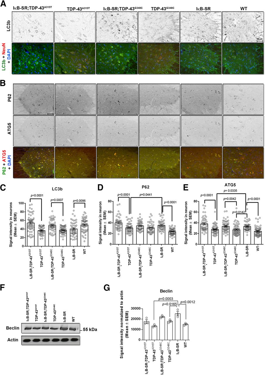
- Figure 7.
Changes in autophagy markers due to IκB super-repression. Spinal cord sections were stained for different markers of autophagy: LC3B, P62, and ATG5. Images in the figure are representative of 3 mice per group. Original magnification ×20. Scale bars: A, B, 50 µm. The signal intensity of these markers in spinal cord neurons with an area > 250 µm2 was measured using Fiji, and the data are represented in tabular form (C-E). Results clearly shows significantly reduced expression of autophagy markers in spinal cord neurons of TDP-43 mutant mice compared with IκB-SR;TDP-43 mutants or WT mice. The levels of P62 and ATG5 were also found to be significantly higher in IκB-SR mice compared with WT. Immunoblot performed from spinal cord lysates to detect Beclin showed an increase in the protein expression in IκB-SR mice compared with WT and TDP-43 mutants. The Beclin expression in IκB-SR;TDP-43 mutant mice was slightly elevated compared with only TDP-43 mutants. Data are representative of 3 mice per group. Data were analyzed by one-way ANOVA with Tukey's multiple comparison post-test. F, G, Adjusted p values.
- Figure 8.
IκB super-repression extends life span of SOD1G93A mice and slows rate of disease progression. On comparing the life span of IκB-SR;SOD1G93A mice with that of only SOD1G93A, a median increase of 15 d was observed. Data were analyzed by the Mantel-Cox log rank test; n = 25 in each group; p = 0.0020 (A). Rotarod analysis clearly showed a decline in performance by IκB-SR;SOD1G93A and SOD1G93A relative to that in IκB-SR and WT mice. The performance of SOD1G93A mice was found to differ significantly from that in IκB-SR;SOD1G93A between 133 and 140 d of age. Data were compared by Student's t test along with Welch's correction at each time point; the p values are denoted in the figure. N = 25 in IκB-SR;SOD1G93A, 15 in SOD1G93A, 8 in IκB-SR, and 10 in WT (B). The grid-hang time of the IκB-SR;SOD1G93A and SOD1G93A mice was also clearly worse than IκB-SR and WT mice. However, no significant differences were observed at any time point between IκB-SR;SOD1G93A and SOD1G93A mice; n = 25 in IκB-SR;SOD1G93A, 15 in SOD1G93A, 8 in IκB-SR, and 10 in WT (C). Hindlimb reflex scoring showed a slower rate of decline in IκB-SR;SOD1G93A mice compared with SOD1G93A between 141 and 161 d of age (denoted by dotted lines). Data were compared by Student's t test along with Welch's correction at each time point, and the p values are listed in Extended Data Table 1-1; n = 20 in IκB-SR;SOD1G93A and 15 in SOD1G93A (D). The actual body weight comparison did not show any difference between IκB-SR;SOD1G93A and SOD1G93A over their life span (E). However, on converting the body weights as percentage of peak weight, a significant difference could be observed between 126 and 147 d of age. Data were compared by Student's t test along with Welch's correction at each time point. p values are denoted in the figure; n = 20 in IκB-SR;SOD1G93A; 15 in SOD1G93A (F).
- Figure 9.
Neuronal IκB super-repression prevents misfolding of SOD1 and extends motor neuron survival in SOD1G93A model. Levels of misfolded SOD1 were detected in spinal cord lysates at presymptomatic, early symptomatic, and end stage by immunoprecipitation. Compared with SOD1G93A, IκB-SR;SOD1G93A always presented lesser amounts of misfolded SOD1. Data are representative of 3 mice per group (A). Quantitative analysis of the immunoblots also reflects this tread. However, the difference in the levels of misfolded SOD1 between IκB-SR;SOD1G93A and SOD1G93A were not statistically significant (B). Spinal cord sections from both IκB-SR;SOD1G93A and SOD1G93A mice were immunostained to detect ChAT. ChAT expression was found to be pronounced in spinal cord of IκB-SR;SOD1G93A mice compared with only SOD1G93A mice. Original magnification ×20. Scale bar, 50 µm. Data are representative of 3 mice per group (C). Motor neuron numbers in spinal cord sections were estimated by staining them with NeuN and counting positively stained cells with an area >250 µm2. Images are representative of 3 mice per group. Original magnification ×20. Scale bar, 50 µm (D). Data were tabulated and represented as box-and-whisker graph showing minimum to maximum range of values. Data were analyzed by one-way ANOVA with Tukey's post-test. E, Adjusted p values.


Tables
- Table 1.
Details of antibodies used for immunoblotting, immunoprecipitation, and immunofluorescence
Antibody against Dilution for immunoblotting/immunoprecipitation Dilution for immunofluorescence Company Actin 1:15,000 Sigma Millipore Atg-5 1:250 Novus Biologicals Beclin-1 1:1000 Novus Biologicals ChAT 1:500 Sigma Millipore FLAG-M2 1:2500
2 µg/300 µg of total proteinaa20 µg/ml Sigma Millipore GAPDH 1:2500 Santa Cruz Biotechnology GFAP 1:300 Cell Signaling Technologies GFP 1:1000 1:300 Thermo Fisher Scientific Iba-1 1:500 Wako Chemicals IkBα 1:2000 Santa Cruz Biotechnology Phospho-IκBα 1:1000 Cell Signaling Technologies LC3-II 1:250 Novus Biologicals NeuN 1:500 Cell Signaling Technologies NF-κB 1:1000
1.5 µg/300 µg of total proteinaaSanta Cruz Biotechnology Phospho-NF-κB (Ser 536) 1:1000 Cell Signaling Technologies SOD1 mAb (misfolded; clone B8H10) 1 μg/300 μg total proteinaa Generated from hybridoma in the laboratory SOD1 pAb 1:5000 Enzo Life Sciences Pan TDP-43 1:2500 Proteintech Human TDP-43 1:1000 Thermo Fisher Scientific Human TDP-43 1:250 Abnova ↵aFor immunoprecipitation.
Table 1-1
Comparison between reflex scoring of IκB-SR;SOD1G93A vs SOD1G93A (Figure 8D). Download Table 1-1, DOCX file
Figure 1-1
Characterization of the IκB-SR mice Spinal cord sections from the 4 founder mice lines, along with non-transgenic littermate, were sectioned and stained with anti-FLAG antibody. Strongest signal was detected from founder line 1397, whose sections were further stained with anti-GFP antibody to confirm presence of transgene. Magnification=20X; scale bar= 100 μm (A). Spinal cord lysates were also immunoblotted and probed with anti-FLAG and anti-GFP antibodies. The protein expressed by the transgene (approximately 63 kDa; denoted by dotted arrow) could be detected in some founders but never in WT (non-transgenic) mice (B). Similar results were obtained from brain lysates. Data is representative of at least 3 mice per group (C). Kidney, liver and spleen from 3 littermate mice from founder line 1397 were lysed and probed for FLAG and GFP. Lack of any specific signal confirmed that the expression of transgene was limited to nervous tissue (D). IκB-SR and WT mice were intraperitoneally challenged with E. coli LPS to induce inflammation and sacrificed after 12h. Spinal cord and spleen were excised and lysed for immunoblotting. When probed for phospho IκBα and total IκBα, transgenic mice showed no induction in spinal cord but robust induction in spleen. However, IκBα was found to be induced in both spinal cord as well as spleen in WT mice (E). Download Figure 1-1, TIF file
Figure 2-2
The IκB-SR plasmid was cloned into a pcDNA3 backbone. Mouse motor neuron-like NSC34 cells were transfected with this plasmid, with or without exposing the cells to recombinant murine TNF-α (10ng/mL). The levels of phospho NF-κB and total NF-κB were estimated from the cell lysates. In un-transfected cells, TNF-α treatment resulted in increased phosphorylation of NF-κB which was not observed in case of cells that were transfected with the IκB-SR plasmid (A). A 40X magnified image of spinal cord neuron, stained for NeuN and hTDP-43 with borders (cellular and nuclear) drawn around it prior to measurement of nuclear and cytosolic signal intensities (B). Body weights of all male and female mice from the six groups showing no significant intergroup variation which could affect performance in motor performance test (C). Download Figure 2-2, TIF file


