


Abstract
Pituitary adenylate cyclase-activating polypeptide (PACAP) is a 38-amino acid C-terminally α-amidated peptide that was first isolated 20 years ago from an ovine hypothalamic extract on the basis of its ability to stimulate cAMP formation in anterior pituitary cells (Miyata et al., 1989. PACAP belongs to the vasoactive intestinal polypeptide (VIP)-secretin-growth hormone-releasing hormone-glucagon superfamily. The sequence of PACAP has been remarkably well conserved during evolution from protochordates to mammals, suggesting that PACAP is involved in the regulation of important biological functions. PACAP is widely distributed in the brain and peripheral organs, notably in the endocrine pancreas, gonads, respiratory and urogenital tracts. Characterization of the PACAP precursor has revealed the existence of a PACAP-related peptide, the activity of which remains unknown. Two types of PACAP binding sites have been characterized: type I binding sites exhibit a high affinity for PACAP and a much lower affinity for VIP, whereas type II binding sites have similar affinity for PACAP and VIP. Molecular cloning of PACAP receptors has shown the existence of three distinct receptor subtypes: the PACAP-specific PAC1-R, which is coupled to several transduction systems, and the PACAP/VIP-indifferent VPAC1-R and VPAC2-R, which are primarily coupled to adenylyl cyclase. PAC1-Rs are particularly abundant in the brain, the pituitary and the adrenal gland, whereas VPAC receptors are expressed mainly in lung, liver, and testis. The development of transgenic animal models and specific PACAP receptor ligands has strongly contributed to deciphering the various actions of PACAP. Consistent with the wide distribution of PACAP and its receptors, the peptide has now been shown to exert a large array of pharmacological effects and biological functions. The present report reviews the current knowledge concerning the pleiotropic actions of PACAP and discusses its possible use for future therapeutic applications.
- α-MSH, α-melanocyte-stimulating hormone
- AC, adenylyl cyclase
- ARC, arcuate nucleus of the hypothalamus
- Bcl-2, B-cell lymphoma 2
- BDNF, brain-derived neurotrophic factor
- cAMP, cyclic adenosine monophosphate
- c-Jun, jun oncogene
- CNS, central nervous system
- CRE, cAMP responsive element
- CREB, cAMP-responsive element-binding protein
- CRH, corticotropin-releasing hormone
- E, embryonic day
- ECL, enterochromaffin-like
- EGL, external granule cell layer
- ERK, extracellular signal-regulated kinase
- FSH, follicle-stimulating hormone
- GH, growth hormone
- GHRH, growth hormone-releasing hormone
- GnRH, gonadotropin-releasing hormone
- hCG, human chorionic gonadotropin
- IGL, internal granule cell layer
- IL, interleukin
- JNK, c-Jun NH2-terminal kinase
- kb, kilobase(s)
- LH, luteinizing hormone
- LI, like immunoreactivity
- MAPK, mitogen-activated protein kinase
- MEK, mitogen-activated protein kinase kinase
- MIP, macrophage inflammatory protein
- MSH, melanocyte-stimulating hormone
- NMDA, N-methyl-d-aspartate
- NO, nitric oxide
- NPY, neuropeptide tyrosine
- P, postnatal day
- PAC1-R, PACAP-specific receptor
- PACAP(6–38), amino acids 6 to 38 of PACAP
- PACAP, pituitary adenylate cyclase-activating polypeptide
- PACAP27, 27-amino acid form of PACAP
- PACAP38, 38-amino acid form of PACAP
- PACAP-LI, PACAP-like immunoreactivity
- PAM, peptidyl glycine α
- -amidating monooxygenase; PC, prohormone convertase
- PCR, polymerase chain reaction
- PHI, peptide histidine-isoleucine
- PI3-K, phosphatidylinositol 3′-OH kinase
- PKA, protein kinase A
- PKC, protein kinase C
- PLC, phospholipase C
- POMC, pro-opiomelanocortin
- PRL, prolactin
- PRP, PACAP-related peptide
- PVN, paraventricular nucleus
- PYY, peptide tyrosine tyrosine
- RO 25–1553, L-threoninamide, N-acetyl-L-histidyl-L-seryl-L-α-aspartyl-L-alanyl-L-valyl-L-phenylalanyl-L-threonyl-L-α-glutamyl-L-asparaginyl-L-tyrosyl-L-threonyl-L-lysyl-L-leucyl-L-arginyl-L-lysyl-L-glutaminyl-L-norleucyl-L-alanyl-L-alanyl-L-lysyl-L-lysyl-L-tyrosyl-L-leucyl-L-asparaginyl-L-α-aspartyl-L-leucyl-L-lysyl-L-lysylglycylglycyl-(25–21)-lactam
- RT, reverse transcription
- SCN, suprachiasmatic nucleus
- Th, T helper
- TRH, thyrotropin-releasing hormone
- TSH, thyroid-stimulating hormone
- VIP, vasoactive intestinal polypeptide
- VPAC1-R, VIP/PACAP receptor, subtype 1
- VPAC2-R, VIP/PACAP receptor, subtype 2
- ZK98299, onapristone
I. Introduction
In October 1989, Akira Arimura and his coworkers published an article, now a citation classic, in which they reported the sequence of a novel regulatory peptide that stimulated adenylyl cyclase (AC1) activity in ante- rior pituitary cells, which they thus called pituitary adenylate cyclase-activating polypeptide (PACAP) (Miyata et al., 1989; Arimura, 2007). At that time, it was unlikely that they could predict the keen interest that this new peptide was going to arouse. Subsequently, it was shown that PACAP and its receptors are broadly expressed in the central nervous system (CNS) and in most peripheral organs. Consistent with this widespread distribution, PACAP has been found to exert pleiotropic effects including control of neurotransmitter release, vasodilation, bronchodilation, activation of intestinal motility, increase in insulin and histamine secretion, immune modulation, and stimulation of cell proliferation and/or differentiation. Twenty years after its discovery, PACAP has become one of the most studied neuropeptides. To date, over 2500 articles dealing directly with PACAP have been published, and the number of articles related to this fascinating polypeptide continues to increase exponentially.
The topic of PACAP was reviewed in this journal in 2000 (Vaudry et al., 2000) and in several other journals (Arimura and Shioda, 1995; Rawlings and Hezareh, 1996; Sherwood et al., 2000; Shioda, 2000). In the last decade, however, significant new knowledge has been gained on both PACAP and its receptors. In 2009, we are celebrating the 20th anniversary of the discovery of PACAP; at this occasion, we thought that it was especially appropriate to comprehensively review the current knowledge regarding PACAP and its receptors.
II. Pituitary Adenylate Cyclase-Activating Polypeptide
PACAP has been originally isolated from an ovine hypothalamus extract on the basis of its ability to stimulate cAMP formation in rat pituitary cells (Miyata et al., 1989). Hypothalamic neurons containing PACAP project toward the median eminence and terminate in the vicinity of the capillary loops of the hypothalamo-pituitary portal system. However, PACAP is widely expressed in numerous extra-hypothalamic regions of the brain as well as in various peripheral tissues.
A. Discovery of Pituitary Adenylate Cyclase-Activating Polypeptide
To isolate novel hypophysiotropic neuropeptides, the group of Arimura has screened fractions from an extract of 4300 ovine hypothalami, by monitoring their stimulatory effect on AC activity in cultured rat anterior pituitary cells. Using this approach, they have isolated in pure form a peptide that markedly increased cAMP formation, which they named pituitary adenylate cyclase-activating polypeptide. Sequencing of the peptide revealed that it comprises 38 amino acid residues (PACAP38) and is C-terminally α-amidated (Fig. 1.) (Miyata et al., 1989. The sequence of PACAP38 encompasses an internal cleavage-amidation site (Gly28-Lys29-Arg30), suggesting that it can generate a 27-residue α-amidated polypeptide fragment or PACAP27.2 Consistent with this hypothesis, Miyata et al. (1990) have subsequently isolated from the ovine hypothalamus another fraction capable of stimulating AC activity in adenohypophysial cells, which, upon characterization, happened to correspond to the N-terminal 27-amino acid portion of PACAP38 (Miyata et al., 1990. The structure of the biologically active region of PACAP, within the PACAP27 sequence, has almost been totally preserved during evolution, from fish to mammals, two phyla that diverged some 380 million years ago (Chartrel et al., 1991; Hoyle, 1998; Sherwood et al., 2000), suggesting that this peptide may exert essential biological functions. The sequence of human PACAP27 shares 68% identity with vasoactive intestinal polypeptide (VIP), identifying PACAP as a member of the VIP- secretin-GHRH-glucagon superfamily (Fig. 1.) (Rosselin et al., 1982; Campbell and Scanes, 1992; Segre and Goldring, 1993).
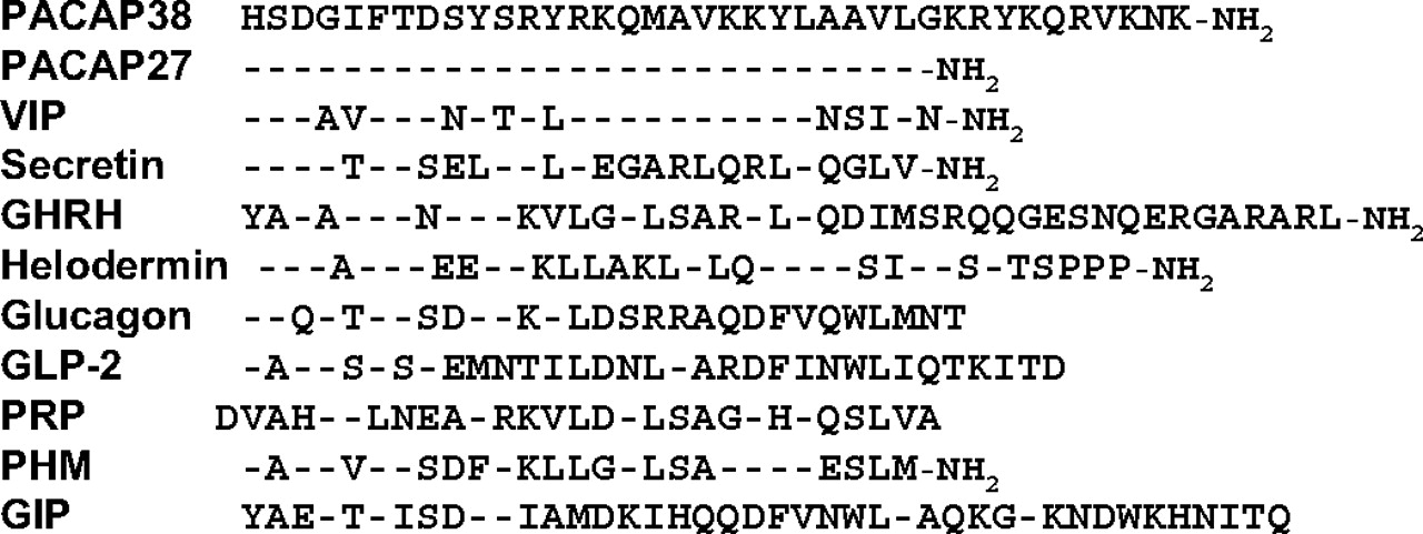
Amino acid sequences of the different members of the PACAP-VIP-secretin-GHRH superfamily in human. -, amino acids identical with those of PACAP.
B. Secondary Structure of Pituitary Adenylate Cyclase-Activating Polypeptide
Conformational analyses by circular dichroism and nuclear magnetic resonance indicate that the secondary structure of PACAP27 is mainly characterized by a helical conformation of various lengths, depending on the medium. In 25% methanol, the disordered eight-amino acid N-terminal sequence is followed by four distinct structured domains (Inooka et al., 1992: the first domain, encompassing residues 9 to 12, forms a β-turn-like conformation, whereas the three others are composed of distinct helical regions that extend from residues 12 to 14, 15 to 20, and 22 to 24 (Inooka et al., 1992). An α-helix spanning residues 9 to 26, with a discontinuity between Lys20 and Lys21, is observed in 50% trifluoroethanol, a solvent that stabilizes helical structures (Wray et al., 1993). In 30% trifluoroethanol, PACAP27 possesses an N-terminal disordered segment followed by a stable α-helical conformation within segment 7 to 27 (González-Muñiz et al., 2001). When PACAP is bound to dodecylphosphocholine micelles, usually used to mimic the membrane environment, the α-helix of PACAP27 extends from the C terminus to residue Ile5 and is preceded by a disordered N-terminal domain (Inooka et al., 2001; Bourgault et al., 2009b). The conformation of PACAP38 mirrors that of PACAP27 and the C-terminal (28⇓⇓⇓⇓⇓⇓⇓⇓⇓–38) extension exhibits a short helix connected by a flexible hinge to the 1-to-27 region (Wray et al., 1993). Grass carp PACAP38, which possesses 89% sequence identity to human PACAP, exhibits a C-terminal α-helix from Arg29 to Arg34, near the central helical core, leading to a ring-like structure (Sze et al., 2007). When the PACAP(6-38) fragment interacts with the isolated N-terminal domain of PAC1-R, the peptide adopts a helical conformation with a bend at residue Ala18 (Sun et al., 2007), whereas the PACAP(1-21) fragment bound to PAC1-R exhibits a single β-coil structure in the residue 3-to-7 region, followed by an α-helix (Inooka et al., 2001).
C. Structure of the Pituitary Adenylate Cyclase-Activating Polypeptide Precursor and Post-Translational Processing
The cDNA encoding the PACAP precursor has been characterized in several vertebrate species (Ogi et al., 1990; Ohkubo et al., 1992; Arimura and Shioda, 1995; Okazaki et al., 1995) and in a protochordate, the ascidian Chelyosoma productum (McRory and Sherwood, 1997). In human, the cDNA encodes a 176-amino acid prepro-protein that comprises a 24-amino acid signal peptide (Hosoya et al., 1992). In all species, the sequence of PACAP38 is located in the C-terminal domain of the precursor (Fig. 2). The cDNA sequences of human (Ohkubo et al., 1992), sheep (Kimura et al., 1990), rat (Ogi et al., 1990), and mouse prepro-PACAP (Okazaki et al., 1995) has revealed the existence of a 29-amino acid peptide, delimited by basic residues at its N- and C-terminal extremities, located upstream of PACAP38 (Fig. 2). This peptide, which exhibits moderate structural homology with PACAP27, has been termed PACAP-related peptide (PRP) (Ogi et al., 1990; Wray et al., 1995; Hoyle, 1998). The overall organization of the PACAP precursor exhibits strong similarities with that of the VIP precursor. In particular, the VIP precursor encompasses a VIP-related peptide, called peptide histidine-methionine amide in human (Itoh et al., 1983; Bodner et al., 1985; Svoboda et al., 1986) or peptide histidine-isoleucine amide (PHI) in sheep (Bounjoua et al.,1991), rat (Nishizawa et al., 1985), mouse (Lamperti et al., 1991), and chicken (McFarlin et al., 1995), which possesses moderate amino acid identity with VIP. The degree of similarity between PACAP27 and PRP (22%) or VIP and PHI (37%) is less than that between PACAP and VIP (68%) or PRP and PHI (44%), respectively. It is thus assumed that intragenomic duplication of a VIP/PACAP ancestor sequence has occurred before duplication of the whole ancestor gene (Ohkubo et al., 1992). A proposed model describing the evolutionary process leading to the generation of distinct precursors for PACAP, VIP, secretin, GHRH, and glucagon in mammals is presented in Fig. 3. In submammalian vertebrates and the tunicate C. productum, the PACAP precursor comprises both PRP and PACAP (Fig. 3) (section II.I).
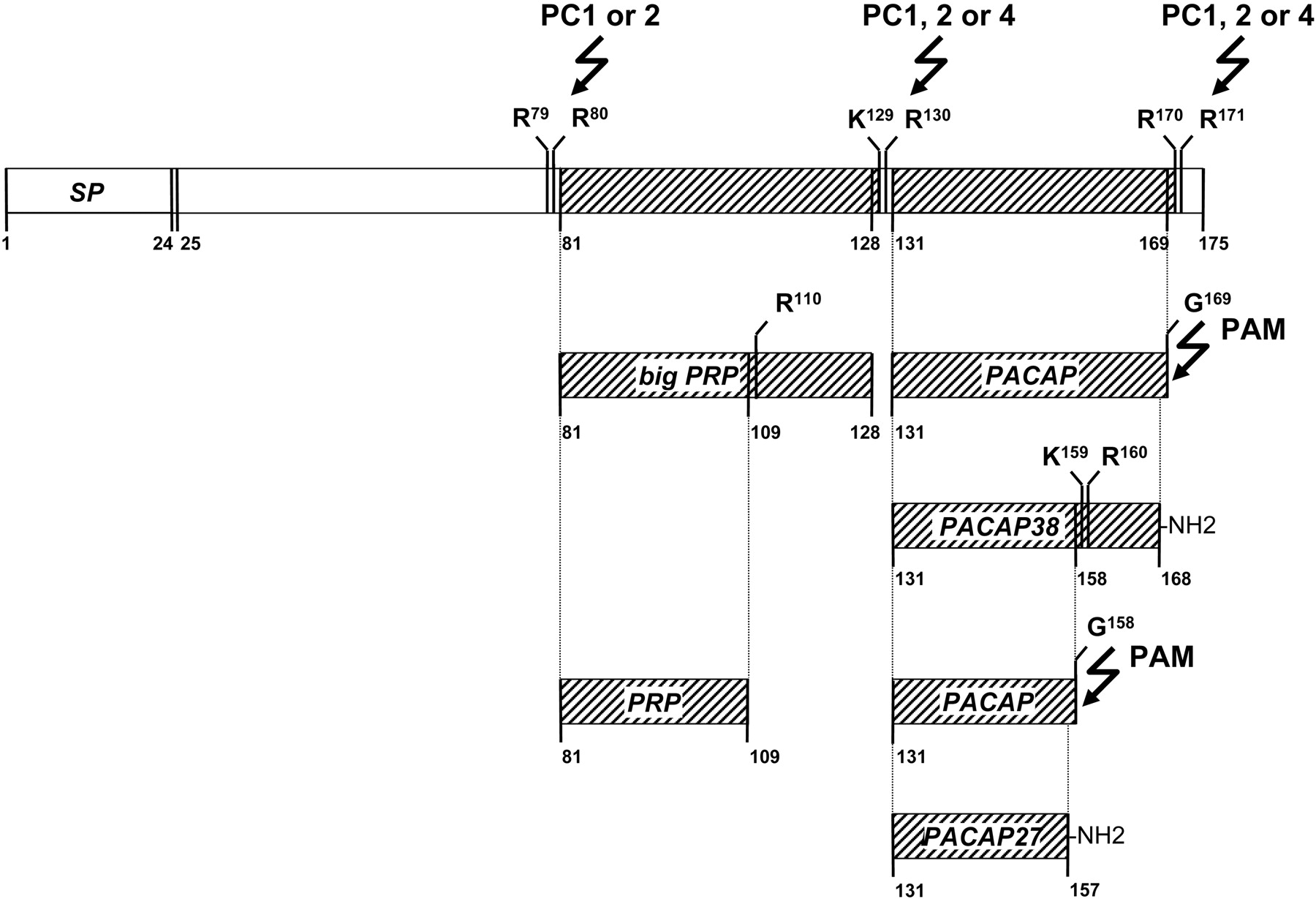
Schematic representation of the post-translational processing of the rat PACAP precursor. The nature and location of each cleavage and amidation site is specified. PAM, peptidyl glycine α-amidating monooxygenase; PC1, -2, or -4, prohormone convertase 1, 2, or 4; SP, signal peptide.

A proposed evolutionary scheme of PHI-VIP, PRP-PACAP, and GHRH genes. Unknown or unclear paths are represented by dotted lines. The unknown secretin genes are noted with question marks. GIP, gastric inhibitory polypeptide; GLP, glucagon-like peptide
In mammals, the primary structure of the PACAP precursor reveals the existence of seven mono- or dibasic residues that can potentially be cleaved by various prohormone convertases (PC) including PC1, PC2, PC4, PC5, PC7, furin, and paired basic amino acid-cleaving enzyme 4 (PACE4) (Seidah et al., 1994, 1998; Seidah and Chrétien, 1999). In the rat, cleavage at three dibasic sites (i.e., Arg79-Arg80, Lys129-Arg130, and Arg170-Arg171) generates a large intermediate precursor of PRP (big PRP) and a glycine-extended form of PACAP38 (Fig. 2). Cleavage at the single Arg110 followed by hydrolysis of this C-terminal Arg residue by carboxypeptidases E, H, or M generates PRP (Rouillé et al., 1995). The Gly169 residue is used by peptidyl glycine α-amidating monooxygenase (Eipper et al., 1992) for the amidation of the Lys168 residue at the C-terminal extremity of PACAP38. Finally, the tripeptide Gly158-Lys159-Arg160 can be cleaved to generate the α-amidated PACAP27 isoform (Fig. 2). Processing of the PACAP precursor has been studied in Chinese hamster ovary-K1 cells transfected with the human PACAP cDNA (Okazaki et al., 1992). Characterization of the various peptides secreted in the incubation medium by high-performance liquid chromatography combined with radioimmunoassay detection has confirmed that processing of the PACAP precursor actually yields to the formation of PACAP38, PACAP27, and PRP (Okazaki et al., 1992).
In the rat hypothalamus, PC1 and/or PC2 are intensively expressed in nuclei enriched with PACAP-immunoreactive neurons, supporting the hypothesis that these two endopeptidases could be involved in the processing of the PACAP precursor (Köves et al., 1994b; Zheng et al., 1994; Dong et al., 1997). Cotransfection experiments in GH4C1 cells have confirmed that both PC1 and PC2 can actually process the rat PACAP precursor to generate mature PACAP38 and PACAP27 (Li et al., 1999). In the rat testis, where PACAP is particularly abundant, PC4 can also process the PACAP precursor to generate both PACAP38 and PACAP27 (Li et al., 1998, Li et al., 2000a, Li et al., 2000b; Basak et al., 1999).
Like most peptides, PACAP released in the blood circulation exhibits poor metabolic stability, and it has been established that the half-life of PACAP38 injected into mice or human is between 2 and 10 min (Zhu et al., 2003; Li et al., 2007). The rapid breakdown of PACAP is attributable at least in part to the activity of the proteolytic enzyme dipeptidyl peptidase IV (Zhu et al., 2003; Bourgault et al., 2008a); hence, inhibition of dipeptidyl peptidase IV extends some of the effects of PACAP (Ahrén and Hughes, 2005). Further investigations are in progress to identify additional enzymes involved in the degradation of PACAP. For example, prolyl oligopeptidase has been reported to degrade PRP but has no effect on PACAP (Tenorio-Laranga et al., 2009).
D. The Pituitary Adenylate Cyclase-Activating Polypeptide Gene
The gene encoding PACAP has been cloned in several species including human (Hosoya et al., 1992) and mouse (Fig. 4) (Yamamoto et al., 1998; Cummings et al., 2002). The PACAP gene is composed of five exons, the sequence of PRP being encoded by exon 4 and that of PACAP by exon 5 (Fig. 4). Northern blot analysis has revealed the presence of a 3-kb PACAP mRNA in the rat hypothalamus (Hosoya et al., 1993; Hannibal et al., 1995a). A shorter transcript with a truncated 5′-untranslated region that uses a testis-specific promoter has been characterized in the rat testis (Hurley et al., 1995; Daniel and Habener, 2000). Likewise, shorter PACAP mRNA has been found in the mouse, bovine, and human testis (Hurley et al., 1995). It has also been reported that another short PACAP transcript is produced in sympathetic neurons (Harakall et al., 1998).

Organization of the human PACAP gene and PACAP mRNA. The five exons are boxed in green and numbered. The untranslated regions of exon 1 and 5 are denoted by a blue dashed line. Exon domains encoding SP, PRP, and PACAP are hatched in red. The locations of binding sites for potential transcriptional factors and polyadenylation has been indicated on the gene. AP-1, activator protein-1; Inr-like, initiator-like element; NRSLE, neural-restrictive silencer-like element; SP, signal peptide; TRE, 12-O-tetradecanoylphorbol-13-acetate response element; TS, transcription start site.
The human PACAP promoter possesses two cAMP-response-like elements (CRE), a 12-O-tetradecanoylphorbol 13-acetate-response element, a pair of sequences homologous to the consensus sequence for pituitary-specific factor growth hormone transactivator factor-1-binding sites, which are known to play a role in the tissue-specific expression of growth hormone (GH), and six binding domains for the thyroid-specific transcription factor-1 (Bodner et al., 1988; Dollé et al., 1990; Castrillo et al., 1991; Kim et al., 2002). Alignment of the human, rat, and mouse genes shows a high level of sequence conservation. In particular, two CRE and growth hormone transactivator factor-1 response elements, a GATA box, and a CT-rich domain with GC boxes are conserved in all three PACAP genes (Fig. 4) (Ohkubo et al., 1992; White et al., 2000). The promoter region of the human PACAP gene does not contain any apparent TATA or CAAT boxes, which are normally required for accurate initiation of transcription (Hampsey, 1998). Investigation of the promoter activity has revealed that PACAP is constitutively expressed and that transcription of the PACAP gene can be enhanced by cAMP, phorbol diester, thyroid-specific transcription factor-1, dexamethasone, progesterone, and even by PACAP itself (Suzuki et al., 1994; Ha et al., 2000; Hashimoto et al., 2000b; Kim et al., 2002; Chi-Wei et al., 2007; Yang et al., 2007). The 5′-flanking region contains two neural-restrictive silencer-like elements 1 and 2, which might be involved in neuron-specific PACAP gene expression (Fig. 4) (Sugawara et al., 2004; Lee et al., 2006).
The structural organization of the PACAP gene is similar to those of the VIP gene (Lamperti et al., 1991) and GHRH gene (Mayo et al., 1985), confirming that all three genes originate from a common ancestral sequence through gene duplication (Fig. 3). In human, the PACAP gene is localized to the P11 region of chromosome 18, which is associated with holoprosencephaly (Hosoya et al., 1992; Chang et al., 1993; Golden, 1998) and psychiatric disorders, suggesting that PACAP might be involved in the control of brain development and/or the etiology of schizophrenia (Ishiguro et al., 2001; Kamnasaran, 2003; Hashimoto et al., 2007; Matsuzaki and Tohyama, 2008).
To investigate the function of PACAP, several mouse lines have been created in which PACAP (Hashimoto et al., 2001; Hamelink et al., 2002) or both PACAP and PRP have been deleted (Gray et al., 2001). Animals with both VIP and PHI gene deletion (Colwell et al., 2003) have also been generated. Interbreeding PACAP(−/−) and VIP(−/−) mice made it possible to generate PACAP/VIP double-knockout animals (Niewiadomski et al., 2008). Although these animals can survive, their growth is significantly reduced, and they exhibit a high rate of mortality after 3 months of age. Finally, transgenic mice overexpressing PACAP in β-islet cells have been used to study the involvement of PACAP in diabetes development (Yamamoto et al., 2003; Tomimoto et al., 2004).
E. Distribution of Pituitary Adenylate Cyclase-Activating Polypeptide in the Central Nervous System
Soon after the characterization of PACAP, the distribution of the peptide has been investigated in the brain of mammals (Arimura et al., 1991; Köves et al., 1991; Vigh et al., 1991; Kivipelto et al., 1992; Ghatei et al., 1993) and amphibians (Yon et al., 1992). In rat, radioimmunoassay measurements have revealed that the highest concentrations of PACAP occur in the hypothalamic area (Arimura et al., 1991; Ghatei et al., 1993). Reversed-phase chromatography analysis showed that PACAP38 is by far the predominant form, PACAP27 representing less than 10% of the total peptide content in brain tissue (Arimura et al., 1991; Ghatei et al., 1993; Masuo et al., 1993; Hannibal et al., 1995a; Piggins et al., 1996). PACAP-containing neurons are not restricted to the hypothalamic area but are widely distributed in various brain regions (Gonzalez et al., 1998).
The mapping of PACAP-expressing neurons has been investigated by immunocytochemistry and in situ hybridization (Table 1). In the rat hypothalamus, PACAP-immunoreactive neurons are located primarily in the parvo- and magnocellular neurons of the paraventricular (PVN) and supraoptic nuclei (Köves et al., 1991, Köves et al., 1994a; Kivipelto et al., 1992; Ando et al., 1994; Kimura et al., 1994; Hannibal et al., 1995a,Hannibal et al., b; Piggins et al., 1996). PACAP mRNA is expressed in the PVN and arcuate nucleus (ARC) (Hannibal et al., 1995b; Murase et al., 1995; Das et al., 2007). A dense accumulation of PACAP-immunoreactive fibers is found in the internal zone of the median eminence and in the vicinity of the capillaries of the hypothalamo-hypophysial portal system (Köves et al., 1990, 1991; Kivipelto et al., 1992; Tamada et al., 1994; Hannibal et al., 1995a,b; Mikkelsen et al., 1995). The concentration of PACAP in rat portal blood (≈ 65 pM) is at least twice as high as in peripheral blood, indicating that the peptide released by hypothalamic nerve terminals is actually transported to the pituitary (Dow et al., 1994). Regional distribution studies reveal that significant amounts of PACAP38 are also found in various extra-hypothalamic regions, including the cerebral cortex, amygdala, hippocampus, pineal gland, substantia nigra, cerebellum, and pons (Ghatei et al., 1993; Hannibal, 2002). In the limbic system, PACAP-immunoreactive fibers are detected in the amygdaloid complex and in the mediodorsal and paraventricular nuclei of the thalamus (Köves et al., 1991; Masuo et al., 1993; Takahashi et al., 1994; Palkovits et al., 1995; Hannibal, 2002). The bed nucleus of the stria terminalis contains high concentrations of PACAP- and VIP-immunoreactive neurons, but no double-labeled cells have been detected (Kozicz et al., 1997). In the lateral septum area, a dense network of immunoreactive fibers innervates blood vessels (Köves et al., 1991). Scattered PACAP mRNA-expressing cell bodies are observed in the cingulate and frontal cortex (Mikkelsen et al., 1994), and immunoreactive cell bodies are found in the olfactory and neocortical area (Hannibal, 2002). In the mesencephalon, PACAP-immunopositive neurons are located in the ventrolateral periaqueductal gray (Das et al., 2007), and PACAP-containing fibers innervate the pretectum and periaquaductal white matter (Tajti et al., 2001; Hannibal, 2002). PACAP and its mRNA have also been detected in the cerebellum (Ghatei et al., 1993; Mikkelsen et al., 1994; Takahashi et al., 1994; Hannibal et al., 1995a; Nielsen et al., 1998b). Specifically, PACAP-like immunoreactivity (PACAP-LI) is localized in the soma and dendrites of Purkinje cells, whose axons directly contact granule cells (Nielsen et al., 1998b; Hannibal, 2002; Cameron et al., 2007). In the myelencephalon, PACAP is found in the brainstem and medulla oblongata (Ghatei et al., 1993; Légrádi et al., 1994). In the brainstem, PACAP-positive cell bodies are located in the locus ceruleus, pontine nucleus, and vagal complex (Tajti et al., 2001; Hannibal, 2002; Farnham et al., 2008), and fibers are found in the lateral parabrachial nucleus (Hannibal, 2002). In the medulla oblongata, the majority of perikarya exhibiting PACAP-LI is found in the commissural and medial subnuclei of the solitary nucleus, the dorsal motor vagal nucleus, the nucleus ambiguus, the ventrolateral medulla, the ventral medullary surface and the caudal raphe nuclei, supporting the view that PACAP may act as a regulator of visceral functions (Légrádi et al., 1994; Hannibal, 2002). In the spinal cord, PACAP mRNA is expressed in a subpopulation of sensory neurons of the dorsal root ganglia (Mulder et al., 1994), and numerous PACAP-immunoreactive fibers are found in the superficial layer of the dorsal horns (Moller et al., 1993; Dun et al., 1996a).
Localization and relative abundance of PACAP mRNA and PACAP–like immunoreactivity in the rat brain
The location of PACAP-containing neurons has also been investigated in the CNS of nonmammalian vertebrates including birds (Peeters et al., 1998; Nowak and Zawilska, 2003), amphibians (Yon et al., 1992, 1993b, 2001) and fishes (Matsuda et al., 1997, 2005b; Montero et al., 1998; Jakab et al., 2004; Mathieu et al., 2004). Overall, the distribution of PACAP-immunoreactive cells exhibits a high degree of similarity with that of mammals. In particular, in the brain of the frog Rana ridibunda, now renamed Pelophylax ridibundus (Conlon et al., 2009), prominent groups of PACAP-containing neurons are located in the hypothalamus [i.e., in the anterior preoptic area, the ventral magnocellular preoptic nucleus, the suprachiasmatic nucleus (SCN), the ventral hypothalamic nucleus, and the posterior tubercle (Yon et al., 1992, 2001)]. Likewise, in the primitive teleost fish Anguilla anguilla, PACAP-containing neurons are primarily located in the parvo- and magnocellular subdivisions of the preoptic nucleus (Montero et al., 1998).
The distributions of PACAP and VIP in the CNS are clearly different (Masuo et al., 1993). For instance, in the thalamus, a few VIP-positive fibers are found running up the wall of the third ventricle, whereas a dense network of PACAP fibers is observed in the central thalamic nuclei (Köves et al., 1991). In the bed nucleus of the stria terminalis, PACAP-containing fibers seem to surround unstained, round-shaped neuronal cell bodies, whereas VIP fibers are homogeneously distributed. Numerous PACAP-immunoreactive fibers are also found in the lateral septum, where only a few VIP fibers are observed (Köves et al., 1991). In magnocellular neurons, PACAP but not VIP is colocalized with oxytocin (Köves et al., 1994a). Consistent with this observation, in the posterior pituitary, PACAP does not colocalize with VIP (Vereczki et al., 2003). In the brainstem, VIP-positive cells are present in the mesencephalic periaqueductal gray and the dorsal and linear raphe nuclei, whereas PACAP neurons are abundant in the PVN and the dorsal vagal complex. In contrast, both PACAP and VIP-immunoreactive fibers seem to innervate the wall of cerebral blood vessels (Jansen-Olesen et al., 1994).
Taken together, these data indicate that although the highest amounts of PACAP occur in the hypothalamus (Arimura et al., 1991), substantial concentrations of the peptide are also found in many other brain regions, including the cerebral cortex, the hippocampus, the thalamus, the striatum, the nucleus accumbens, the substantia nigra, the locus ceruleus, and the pineal gland (Table 1) (Köves et al., 1991; Ghatei et al., 1993; Palkovits et al., 1995).
F. Distribution of Pituitary Adenylate Cyclase-Activating Polypeptide in Peripheral Organs
In peripheral tissues, as in the brain, PACAP38 is by far the major molecular form, but the proportions of PACAP27 and PACAP38 vary between the different organs (Arimura et al., 1991). For instance, in the colon, PACAP27 represents 30% of the total immunoreactivity, whereas in the testis, PACAP27 is hardly detectable (Arimura et al., 1991). The occurrence of different proportions of the two peptides in various tissues is probably attributable to the existence of different sets of PC enzymes.
The presence of PACAP mRNA and PACAP has been detected in most endocrine glands in rat (Table 2). In particular, PACAP is found in the different lobes of the pituitary gland (Rawlings and Hezareh, 1996; Arimura, 1998). In the anterior pituitary, PACAP is observed in a subpopulation of gonadotrope cells (Mikkelsen et al., 1995; Köves et al., 1998). In the ventral part of the neural lobe, PACAP is contained in nerve fibers with large terminal boutons (Mikkelsen et al., 1995). At the ultrastructural level, PACAP-LI seems to be located in dense-core granules contained in neurosecretory fibers (Kimura et al., 1994). PACAP-immunoreactive elements are also found in the gonads (Shioda et al., 1994; Hannibal and Fahrenkrug, 1995), adrenal gland (Arimura et al., 1991; Mazzocchi et al., 2002), parathyroid (Luts and Sundler, 1994), and endocrine pancreas (Table 2) (Arimura and Shioda, 1995; Love and Szebeni, 1999). In rat, the highest amounts of PACAP are found in the testis. In fact, the concentration of PACAP in the testis is higher than in the whole brain and exceeds the concentration of any other known peptides (Arimura et al., 1991). In situ hybridization studies have shown that PACAP mRNA is present in germ cells and not in Sertoli or Leydig cells (Shioda et al., 1994; Hannibal and Fahrenkrug, 1995). Electron microscopic studies indicate that PACAP is located in acrosoma caps and granules of primary spermatocytes, and later on in mature spermatids (McArdle, 1994; Shioda et al., 1994; Hannibal and Fahrenkrug, 1995; Hannibal et al., 1995b; Li et al., 2004). The expression of PACAP in germ cells decreases after ethanol exposure (Koh et al., 2006). In the ovary, the concentration of PACAP is much lower than in the testis, and the peptide seems to be contained in nerve fibers (Steenstrup et al., 1995). In the uterus, decidual endometrium contains significant amounts of PACAP mRNA (Spencer et al., 2001). The occurrence of PACAP and PACAP mRNA has been reported in both rat and human placenta. In human, PACAP-LI is associated with stromal cells of both stem and terminal placental villi (Scaldaferri et al., 2000). In rat, PACAP-containing cells are present in the placental labyrinth and in the villous-like structures of the intraplacental yolk sac (Scaldaferri et al., 2000). In the human placenta, moderate concentrations of PACAP mRNA are expressed in stroma cells of stem and terminal villi at 7 and 14 weeks of gestation, and the density of PACAP mRNA gradually increases as pregnancy progresses (Koh et al., 2005). In the rat placenta, as gestation advances, the expression of PACAP mRNA gradually declines in decidual cells and increases in chorionic vessels and stromal cells of chorionic villi within the labyrinth zone (Koh et al., 2003).
Localization and relative abundance of PACAP mRNA and PACAP-like immunoreactivity in rat peripheral tissues
The adrenal gland contains a high concentration of PACAP (Arimura et al., 1991; Watanabe et al., 1992; Ghatei et al., 1993). In mammals, PACAP is found in the adrenal medulla (Shiotani et al., 1995), where it is contained both in chromaffin cells (Holgert et al., 1996) and in fibers (Frödin et al., 1995; Moller and Sundler, 1996; Tornøe et al., 2000). In the Italian wall lizard, Podarcis sicula, PACAP and its mRNA are detected in chromaffin cells, whereas in the frog adrenal gland, PACAP-LI is restricted to nerve fibers that contact either chromaffin cells or steroid-producing cells (Yon et al., 1993a; Valiante et al., 2008). It has been suggested that in the rat and dog adrenal gland, PACAP released from nerve endings contributes to neurally evoked catecholamine release (Fukushima et al., 2001a; Lamouche and Yamaguchi, 2003). Likewise, the parathyroid gland and the intrapancreatic ganglia are innervated by PACAP-containing fibers (Luts and Sundler, 1994; Filipsson et al., 1998a; Love and Szebeni, 1999).
Large amounts of PACAP-LI are found in all parts of the gastrointestinal tract (Arimura et al., 1991; Hauser-Kronberger et al., 1992; Ghatei et al., 1993; Mao et al., 1998; Vincze et al., 1999). The presence of PACAP-immunoreactive cell bodies has been observed in the myenteric ganglia throughout the gastrointestinal tract, and the existence of intrinsic neurons has been confirmed by in situ hybridization (Shen et al., 1992; Hannibal et al., 1998). Numerous PACAP-containing nerve fibers have been visualized along the circular muscle fibers and in the longitudinal smooth muscle layer of the esophagus (Uddman et al., 1991a; Köves et al., 1993; Olsson and Holmgren, 1994). PACAP-LI has also been detected in various exocrine glands of the alimentary canal (e.g., the parotid and submandibular glands, the liver, and the exocrine pancreas) (Arimura et al., 1991; Fridolf et al., 1992; Moller et al., 1993; Luts and Sundler, 1994). In the urinary bladder, networks of PACAP-immunoreactive fibers are found in the vicinity of blood vessels (Moller et al., 1993; Fahrenkrug and Hannibal, 1998; Zvarova et al., 2005). In the airways, PACAP-containing fibers innervate smooth muscle bundles and blood vessels in the trachea as well as small bronchioles in the lung (Cardell et al., 1991; Uddman et al., 1991b; Hauser-Kronberger et al., 1996; Shigyo et al., 1998). In the immune system, PACAP is expressed in various lymphoid tissues, including the thymus, spleen and duodenal mucosa (Gaytan et al., 1994; Abad et al., 2002) and in peritoneal macrophages (Pozo et al., 1997). The occurrence of PACAP mRNA has been demonstrated in the superior cervical ganglion (Nogi et al., 1997b). Depolarization of these neurons stimulates the release of PACAP27 and PACAP38 and causes a concomitant increase of PACAP mRNA and peptide (Brandenburg et al., 1997). A few PACAP-positive perikarya are also present in the sphenopalatine and otic ganglia (Uddman et al., 1991b, 1999). In the eye, PACAP-LI is present in fibers innervating the iris sphincter and in cell bodies scattered in the ciliary ganglia (Wang et al., 1995; Elsås and White, 1997; Olianas et al., 1997; Samuelsson-Almén and Nilsson, 1999). In the retina, PACAP is found in fibers of the ganglion cell and nerve fiber layer (Hannibal et al., 1997; Seki et al., 1997, 2000) and in amacrine cells in the inner nuclear layer (Seki et al., 2000).
In peripheral organs, in contrast to the CNS, PACAP and VIP often seem to be coexpressed by the same cells. For instance, colocalization of PACAP and VIP has been demonstrated in cell bodies and nerve fibers in the human and sheep esophageal sphincter (Uddman et al., 1991a; Ny et al., 1995), in the human and chicken gut (Sundler et al., 1992), and in the ovine respiratory tract (Uddman et al., 1991b). Nerve fibers containing both PACAP and VIP are also found in other tissues, notably in the parathyroid glands of cat and sheep (Luts and Sundler, 1994) and in the gill arch of the goldfish C. auratus (de Girolamo et al., 1998).
To summarize, in peripheral organs, the highest concentrations of PACAP are found in the testis, the adrenal gland, the gastrointestinal tract, and the lymphoid tissues (Arimura et al., 1991). PACAP is frequently found in sensory and parasympathetic neurons (Mulder et al., 1995). PACAP38 is much more abundant than PACAP27 in all tissues. Although PACAP is often localized in nerve cell bodies and fibers, PACAP is also detected in non neuronal cells such as lymphocytes (Delgado et al., 2002c) or germ cells (Fahrenkrug et al., 2003).
G. Pituitary Adenylate Cyclase-Activating Polypeptide in Tumor Cells
The PACAP gene is differentially expressed in brain tumors. PACAP mRNA is present in most gliomas but is detected in only one fifth of meningiomas (Vertongen et al., 1995a; Jaworski, 2000). PACAP mRNA and PACAP-LI are abundant in human neuroblastomas and breast carcinoma (Suzuki et al., 1993; Takahashi et al., 1993a; Vertongen et al., 1997a; Waschek et al., 1997; García-Fernández et al., 2004; Isobe et al., 2004). PACAP and VIP are frequently colocalized and intensely expressed in pancreatic carcinoma, neuroblastoma, and pheochromocytoma (Fahrenkrug et al., 1995). VIP is known to exert an autocrine stimulation of neuroblastoma cell growth and differentiation (Pence and Shorter, 1993; Lelièvre et al., 1998b). The presence of PACAP suggests that it could also control neuroblastoma tumor cell proliferation (O'Dorisio et al., 1992; Pence and Shorter, 1992). Most pituitary tumors contain large amounts of PACAP (Takahashi et al., 1993a; Takahashi et al., 1993b). Because pituitary cells are programmed to proliferate in response to cAMP (Lin et al., 1992), it is conceivable that in pituitary adenomas, PACAP may contribute to tumorigenesis (Spada et al., 1996). Overexpression of PACAP has also been reported in ovarian tumors (Odum and Fahrenkrug, 1998), pheochromocytomas (Takahashi et al., 1993b), and prostate cancer cell lines (Gutiérrez-Cañas et al., 2003).
H. Ontogenesis of Pituitary Adenylate Cyclase-Activating Polypeptide
The content of PACAP during development has been studied in detail in the CNS of rodents (Fig. 5) (Shuto et al., 1996; Waschek et al., 1998; Skoglösa et al., 1999b,c; Jaworski and Proctor, 2000; Watanabe et al., 2007). In the mouse embryo, PACAP mRNA is present in the brain as early as embryonic day (E) 9.5, and the mRNA level increases during the prenatal period to reach a maximum at birth (Shuto et al., 1996; Waschek et al., 1998). The PACAP gene is widely expressed in the mouse neural tube at E10.5 (Shuto et al., 1996; Waschek et al., 1998). PACAP mRNA is observed in differentiating neurons, suggesting that PACAP may control proliferation or differentiation of neuroblasts in the neural tube. In the brain of the rat embryo, PACAP mRNA is detected as early as E12 in the anterior mesencephalic tegmental neuroepithelium. At E14, a high density of PACAP mRNA is observed throughout the neuraxis, notably in the hypothalamic neuroepithelium. By E18, the PACAP gene is expressed in the pituitary, in discrete thalamic and brainstem nuclei, and in the spinal cord (Fig. 5A) (Jaworski and Proctor, 2000). After birth, high concentrations of PACAP mRNA are present in the hippocampus, hypothalamus, and pontine gray nucleus (Fig. 5B) (Jaworski and Proctor, 2000). PACAP is readily measurable by radioimmunoassay in the rat brain at E14 (Masuo et al., 1994; Tatsuno and Arimura, 1994; Tatsuno et al., 1994). Immunoreactive nerve fibers are observed in the spinal cord and ganglia at E16 (Nielsen et al., 1998a). In the septum and hypothalamus, the PACAP content increases gradually from birth to postnatal day (P) 60. In the cortex, hippocampus, thalamus, and midbrain, PACAP levels increase more rapidly from P10 to P20 and reach a plateau by P30 (Masuo et al., 1994). In the striatum and cerebellum, PACAP content is very high at birth and during the first postnatal weeks and then decreases gradually from P20 to adulthood. In the developing rat and mice cerebellum, PACAP is expressed in Purkinje cells (Nielsen et al., 1998b; Skoglösa et al., 1999b; Cameron et al., 2007), which are known to regulate granule neurons survival.

Microphotographs showing PACAP and PAC1-R mRNA expression in the CNS during development and in adulthood. A, sagittal sections of E18 rat embryos. Intense PACAP expression is observed in postmitotic cells in the cerebral aqueduct (CA); pituitary (Pit), discrete thalamic and brainstem nuclei, and the spinal cord. PAC1-R expression is observed in the olfactory bulb (OB), thalamus (Thl), cerebellar primordium, the ganglionic eminence, and the neuroepithelium surrounding the lateral (LV) and third (3v) ventricles. B, sagittal sections of P14 and P60 rat brains. PACAP expression peaks at P14, whereas at the same age, PAC1-R expression starts to decline, except in the dentate gyrus and migratory path of the olfactory bulb. AON, olfactory nucleus; GCL, granule cell layer; Hy, hypothalamus; IC, inferior colliculus; IO, inferior olivary complex; NST, nucleus of the solitary tract; PN, pontine nuclei; RMS, migratory path to the olfactory bulb; SC, superior colliculi; SeN, septal nuclei; SuN, substantia nigra; Vm, motor trigeminal, VsP, spinal trigeminal nucleus. [Reprinted from Jaworski DM and Proctor MD (2000) Developmental regulation of pituitary adenylate cyclase-activating polypeptide and PAC(1) receptor mRNA expression in the rat central nervous system. Brain Res Dev Brain Res 120:27–39. Elsevier Science.]
PACAP is expressed at high levels in the fetal pituitary, where it could stimulate LH secretion and restrain FSH synthesis (Moore et al., 2009). PACAP levels would then decline after birth to allow FSH and GnRH increase. The presence of PACAP has also been reported in the human foregut derivates during ontogenesis (Vincze et al., 2001). In 18- and 20-week-old fetuses, PACAP-LI is present in the developing Lieberkühn's glands and epithelial cells of the stomach (Vincze et al., 2001). The presence of PACAP in the growing end of the epithelial invaginations suggests that the peptide could play a role in proliferation and/or differentiation of foregut derivates.
In conclusion, the early expression of PACAP in numerous tissues during development supports the concept that PACAP plays crucial roles in the histogenesis of various organs. In particular, the occurrence of PACAP in postmitotic parenchyma during embryonic and early postnatal development is consistent with the functions that the peptide exerts in the control of proliferation and/or differentiation of neuroblasts (section IV.A).
I. Phylogenetic Evolution of Pituitary Adenylate Cyclase-Activating Polypeptide
The primary structure of PACAP has been determined in several mammalian species (Fig. 6), including sheep (Miyata et al., 1989), rat (Ogi et al., 1990), human (Ohkubo et al., 1992), mouse (Okazaki et al., 1995), pig (Kollers et al., 2006), and cattle (Sayasith et al., 2007). The sequence of PACAP has also been established in representative species of nonmammalian vertebrates, notably the chicken Galus domesticus (McRory and Sherwood, 1997), the lizard Podarcis sicula (Valiante et al., 2007), the frogs R. ridibunda (Chartrel et al., 1991) and Xenopus laevis (Hu et al., 2000), the lungfish Protopterus dolloi (Lee et al., 2009), the salmon Oncorhynchus nerka (Parker et al., 1993), the catfish Clarias macrocephalus (McRory et al., 1995), the stargazer Uranoscopus japonicus (Matsuda et al., 1997), the channel catfish Ictalurus punctatus (Small and Nonneman, 2001), the Arctic grayling Thymallus arcticus, the yellowtail flounder Pleuronectes ferrugineu, the Atlantic halibut Hippoglossus hippoglossus, the Atlantic cod Gadus morhua (Xu and Volkoff, 2009), the sturgeon Ascipenser transmontanus (Adams et al., 2002), the zebrafish Danio rerio (Krueckl et al., 2003; Wang et al., 2003), the seabream Sparus aurata (Cardoso et al., 2007a), the fugu Takifugu rubripes (Cardoso et al., 2007a), the African cichlid fish Haplochromis burtoni (Grone et al., 2007), and the grass carp Ctenopharyngodon idella (Sze et al., 2007). The N-terminal 1-to-27 region of PACAP, which is responsible for the biological activity of the peptide, has been fully conserved in all vertebrate species, except the chicken, sturgeon, and stargazer/flounder/halibut, with one amino acid substitution at positions 2, 15, and 20, respectively (Fig. 6). In contrast, the C-terminal portion of PACAP, which is not crucial for the biological activity of the peptide, is more variable (Fig. 6). The fact that evolutionary pressure has acted to strongly preserve the bioactive sequence of PACAP clearly indicates that the peptide must exert important physiological functions.

Comparison of the amino acid sequences of PACAP from various vertebrate species and a protochordate. Percentages indicate amino acid identity between PACAP38 from different nonmammalian vertebrates and mammalian PACAP38 and between tunicate PACAP27 and mammalian PACAP27. —, amino acids identical to those of human, cattle, sheep, pig, mouse, rat, and guinea pig PACAP. The potential cleavage-amidation sites are underlined.
Until recently, GHRH genes had been identified only in mammals and it was thus hypothesized that nonmammalian GHRH-like peptides were encoded in the same gene with PACAP (Hoyle, 1998; Montero et al., 2000; Sherwood et al., 2000). Recent data, however, indicate that in nonmammalian vertebrates, as in mammals, GHRH is encoded by a separate gene distinct from the PACAP gene (Fig. 3) (Lee et al., 2007). The GHRH-like peptides previously identified in several species of fish are therefore orthologs of mammalian PRPs. Based on chromosome synteny comparisons and gene prediction from various genome projects, it has been proposed that the PACAP/VIP/GHRH peptides were evolved from two to three rounds of genome duplication that were coincident with the diversification of species in early vertebrate evolution (Lee et al., 2007). According to this scenario, after the first and second rounds of gene duplication (1R and 2R), which are estimated to have occurred between approximately 800 and 500 million years ago (Flajnik and Kasahara, 2001; Vandepoele et al., 2004), the ancestral gene gave rise to four paralogous genes (i.e., PRP-PACAP, PHI-VIP, GHRH, and secretin) (Fig. 3). The duplicated PRP-PACAP and PHI-VIP genes found in many fish species were produced by a teleost-specific genome third round of duplication (3R) that occurred approximately 320 million years ago (Fig. 3) (Van de Peer et al., 2003; Meyer and Van de Peer, 2005). Although there is no published sequence of secretin in fish, secretins and their receptors have been recently identified in two frog species, X. laevis and Rana tigrina rugulosa (B. K. C. Chow, unpublished data). Fish PRPs (previously known as GHRH-like peptides) can structurally be classified into PRPsalmon-like and PRPcatfish-like (Tam et al., 2007); it is noteworthy that a receptor highly specific for the PRPsalmon-like peptide is present in goldfish (Chan et al., 1998). Because the PRPsalmon-like receptor is expressed in a tissue-specific manner, notably in the pituitary, at least in goldfish (Chan et al., 1998), it is highly possible that the PRPsalmon-like peptide in nonmammalian vertebrates is functional (Tam et al., 2007), although the physiological importance of this peptide remains to be determined. In contrast, in mammals, PRP is substantially shorter than fish PRPs, and no PRP-like receptor has been identified in mammalian genomes (Lee et al., 2007), suggesting that PRP has lost its function in the mammalian lineage.
Taken together, phylogenetic studies have revealed the presence of novel GHRHs in nonmammalian vertebrates and, based on that, a revised scheme for evolution of PACAP, VIP, and GHRH was proposed. Moreover, the remarkable conservation of the primary structure of PACAP in the vertebrate lineage suggests that this peptide must be involved in some vital biological functions (section IV).
III. Pituitary Adenylate Cyclase-Activating Polypeptide Receptors
The high degree of sequence homology between PACAP and VIP suggested that the biological effects of the two peptides could be mediated through common receptors. But in fact the situation is more complex because three PACAP receptors have been cloned in vertebrates: one that binds PACAP with high affinity and has a much lower affinity for VIP, and two that recognize PACAP and VIP equally well. So numerous studies have now been conducted to determine the spatiotemporal expression of these three receptors in the CNS and in peripheral organs and to identity the signaling pathways that are activated by PACAP.
A. Pharmacological Characterization of Pituitary Adenylate Cyclase-Activating Polypeptide Receptors
Two classes of PACAP binding sites have been characterized on the basis of their relative affinities for PACAP and VIP (Table 3). Type I binding sites, which have been originally characterized in the anterior pituitary and hypothalamus using 125I-PACAP27 as a radioligand, exhibit high affinity for PACAP38 and PACAP27 (Kd ≈ 0.5 nM) and much lower affinity for VIP (Kd > 500 nM) (Cauvin et al., 1990; Gottschall et al., 1990, 1991; Lam et al., 1990; Suda et al., 1992). Type II binding sites, which are abundant in various peripheral organs, including the lung, duodenum, and thymus, possess similar affinity for PACAP and VIP (Kd ≈ 1 nM) (Gottschall et al., 1990; Lam et al., 1990). Subtle differences in the ability of PACAP38 and PACAP27 to displace 125I-PACAP27 from its recognition sites in the CNS suggest that the C-terminal extremity of PACAP must contribute to the binding of the peptide to its receptors (Cauvin et al., 1991; Robberecht et al., 1991b). Likewise, type II binding sites have been subdivided into two classes depending on their affinity for secretin (Hubel, 1972) and helodermin (Christophe et al., 1986): classic VIP binding sites exhibit low affinity for secretin (Christophe et al., 1981; Robberecht et al., 1982, 1988), whereas helodermin-preferring binding sites possess higher affinity for helodermin than for VIP or PACAP and no affinity for secretin (Robberecht et al., 1984, 1988; Gourlet et al., 1991a; Shima et al., 1996; Solano et al., 1996; Laburthe and Couvineau, 2002; Laburthe et al., 2007). Characterization of 125I-PACAP27 binding on membrane preparations indicated that the expression of type I and II binding sites is not cell-specific and that most of the tissues possess various proportions of each receptor subtype (Tatsuno et al., 1990; Robberecht et al., 1991a; Nguyen et al., 1993).
Pharmacological characteristics and transduction mechanisms associated with PACAP receptors
B. Biochemical Characterization of Pituitary Adenylate Cyclase-Activating Polypeptide Receptors
Type I PACAP binding sites were first isolated from a tumoral cell line derived from the rat exocrine pancreas (Buscail et al., 1990). Cross-linking of 125I-PACAP27 to cell membrane preparations made it possible to isolate a 65-kDa protein (Buscail et al., 1990). In the porcine brain, type I PACAP binding sites exhibit an apparent molecular mass of 60 kDa (Schäfer and Schmidt, 1993; Schäfer et al., 1994). The extent of N-glycosylation of type I PACAP binding sites seems to be rather low compared with other glycosylated receptors (Klueppelberg et al., 1989; Feldman et al., 1990), though it is similar to those of type II PACAP or glucagon receptors (Iwanij and Hur, 1985; Raymond and Rosenzweig, 1991). In the bovine brain, type I PACAP binding sites have a molecular mass of 57 kDa and are coupled to a Gs protein (Ohtaki et al., 1990, 1993). Type I PACAP binding sites purified from bovine brain membranes were used to sequence the N-terminal portion of the protein (Ohtaki et al., 1993). The amino acid sequence was subsequently used to clone the type I PACAP receptor (section III.C).
Type II PACAP binding sites have been isolated in pure form from bovine brain membranes (Ohtaki et al., 1990). The protein has an apparent molecular mass of 45 kDa, very similar to that previously reported for the VIP receptor (Couvineau et al., 1986a,b). Biochemical characterization revealed differences in the degree of N-glycosylation of type II binding sites according to tissues or species (Fabre et al., 1993; Laburthe et al., 1996).
C. Cloning of Pituitary Adenylate Cyclase-Activating Polypeptide Receptors
Three PACAP receptors have been cloned so far and have been termed PAC1, VPAC1, and VPAC2 receptors (Table 3) by the International Union of Pharmacology according to their relative affinity for PACAP and VIP (Harmar et al., 1998).
The PAC1 receptor (PAC1-R) cDNA sequence has been first determined from a pancreatic acinar carcinoma cell line (Pisegna and Wank, 1993). This PAC1-R cDNA, which encodes a 495-amino acid protein with seven putative membrane spanning domains, exhibits a high degree of sequence identity with the glucagon, secretin, and calcitonin receptor cDNAs. PAC1-R have subsequently been cloned in human (Ogi et al., 1993; Pisegna and Wank, 1996;Pisegna et al., 1996), bovine (Miyamoto et al., 1994), rat (Hashimoto et al., 1993; Hosoya et al., 1993; Morrow et al., 1993; Spengler et al., 1993; Svoboda et al., 1993), and mouse (Hashimoto et al., 1996b). The PAC1-R has also been cloned in several nonmamalian species (Wong et al., 1998; Alexandre et al., 1999; Hu et al., 2000; Cardoso et al., 2007b). Five variants resulting from alternative splicing in the third intracellular loop region have been identified in rat (Spengler et al., 1993). The splice variants are characterized by the absence (short variant, S) or presence of either one or two cassettes of 28 amino acids (hip or hop1 variant) or 27 amino acids (hop2 variant; Journot et al., 1994). The presence of the hip cassette impairs AC stimulation and abolishes phospholipase C (PLC) activation, suggesting that the various cassettes are involved in the differential coupling to second messengers (Table 3). PAC1-R can also activate other intracellular messengers, such as phospholipase D (McCulloch et al., 2001; Dickson and Finlayson, 2009). A very short splice variant of PAC1-R, characterized by a 21-amino acid deletion in the N-terminal extracellular domain (Versus), has also been characterized (Pantaloni et al., 1996; Dautzenberg et al., 1999; Lutz et al., 2006). The existence of this 21-amino acid sequence influences the receptor selectivity for the PACAP38 and PACAP27 isoforms and determines the relative potencies of the two peptides in stimulating PLC. Another PACAP receptor variant termed PAC1-R transmembrane domain 4 has been cloned in the rat cerebellum (Chatterjee et al., 1996). This latter receptor differs from the short variant of the PAC1-R by discrete sequence substitutions located in transmembrane domains II and IV. Surprisingly, activation of PAC1-R transmembrane domain 4 has no effect on AC or PLC activity but causes calcium influx through L-type voltage-sensitive calcium channels (Table 3). Other variants that exhibit altered AC activation have also been reported in frog (Alexandre et al., 2002). Several reports indicate that PAC1-R undergoes rapid desensitization in particular through activation of the protein kinase C (PKC) pathway (Taupenot et al., 1999; Shintani et al., 2000; Dautzenberg and Hauger, 2001; Niewiadomski et al., 2002). Some processes such as receptor internalization or coupling to second messengers may also be modulated by the interaction with receptors modifying proteins (Sexton et al., 2006). The mouse PAC1-R gene spans more than 50 kb and is divided into 18 exons (Aino et al., 1995). The proximal promoter region has no apparent TATA box but contains a CCAAT box and two potential Sp1-binding sites that act as transcriptional activators (Dynan and Tjian, 1983; Skak and Michelsen, 1999). The activity of the promoter is also controlled by negative regulatory cis-elements and trans-acting factors such as Zac1 and estrogen receptor α (Rodríguez-Henche et al., 2002). The rat PAC1-R gene is localized on chromosome 4 (Cai et al., 1995) and spans 40 kb with 15 exons (Chatterjee et al., 1997), whereas the human PAC1-R gene is located in region p15 of chromosome 7 (Brabet et al., 1996). The intron/exon organization of the PAC1-R gene is very similar to that of the other members of the secretin receptor family. Alternative splicing of the PAC1-R gene also occurs in the untranslated region and could represent a regulatory mechanism involved in tissue-selective expression of the gene and/or in mRNA stability.
The VPAC1 receptor (VPAC1-R) has first been cloned from a rat lung cDNA library by cross-hybridization with a secretin receptor cDNA. The rat VPAC1-R cDNA encodes a 459-amino acid protein (Ishihara et al., 1992) and exhibits 50% amino acid sequence identity with the rat PAC1-R (Pisegna and Wank, 1993). The human VPAC1-R cDNA has been characterized from a HT29 human colonic adenocarcinoma cell line library. The human VPAC1-R comprises 457 amino acids and possesses 84% sequence identity with the rat VPAC1-R (Sreedharan et al., 1993). The VPAC1-R gene spans 22 kb and is composed of 13 exons ranging in size from 42 to 1400 bp (Sreedharan et al., 1995; Pei, 1997). The promoter region encompasses several potential binding sites for nuclear factors including Sp1, activator protein-2, or autotumorolytic fraction and contains GC-rich sequences (Couvineau et al., 2000). The human VPAC1-R gene is located on region p22 of chromosome 3 (Sreedharan et al., 1995). Selective substitution of amino acids His178→Arg and Thr343→Lys, Pro, or Ala by directed mutagenesis results in constitutive activation of VPAC1-R with respect to cAMP production (Gaudin et al., 1998, 1999). The VPAC1-R has also been cloned in the goldfish C. auratus (Chow et al., 1997) and the frog R. ridibunda (Alexandre et al., 1999). The fact that the frog VPAC1-R exhibits pharmacological characteristics of both VPAC1-R and VPAC2 receptor (VPAC2-R) in mammals should help to decipher the structure-activity relationships of the VIP/PACAP receptor family.
The VPAC2-R has initially been cloned from a rat pituitary cDNA library (Lutz et al., 1993) and subsequently from a mouse β-cell line (Inagaki et al., 1994) and a human placenta (Adamou et al., 1995) cDNA library. The rat and human VPAC2-R proteins exhibit 87% amino acid identity (Gagnon et al., 1994; Svoboda et al., 1994; Adamou et al., 1995). Two VPAC2-R mRNAs of 2.3 and 4.6 kb are expressed in the human skeletal muscle, heart, brain, placenta, and pancreas (Adamou et al., 1995). The VPAC2-R gene is located in region q36.3 of chromosome 7 in human (Mackay et al., 1996) and on chromosome 4 in rat (Cai et al., 1995). The human VPAC2-R is encoded by 13 exons, and the human gene spans 117 kb (Lutz et al., 1999). Although VPAC1-R and VPAC2-R are established to be seven-transmembrane receptors, a five-transmembrane form resulting from alternative splicing has also been characterized (Bokaei et al., 2006). VPAC1-R and VPAC2-R exhibit a similar efficacy to activate AC after stimulation with either VIP or PACAP (Shioda et al., 2003). In addition, the two VPAC receptors may induce the formation of other second messengers, notably cyclic GMP (Murthy et al., 1997).
The diversity of PACAP receptor variants and the versatility of the signaling pathways that they can activate, depending on the cell type in which they are expressed, probably account for the wide spectrum of biological responses evoked by the peptide, and may explain some apparently contradictory results. Further studies on the temporal expression of PACAP receptor variants at the cellular level, and the development of new pharmacological agents that can discriminate among the various receptor subtypes will help to decipher the function of PACAP in each cell type. Because selective PACAP agonists and antagonists are still limited, animals lacking either PAC1-R (Jamen et al., 2000a; Otto et al., 2001) or VPAC2-R (Goetzl et al., 2001) remain the best models to determine the functional implication of each receptor. Likewise, studies have shown that mice overexpressing PAC1-R suffer from hydrocephalus (Lang et al., 2006) and exhibit a marked decline in visual acuity (Lang et al., 2009), whereas overexpression of VPAC2-R in the SCN alters the rhythmicity of the circadian clock (Shen et al., 2000).
D. Structure-Activity Relationships
A number of PACAP analogs have been synthesized to identify the molecular determinants responsible for the recognition and activation of the receptors (Fig. 7) (Bourgault et al., 2009a). As previously reported for other members of the VIP-glucagon-secretin-GHRH superfamily, the N-terminal region of PACAP seems to play a crucial role for the biological activity of the peptide. For instance, it has been shown that the deletion of the His1 residue decreases by 50-fold the affinity of PACAP27 for rat and human PAC1-R (Gourlet et al., 1991b; Bitar and Coy, 1993). Suppression of the His1 and Ser2 residues reduces by 3000-fold the potency of PACAP27 to stimulate AC in AR4–2J rat pancreatic acinar cells (Robberecht et al., 1992a). Gradual deletion of the N-terminal residues of PACAP38 showed that PACAP(6⇓⇓⇓⇓⇓⇓⇓⇓⇓⇓⇓⇓⇓⇓⇓⇓⇓⇓⇓⇓⇓⇓⇓⇓⇓⇓⇓⇓⇓⇓⇓–38) is a potent antagonist (Robberecht et al., 1992b). Oddly enough, shorter analogs such as PACAP(14⇓⇓⇓⇓⇓⇓⇓⇓⇓⇓⇓⇓⇓⇓⇓⇓⇓⇓⇓⇓⇓⇓⇓–38) retain some AC-stimulating potency (Fig. 6) (Vandermeers et al., 1992). Replacement of the Ser2 residue by an Ala moiety has little effect, whereas substitution of Ser2 by Phe or Arg decreases by 1000-fold the ability of PACAP27 analogs to stimulate AC (Hou et al., 1994; Bourgault et al., 2009b). Ala scanning of the N-terminal segment revealed that residues Asp3 and Phe6 are key pharmacophore elements of the PAC1-R (Bourgault et al., 2009b). Besides, C-terminally truncated PACAP27 analogs, from PACAP(1⇓⇓⇓⇓⇓⇓⇓⇓⇓⇓⇓⇓⇓⇓⇓⇓⇓⇓⇓⇓⇓⇓⇓⇓–26) to PACAP(1⇓⇓⇓⇓⇓⇓⇓⇓⇓⇓⇓⇓⇓⇓⇓⇓⇓⇓⇓⇓⇓⇓–24), act as full agonists of PAC1-R, although with reduced binding affinity (Gourlet et al., 1996b). Additional truncation of the C-terminal domain of PACAP27, from residues Ala24 to Lys20, gradually decreases both the affinity and the potency of the peptide (Bourgault et al., 2008b). Although PACAP27 and PACAP38 are both potent agonists on PACAP/VIP receptors, the C-terminal domain of PACAP38 seems to facilitate the recognition of the binding sites. For instance, N-terminally truncated or substituted analogs derived from PACAP38 exhibit higher activity than their PACAP27 counterparts (Vandermeers et al., 1992; Bourgault et al., 2009b). A chimeric peptide formed by adding the PACAP(28⇓⇓⇓⇓⇓⇓⇓⇓⇓–38) sequence to the VIP moiety exhibits an affinity 100-fold higher than VIP for PAC1-R (Gourlet et al., 1996a, 1997b), which provides additional evidence that the C-terminal region of PACAP38 reinforces the binding efficiency of the peptide. Furthermore, in human plasma, a factor identified as ceruloplasmin has been reported to bind PACAP38 but not PACAP27, suggesting that the 28-to-38 extension is important for blood transport of PACAP (Tams et al., 1999). In the same way, the segment 28-to-38 seems to be essential to allow the recognition of PACAP by the blood-brain barrier transporter PTS-6 (Banks et al., 1993). The observation that PACAP27 is relatively resistant to degradation in human plasma in vitro, whereas the 38-residue isoform displays a half-life of less than 5 min in isolated human plasma (Bourgault et al., 2008a), suggests that the 28-to-38 region is essential for the degradation of PACAP by plasma endopeptidases.

Primary structure of PACAP38 indicating domains responsible for recognition, activation, and selectivity of the receptors inferred from structure-activity relationship studies.
Structure-activity relationship data are consistent with the two-domain model mechanism described for peptide-ligand interaction with class B G protein-coupled receptors (Hoare, 2005). According to this model, the central and C-terminal helical segments of PACAP bind to the N-terminal domain of the receptor, and the disordered N-terminal region of the peptide ligand interacts with the juxtamembrane domain of the receptor to stimulate intracellular signaling (Hoare, 2005). In this respect, the integrity of the helical conformation seems crucial for the binding of PACAP to PAC1-R (Bourgault et al., 2009a). For instance, breaking-helix structural modifications, such as the incorporation of a Gly residue at positions 20 and 21, substitution of the peptide bond between residues 21 and 22 by a CH2-NH surrogate, or incorporation of d- or N-methyl-amino acids at positions 5 to 7, cause a significant loss of binding affinity (Robberecht et al., 1992a; Bourgault et al., 2008a, 2009b). Moreover, the N-terminal domain (His1-Ser2-Asp3-Gly4) seems to adopt a precise bioactive conformation, similar to an Asx-Pro turn, when PACAP interacts with the PAC1-R (Bourgault et al., 2009b).
PACAP27 and VIP possess a high degree of sequence homology (68%). However, VIP is not able to bind to PAC1-R efficiently. Because sequence differences between VIP and PACAP are restricted to regions 4 to 13 and 24 to 28, the PAC1-R selectivity should reside within these two regions. Synthesis and pharmacological characterization of VIP/PACAP chimeras showed that the selectivity of PAC1-R toward PACAP implicates not the C-terminal domain but rather the chemical motifs of the 4-to-13 region (Schäfer et al., 1999; Onoue et al., 2001).
A natural 61-amino acid polypeptide called maxadilan has been isolated from the salivary gland of the blood-feeding sand fly Lutzomia lingipalpis on the basis of its vasodilatory activity (Lerner et al., 1991) and has been characterized as a potent selective agonist of PAC1-R (Table 4) (Moro and Lerner, 1997; Lerner et al., 2007). Because maxadilan possessed no significant sequence identity with PACAP, this is a unique example of functional convergence between two peptides that do not share structural similarities. A shortened maxadilan synthetic analog, termed M65, in which the amino acid sequence 25 to 41 has been deleted, acts as a specific antagonist of PAC1-R (Uchida et al., 1998; Moro et al., 1999).
Major pharmacological tools available for the study of PAC1, VPAC1, and VPAC2 receptors
Most of the structure-activity relationship studies focusing on type II receptor so far have been carried out with VIP derivatives and have contributed to the development of pharmacological tools that discriminate between VPAC1-R and VPAC2-R (Table 4) (Robberecht et al., 2003; Laburthe and Couvineau, 2002; Laburthe et al., 2003; Couvineau et al., 2006). N-terminally truncated analogs of PACAP show a preference for VPAC2-R. For instance, the PACAP(6⇓⇓⇓⇓⇓⇓⇓⇓⇓⇓⇓⇓⇓⇓⇓⇓⇓⇓⇓⇓⇓⇓⇓⇓⇓⇓⇓⇓⇓⇓⇓–38) fragment exhibits a 15-fold higher affinity for VPAC2-R than for VPAC1-R (Gourlet et al., 1995), whereas PACAP(1⇓⇓⇓⇓⇓⇓⇓⇓⇓⇓⇓⇓⇓⇓⇓⇓⇓⇓⇓⇓⇓⇓⇓–25) possesses a 66-fold higher affinity for VPAC1-R than for VPAC2-R (Gourlet et al., 1998). The VIP analog RO 25-1553, that possesses a C-terminally extended tail and an α-helix-stabilizing lactam bridge between residues 21 and 25, behaves as a selective VPAC2-R agonist (Table 4) (Bolin et al., 1995; Gourlet et al., 1997c). Together, these data suggest that the C-terminal helical domains of PACAP and VIP are important for the binding affinity toward VPAC2-R, whereas, conversely, VPAC1-R seems tolerant to deletion at the C terminus.
Further structure-activity relationship studies are now required to precisely identify the pharmacophores involved in the binding of PACAP and the activation of its receptors. A better understanding of the mechanism of activation of the PAC1-R will also be very helpful for the design of new analogs specifically activating this receptor, and a new mode of action may emerge. For instance, in investigating the antiparasitic activity of PACAP against the African trypanosome Trypanosoma brucei, it has been suggested that PACAP, based on its cationic and α-helical amphipathic structure, could cause the destruction of the infective form of the parasite through a mechanism involving its direct entry and accumulation into the cytosol (Gonzalez-Rey et al., 2006).
E. Distribution of Pituitary Adenylate Cyclase-Activating Polypeptide Receptors in the Central Nervous System
The localization of PACAP binding sites and PACAP receptor mRNAs has been thoroughly investigated in the rat brain (Masuo et al., 1991; Schäfer et al., 1991; Masuo et al., 1992; Hashimoto et al., 1996a; Nomura et al., 1996; Shioda et al., 1997a; Vertongen et al., 1997b; Basille et al., 2000b). The distribution and relative density of type I (PACAP specific) and type II (PACAP/VIP) binding sites are compared in Table 5.
Localization and relative abundance of type I and type II PACAP binding sites in the rat brain
In the rodent and primate brain, high concentrations of type I binding sites occur in many brain structures, including the olfactory bulb, the cerebral cortex, the septum and amygdala, the hippocampus, the thalamus, the hypothalamus, and the substantia nigra (Table 5; Fig. 8) (Cauvin et al., 1991; Masuo et al., 1991; Suda et al., 1991; Masuo et al., 1992; Hou et al., 1994; Zawilska et al., 2003; Jolivel et al., 2009). Significant densities of type I binding sites are also present in the cerebellum (Basille et al., 1993, Basille et al., 1994) and pons (Cauvin et al., 1991; Masuo et al., 1992). In the rat CNS, type II binding sites are mainly located in the olfactory bulb, the cerebral cortex, the dentate gyrus, the pineal gland, and the thalamus (Table 5) (Besson et al., 1984, 1986; Martin et al., 1987; Vertongen et al., 1998). In contrast, the concentration of type II binding sites is much lower than that of type I sites in many other brain regions such as the medial nucleus of the amygdaloid complex, the frontal cortex, the lateral hypothalamic nucleus, and the cerebellum (Table 5) (Masuo et al., 1992; Basille et al., 1993). In the human brain, VIP/PACAP binding sites are primarily found in the cortex, the basal ganglia, the hypothalamus, the cerebellum, and the brainstem (Suda et al., 1991). These sites exhibit an affinity for PACAP 10 to 20 times higher than that for VIP (Suda et al., 1992). The occurrence of type I and II binding sites on cultured rat astrocytes (Tatsuno et al., 1990) suggested that PACAP and/or VIP receptors are not only present on neurons but can also be expressed in glial cells (Martin et al., 1992).

Autoradiographic distribution of 125I-PACAP27 binding sites in the brain of the primate Jacchus callithrix (marmoset). The localization of the anatomical structures, at the A3 level, is indicated on the left hemisection (cresyl violet staining), and the distribution of PACAP binding sites is illustrated on the right hemisection. GC, central gray matter; GM, medial geniculate body; Hb, habenula; Hf, hippocampal formation; PCx, parietal cortex; Pu, pulvinar thalami; TCx, temporal cortex. Scale bar, 1 mm. [Reprinted from Jolivel V, Basille M, Aubert N, de Jouffrey S, Ancian P, Le Bigot JF, Noack P, Massonneau M, Fournier A, Vaudry H, Gonzalez BJ, and Vaudry D (2009) Distribution and functional characterization of pituitary adenylate cyclase-activating polypeptide receptors in the brain of non-human primates. Neuroscience 160:434–451. Copyright © 2009 Elsevier Science.]
The distribution and relative density of PAC1-R, VPAC1-R, and VPAC2-R mRNAs are compared in Table 6. On the whole, in the CNS, PAC1-R transcript is much denser than VPAC1-R and VPAC2-R transcripts (Basille et al., 2000b). The expression of PAC1-R mRNA is particularly intense in the olfactory bulb, the dentate gyrus of the hippocampus, the supraoptic nucleus of the hypothalamus, the cerebellar cortex, and the area postrema (Fig. 5) (Hashimoto et al., 1996a; Nomura et al., 1996; Shioda et al., 1997a; Otto et al., 1999; Zhou et al., 2000). High levels of PAC1-R mRNA are also observed in the cingulate, entorhinal, and piriform cortices; pyramidal and nonpyramidal cells of the hippocampal formation; the amygdaloid nuclei; the centromedial, mediodorsal, and ventromedial nuclei of the thalamus; the hypothalamus; the central gray; the raphe nuclei; and the superior colliculus (Hashimoto et al., 1996a; Shioda et al., 1997a; Zhou et al., 2000). In the brain, the localization of PAC1-R transcript correlates well with the distribution of type I binding sites (Basille et al., 1993; Shioda et al., 1997a). The major splice variant of PAC1-R in the rat brain is the short isoform that does not contain either hip or hop cassette (Zhou et al., 2000). Although the PAC1-R gene is predominantly expressed in neurons, PAC1-R transcript is also detected in glial cells, including activated astrocytes (Tatsuno et al., 1991b; Suzuki et al., 2003). In neurons, PAC1-R-LI is mainly located on cell bodies and dendrites (Shioda et al., 1997a). At the ultrastructural level, accumulation of PAC1-R-immunoreactive material is observed on the plasma membrane, notably at synaptic formations (Shioda et al., 1997a). Moderate levels of PAC1-R are observed in Bergmann glial cells in the rat cerebellar cortex (Ashur-Fabian et al., 1997). Characterization of PACAP receptor mRNA indicates that cultured glial cells express the hop1 splice variant of PAC1-R (Hashimoto et al., 1996b; Grimaldi and Cavallaro, 1999).
Localization and relative abundance of PACAP receptor mRNAs in the rat brain
Anatomical mapping of VPAC1-R and VPAC2-R mRNAs indicates that the two receptor transcripts have completely different and apparently complementary distribution in the rat CNS (Ishihara et al., 1992; Usdin et al., 1994). Thus, VPAC1-R mRNA is expressed mainly in the cerebral cortex and the hippocampus (Usdin et al., 1994; Sheward et al., 1995), whereas VPAC2-R mRNA is expressed in the thalamus, the SCN, the central nucleus of the amygdala, and the pontine nucleus (Usdin et al., 1994; Sheward et al., 1995). The distribution of VPAC2-R overlaps with that of VPAC1-R only in the hippocampus (Usdin et al., 1994). In the olfactory bulb, VPAC1-R and VPAC2-R mRNAs are differentially distributed; i.e., VPAC1-R mRNA is present in the external plexiform layer, whereas VPAC2-R mRNA is expressed in the internal granular layer (Usdin et al., 1994). In the cerebral cortex, VPAC1-R mRNA is abundant in layers III and V, whereas VPAC2-R mRNA is exclusively localized in layer VI (Usdin et al., 1994). Both VPAC1-R and VPAC2-R mRNAs have been characterized by RT-PCR on glial cells (Grimaldi and Cavallaro, 1999). The distribution patterns of PACAP receptors in the brains of marmoset and macaque, as well as in the human cerebellum, are very similar to those described in mice or rats, suggesting that PACAP probably exerts the same effects in the brain of primates as in rodents (Basille et al., 2006a, Basille et al., 2006b; Aubert et al., 2007; Jolivel et al., 2009). In the murine superior cervical ganglion, intense expression of PAC1-R mRNA is observed in all neurons, but neither VPAC1-R nor VPAC2-R mRNAs are present (Moller et al., 1997a,b; Nogi et al., 1997b; Braas and May, 1999). In the retina, type I PACAP binding sites predominate, whereas in the choroid, both type I and II PACAP binding sites are expressed (Nilsson, 1994; D'Agata and Cavallaro, 1998). Immunocytochemical and in situ hybridization studies indicate that PAC1-R is actively expressed in ganglion cells and amacrine cells as well as in the inner plexiform layer of the retina (Seki et al., 1997).
To conclude, in the CNS, PAC1-R is generally more abundant and widely distributed compared with VPAC1-R and VPAC2-R. In the adult brain, the expression of PAC1-R is particularly high in neurogenic areas such as the subventricular zone of the olfactory bulb or the dentate gyrus of the hippocampus. The expression of VPAC-R is rather found in the olfactory bulb, cortex, dentate gyrus, pineal gland, and thalamus.
F. Distribution of Pituitary Adenylate Cyclase-Activating Polypeptide Receptors in Peripheral Organs
PACAP binding sites and/or receptor mRNAs have been identified in most endocrine glands (Tables 7 and 8). Type I PACAP binding sites have been characterized on rat and frog anterior pituitary membranes (Gottschall et al., 1990; Lam et al., 1990; Jeandel et al., 1999). Cytochemical labeling using biotinylated PACAP has revealed that all cell types of the adenohypophysis possess PACAP recognition sites (Vigh et al., 1993; Rawlings, 1996). RT-PCR amplification on single pituitary cells indicated that gonadotrophs express the short and hop splice variant isoforms of PAC1-R (Bresson-Bépoldin et al., 1998). VPAC2-R mRNA is widely distributed in the anterior pituitary, whereas VPAC1-R mRNA is not expressed (Usdin et al., 1994). In the posterior pituitary, both the neural lobe (Hashimoto et al., 1996a) and the intermediate lobe (René et al., 1996) contain moderate concentrations of PAC1-R mRNA. In the pancreas, insulin-producing cells express both PAC1-R and VPAC2-R mRNAs (Usdin et al., 1994; Wei and Mojsov, 1996a,b; Filipsson et al., 1998a; Torii et al., 1998), whereas VPAC1-R mRNA is found only in the walls of blood vessels (Usdin et al., 1994). In the rat adrenal gland, type I PACAP binding sites have been characterized in medullary chromaffin cells and ganglion cells by cytoautoradiography (Shivers et al., 1991; Shioda et al., 2000) and immunocytochemistry (Moller and Sundler, 1996). In situ hybridization studies indicate that adrenochromaffin cells actively express both the hop1 splice variant of PAC1-R (Nogi et al., 1997a) and VPAC1-R (Usdin et al., 1994). In contrast, the expression level of VPAC2-R in the adrenal medulla is much lower (Usdin et al., 1994). In the frog adrenal gland, type I PACAP binding sites are expressed on both adrenocortical and chromaffin cells (Yon et al., 1994). In the rat ovary, the presence of PAC1-R and VPAC2-R mRNAs has been reported (Usdin et al., 1994; Scaldaferri et al., 1996; Kotani et al., 1997, 1998). Granulosa cells of the developing follicle express VPAC2-R mRNA (Usdin et al., 1994), whereas the corpus luteum contains PAC1-R mRNA (Kotani et al., 1997). In the placenta, PAC1-R is expressed in decidual cells, chorionic vessels, and stromal cells of the chorionic villi, at variable intensity, depending on the day of gestation (Koh et al., 2003). Besides, both VPAC1-R and VPAC2-R mRNAs occur in the placenta, as shown by Northern blot analysis (Adamou et al., 1995; Sreedharan et al., 1995). In the human and rat placenta, PAC1-R mRNA colocalizes with PACAP mRNA, and the two transcripts exhibit the same kinetics of expression throughout pregnancy (Koh et al., 2003, 2005). In the testis, type I PACAP binding sites are found in germ cells (Shivers et al., 1991), Leydig cells (Romanelli et al., 1997), and Sertoli cells (Heindel et al., 1992; Daniel et al., 2001). In situ hybridization and Northern blot analyses indicate that PAC1-R and VPAC2-R mRNAs, but not VPAC1-R mRNA, are expressed in germ cells (Usdin et al., 1994; Krempels et al., 1995; El-Gehani et al., 1998a,b; Koh and Won, 2006). Spermatids contain both PAC1-R and VPAC2-R (Li et al., 2004). In the healthy and tumoral prostate, all PACAP receptors are expressed (Juarranz et al., 1999; Solano et al., 1999; García-Fernández et al., 2003).
Localization and relative abundance of type I and type II PACAP binding sites in rat peripheral tissues
In the digestive system, PACAP/VIP receptors are found both in the alimentary canal and accessory glands. In the human labial and submandibular gland, type II sites are found in acinar cells (Törnwall et al., 1994; Kusakabe et al., 1998). In the guinea pig stomach, type II binding sites are present in chief cells (Felley et al., 1992), whereas in the rabbit stomach, type II sites are borne by smooth muscle cells (Murthy et al., 1997). Characterization of the receptor mRNAs confirmed that only the VPAC2-R gene is expressed in the rat, guinea and rabbit stomach (Usdin et al., 1994; Teng et al., 1998). Type II binding sites are also present at different levels of the intestine (Prieto et al., 1981; Zimmerman et al., 1988; Zimmerman et al., 1989). In the human colon, type II sites are located on epithelial cells (Broyart et al., 1981; Salomon et al., 1993). Type II binding sites are found on liver membranes (Guijarro et al., 1992, 1995; Gagnon et al., 1994). Characterization of the receptor mRNAs by in situ hybridization and real-time PCR indicates that the VPAC1-R gene is predominantly expressed in the rat liver and gallbladder epithelial cells (Usdin et al., 1994; Chignard et al., 2005).
The presence of PACAP/VIP receptors has been reported in various components of the immune system (Xin et al., 1994; Ganea, 1996). The PAC1-R gene is expressed in rat peritoneal macrophages but not in peritoneal lymphocytes (Delgado et al., 1996a; Pozo et al., 1997). VIP-preferring sites are present in human blood mononuclear cells (Guerrero et al., 1981) and in murine splenocytes (Tatsuno et al., 1991b). The VPAC1-R gene is constitutively expressed in T-lymphocytes and thymocytes (Waschek et al., 1995a; Delgado et al., 1996c,d; Johnson et al., 1996). Stimulation through the T-cell receptor-associated CD3 complex induces the expression of the functional VPAC2-R in T lymphocytes (Delgado et al., 1996a; Miller et al., 2006).
PACAP/VIP receptors are found at all levels of the respiratory tract. In the human trachea, type II binding sites are localized in acini and excretory ducts of submucosal glands (Fischer et al., 1992). High densities of type II binding sites are also present in the lung (Lam et al., 1990; Shivers et al., 1991; Bitar and Coy, 1993; Sreedharan et al., 1995). VPAC1-R mRNA is highly expressed in the epithelium of large bronchi, whereas VPAC2-R mRNA is present in small terminal bronchioles (Ishihara et al., 1992; Sreedharan et al., 1993; Usdin et al., 1994).
PACAP receptors are expressed in various components of the cardiovascular system. In the heart, PAC1-R, VPAC1-R, and VPAC2-R mRNAs have been characterized by Northern blot analysis (Gagnon et al., 1994; Adamou et al., 1995; Wei and Mojsov, 1996a,b; Wong et al., 1998). Various isoforms of PAC1-R mRNA and VPAC2-R mRNA are located in cardiac ganglia (Gagnon et al., 1994; Braas et al., 1998). The aortic tissue expresses mRNAs for all PACAP receptors (Miyata et al., 1998). However, in de-endothelialized aortic tissue and cultured vascular smooth muscle cells, only VPAC2-R mRNA is detected, suggesting that VPAC2-R may mediate the vasodilatory effects of PACAP (Miyata et al., 1998).
Transcripts of VPAC2-R are found in a number of other peripheral tissues, such as the skeletal muscle (Wei and Mojsov, 1996a,b), the loops of Henle and the collecting tubules of the renal medulla (Usdin et al., 1994), and the white fat (Wei and Mojsov 1996a,b).
To conclude, PACAP receptor subtypes exhibit a distinct distribution pattern in peripheral organs. PAC1-R is mainly found in the pituitary, adrenal medulla, and placenta. VPAC1-R is highly expressed in lung, intestine, pancreas and adrenal medulla, whereas VPAC1-R is more located in pituitary, testis and ovary, spleen, and adrenal cortex. This widespread expression supports the view that PACAP exerts a large array of biological functions (section IV). However, it also indicates that adverse side effects may preclude the development of therapeutic agents targeting PACAP receptors.
G. Pituitary Adenylate Cyclase-Activating Polypeptide Receptors in Tumor Cells
Neoplastic cells from breast, lung, and prostate, as well as pancreatic, colonic, and hepatocellular carcinoma, often express type II PACAP/VIP binding sites (Reubi, 1995; Moody et al., 1998; Reubi et al., 1999a,b; Busto et al., 1999, 2003; Germano et al., 2004; Schulz et al., 2004; Collado et al., 2005; García-Fernández et al., 2005; Mammi et al., 2006; Moretti et al., 2006). The presence of type II recognition sites also occurs in human pituitary adenoma (Robberecht et al., 1993; Oka et al., 1998) and brain glioma (Robberecht et al., 1994; Vertongen et al., 1995a; Sokolowska and Nowak, 2006). Therefore, attempts have been made to use PACAP or VIP radioligands to localize tumor cells by scintigraphy in various tissues (Moody et al., 1998; Raderer et al., 1998; Virgolini et al., 1998; Reubi, 2000; Igarashi et al., 2005; Zhang et al., 2007a), and VIP derivatives mixed with nanomicelles are currently developed as a possible delivery platform to target breast cancer cells (Ashok et al., 2004; Rubinstein et al., 2008; Onyüksel et al., 2009a, Onyüksel et al., b). In vitro studies have confirmed that a number of tumor cell lines express PACAP/VIP receptors. Type I binding sites have been characterized in the rat pancreatic acinar AR4–2J (Buscail et al., 1990) and medullary carcinoma 6/23 cell lines (Vertongen et al., 1994), the human neuroblastoma NB-OK cell line (Cauvin et al., 1990; Vertongen et al., 1997a), neuroendocine BON cells (Lieu et al., 2006), and oligodendrogliomas (Jaworski, 2000). The hypothalamic GnRH neural cell line GT1–7 expresses the VPAC2-R gene (Olcese et al., 1997). Functional PACAP receptors have also been characterized in adrenal pheochromocytoma PC12 cells (Watanabe et al., 1990) and adrenocortical NCI-H295 cells (Haidan et al., 1998). Tumoral breast and intestinal cell lines exhibit VPAC1-R mRNA, whereas neuroectodermal and pancreatic cell lines express both VPAC1-R and VPAC2-R mRNAs (Waschek et al., 1995b; Jiang et al., 1997; Madsen et al., 1998; Dagar et al., 2001). It is noteworthy that receptor subtypes expressed in rat pituitary tumor cells are reportedly different from those found in normal adenohypophysial cells (Rawlings, 1994; Rawlings et al., 1994; Vertongen et al., 1996), suggesting a possible involvement of PACAP in the tumorigenic process.
H. Ontogenesis of Pituitary Adenylate Cyclase-Activating Polypeptide Receptors
The distribution and density of PACAP/VIP receptors has been thoroughly investigated in the developing brain and adrenal gland. In the rat CNS, type I PACAP binding sites are detected as early as E14, and their density gradually increases during development to reach a plateau between 1 and 4 months (Tatsuno et al., 1994). The highest concentrations of type I PACAP binding sites are found in discrete regions of the germinative neuroepithelia at the level of the metencephalon and myelencephalon (Hill et al., 1994; Basille et al., 2000a,Basille et al., 2000b. PAC1-R mRNA is first detected in the neural tube in 9.5-day-old mouse and rat embryos (Sheward et al., 1996, 1998; Waschek et al., 1998; Zhou et al., 1999a). From E9.5 to E12, the density of PAC1-R mRNA increases in the neuroepithelia of the mesencephalon and rhombencephalon (Sheward et al., 1996, 1998; Shuto et al., 1996; Zhou et al., 1999a; Watanabe et al., 2007). At E13 or E14, PAC1-R is expressed in the basal telencephalon and in the neuroepithelia of the hippocampal formation, cerebral cortex, and cerebellum (Zhou et al., 1999a; Jaworski and Proctor, 2000). In infant rats, PAC1-R mRNA is intensely expressed in the olfactory bulb and the hippocampus (Fig. 5) (Zhou et al., 1999a; Jaworski and Proctor, 2000). In the human brain, the PAC1-R-null and PAC1-R isoforms lacking exons 5 and 6 are the major variants expressed, and it has been suggested that during brain maturation, a switch between functionally distinct isoforms may occur (Lutz et al., 2006). The ontogeny of type I binding sites has been investigated in detail in the rat cerebellum during postnatal development (Basille et al., 1994). In the external granule cell layer (EGL) and medulla, the density of sites is high from birth to P8 and decreases markedly from P8 to P25. In the internal granule cell layer (IGL) and molecular layer, binding sites are first detected at P8, and the density then gradually decreases from P8 to P25 (Basille et al., 1994). These binding sites correspond to PAC1-R, and their expression in granule cells can be stimulated by neurotrophins (notably nerve growth factor) in a mitogen-activated protein kinase (MAPK)-dependent manner (Jamen et al., 2002a). The presence of functional PACAP receptors in a germinative matrix such as the EGL (Basille et al., 1993, Basille et al., 1995; D'Agata et al., 1996; Gonzalez et al., 1996) suggests that PACAP may act as a trophic factor during development (section IV.A.4). In the P10 mice, PAC1-R is also actively expressed in the neurogenic region of the rostral migratory stream, from the apical subventricular zone to the olfactory bulb (Matsuno et al., 2008). Comparative distribution of PACAP and PACAP receptors in the developing rat brain reveals the existence of a good correlation between the localization of the peptide and its receptors in all germinative neuroepithelia, providing additional support for the involvement of PACAP as a neurotrophic factor (Masuo et al., 1994; Tatsuno et al., 1994; Sheward et al., 1996, 1998; Shuto et al., 1996; Lindholm et al., 1998; Waschek et al., 1998; Skoglösa et al., 1999c).
Type II PACAP binding sites are also found in the CNS of rodents at early embryonic stages, and the density of binding sites increases during postnatal development (Roth and Beinfeld, 1985). The distribution pattern of VPAC1-R mRNA exhibits striking similarities to that of PAC1-R transcript, although the expression level of the former is much lower than that of the latter (Pei, 1997; Basille et al., 2000a,Basille et al., 2000b). In rat, VPAC1-R mRNA is expressed from E14 to birth in the neuroepithelia bordering the ventricles (Pei, 1997; Basille et al., 2000b). Likewise, in the mouse brain, VPAC2-R mRNA is present at E14 (Waschek et al., 1996). From E21 to adulthood, VPAC2-R mRNA is observed mainly in the SCN in the hypothalamus and the ventrolateral nucleus of the thalamus (Basille et al., 2000b).
In the developing human cerebellum, the PAC1-R and VPAC1-R genes are expressed from 15-week-old fetuses to 22-year-old subjects (Basille et al., 2006a). In human fetuses and infants, as in rodents, PAC1-R and VPAC1-R mRNAs and PACAP binding sites are present in the EGL and IGL (Basille et al., 2006a,Basille et al., 2006b), suggesting that PACAP may exert neurodevelopmental functions in the cerebellum.
The presence of PACAP receptors has been studied in the rat and human adrenal gland during development. In newborn rat, PAC1-R mRNA is expressed in the adrenal medulla (Moller and Sundler, 1996), and exposure of cultured neonatal rat chromaffin cells to PACAP stimulates neurite outgrowth (Wolf and Krieglstein, 1995). In 14- to 20-week-old human fetuses, PACAP binding sites are observed in the adrenal medulla (Breault et al., 1998; Yon et al., 1998); in cultured human adrenochromaffin cells, PACAP stimulates AC activity, indicating that the binding sites found in the fetal human adrenal medulla actually correspond to functional receptors (Yon et al., 1998; Breault et al., 2000). Because, during the second trimester of gestation, cells derived from the ectoderm migrate inside the fetal cortical zone to form the medulla (Cooper et al., 1990; Ehrhart-Bornstein et al., 1997), these observations suggest that PACAP may play a crucial role in the ontogenesis of the adrenal gland.
In summary, PACAP receptors are detected as early as E9.5 in the brains of mouse and rat embryos, and their density gradually increases throughout development. The expression of the PAC1-R during ontogenesis is particularly high in germinative areas. Although VPAC1-R is expressed at lower levels, its distribution pattern in the developing brain is very similar to that of PAC1-R. In contrast, the distribution pattern of VPAC2-R is quite different as these receptors are rather detected in postmitotic areas.
I. Phylogenetic Evolution of Pituitary Adenylate Cyclase-Activating Polypeptide Receptors
Phylogenetic analysis of receptors for VIP, PACAP, and related peptides in vertebrates shows a tree topology containing five sub-branches, including PAC1, VPAC1, VPAC2/PHI, PRP, and GHRH receptors, that were evolved from a common ancestral gene (Fig. 9). In addition, a teleost-specific duplication has occurred (Cardoso et al., 2007a) that is in line with the proposed partial or whole genome duplication event in fish (3R).

Phylogenetic relationships of PAC1-R, VPAC1-R, VPAC2-R, PHI-R, GHRH-R, PRP-R, and glucagon-R. Glucagon-R was used as an outgroup. Analysis was performed with MEGA 3.1 (http://www.megasoftware.net/) using the neighbor-joining method. Bootstrap is provided on each branching point as test of inferred phylogeny using 103 replications.
PAC1-R cDNAs have been cloned in the goldfish C. auratus (Wong et al., 1998), the fugu Fugu rubripes (isoforms A and B; Cardoso et al., 2004), the sea bream Sparus auratus (isoforms A and B, and a hop-1 variant from isoform A; Cardoso et al., 2007b), the zebrafish D. rerio (Wu et al., 2008), the frog R. ridibunda (Alexandre et al., 1999), and the chicken Gallus domesticus (Peeters et al., 1999). Consistent with the idea that PAC1-R is a PACAP-specific receptor, both goldfish and sea bream PAC1-Rs are stimulated by PACAP27 and PACAP38, whereas VIP is a much weaker agonist. The two seabream PAC1-Rs, which probably result from 3R, show very different expression patterns as determined by RT-PCR, suggesting distinct functions for these isoforms in fish.
VPAC1-R cDNAs have been cloned in the goldfish (Chow et al., 1997), the fugu (2 isoforms A and B: Cardoso et al., 2004), the dogfish Squalus acanthias (Bewley et al., 2006), the zebrafish (Wu et al., 2008) and the frog R. ridibunda (Alexandre et al., 1999). In contrast to mammals, in which VPAC1-R interacts with VIP and PACAP with similar affinities, VPAC1-R from goldfish and dogfish show higher affinities toward VIP than PACAP. For example, the dogfish VPAC1-R mediates chloride secretion in the rectal gland with affinity VIP > PHI > PACAP > secretin. In frog, however, VPAC1-R is able to bind both VIP and PACAP (Alexandre et al., 2000a), suggesting that the receptor ability to interact with PACAP could have emerged only after the divergence giving rise to the tetrapod lineage. In fact, based on gene prediction of the class II B receptor family in teleosts, it has been proposed that the VPAC1-R gene is the ancestral form of the receptor (Cardoso et al., 2005). Thus, the first VIP/PACAP receptor possibly interacted specifically with VIP but not with PACAP.
VPAC2-R cDNAs have been cloned in the goldfish (Tse et al., 2002) and the zebrafish (Wu et al., 2008). Although these receptors are structurally similar to mammalian VPAC2-R, they exhibit highest affinity to PHI and peptide histidine valine (Tse et al., 2002). In contrast, the pharmacological profile of the frog VPAC2-R characterized in R. tigrina rugulosa is similar to that of the mammalian VPAC2-R (Hoo et al., 2001). These findings suggest that the common ancestral receptor for VPAC2-R/PHI-R was originally a functional PHI/peptide histidine valine receptor in early vertebrates and that this receptor has evolved to become a VIP/PACAP receptor only after divergence of the tetrapod lineage. Alternatively, it is also possible that the specificity of VPAC2-R has changed to bind PHI after the teleost/tetrapod split in teleosts. Functional characterization of VPAC2-Rs in fish or ancient extant vertebrate species such as lamprey and hagfish should provide clues to understand the evolution of VPAC2-R and other VIP/PACAP receptors in vertebrates.
IV. Biological and Pharmacological Effects of Pituitary Adenylate Cyclase-Activating Polypeptide
The widespread distribution of PACAP and its receptors indicates that the peptide may exert pleiotropic physiological functions. As a matter of fact, PACAP has now been shown to act as a hormone, a neurohormone, a neurotransmitter, and a trophic factor in a number of tissues.
A. Effects of Pituitary Adenylate Cyclase-Activating Polypeptide on the Central Nervous System
1. Actions on the Hypothalamus.
The most abundant population of PACAP-containing neurons and the highest density of PACAP binding sites are found in the hypothalamus (Tables 1 and 4) (Arimura, 1992; Arimura and Shioda, 1995). In particular, a dense accumulation of PACAP-immunoreactive neurons and PACAP receptors is present in the magnocellular region of the PVN and supraoptic nucleus, where the neurosecretory perikarya producing oxytocin and vasopressin are located (Köves et al., 1990, 1991; Masuo et al., 1992; Kimura et al., 1994; Tamada et al., 1994; Hannibal et al., 1995a,b; Shioda et al., 1997b; Légrádi et al., 1998). Intracerebroventricular injection of PACAP causes a robust enhancement of Fos-LI in these two hypothalamic nuclei (Nomura et al., 1999). Exposure of rat brain slices to PACAP increases the firing rate activity and causes membrane depolarization of magnocellular neurons in the PVN (Uchimura et al., 1996) and supraoptic nucleus (Shibuya et al., 1998a,b). Intracerebroventricular and intracisternal injection of PACAP induces a dose-dependent elevation of plasma vasopressin concentration (Murase et al., 1993; Seki et al., 1995b). In addition, prolonged dehydration increases immunoreactivity for PACAP27, PACAP38, and PAC1-R and stimulates local release of PACAP in the supraoptic nucleus (Gillard et al., 2006). In the neural lobe of the pituitary, PACAP stimulates the release of oxytocin and vasopressin through activation of the cAMP/protein kinase A (PKA) signaling pathway (Lutz-Bucher et al., 1996). Collectively, these observations indicate that PACAP is a potent modulator of hypothalamic magnocellular neurons.
PACAP modulates the activity of various other hypothalamic neuronal populations. For instance, central administration of PACAP provokes significant increases in GnRH, somatostatin, and CRH gene expression that are prevented by concomitant injection of the PACAP antagonist PACAP(6⇓⇓⇓⇓⇓⇓⇓⇓⇓⇓⇓⇓⇓⇓⇓⇓⇓⇓⇓⇓⇓⇓⇓⇓⇓⇓⇓⇓⇓⇓⇓–38) (Li et al., 1996; Grinevich et al., 1997; Kageyama et al., 2007). Intracerebroventricular injection of PACAP stimulates the expression of prolactin (PRL) mRNA in the rat hypothalamus (Bredow et al., 1994). PACAP injection also induces phosphorylation of the transcription factor CREB and stimulates Fos expression in the majority of CRH neurons in the PVN, leading to a substantial increase in plasma corticosterone concentration and enhanced behavioral stress responses (Agarwal et al., 2005; Norrholm et al., 2005). Intracerebroventricular injection of PACAP increases the level of the dopamine metabolite 3,4-dihydroxyphenylacetic acid in the sheep medial basal hypothalamus (Anderson and Curlewis, 1998). In the ovariectomized ewe, infusion of PACAP in the ARC reduces plasma PRL concentration (Anderson et al., 1996). Likewise, injection of PACAP in the medial basal hypothalamus suppresses luteinizing hormone (LH) secretion, LH pulse frequency, and ovulation (Anderson et al., 1996). Single administration of PACAP to neonatal female rats delays the onset of puberty by influencing the GnRH neuronal system through PAC1-R (Choi et al., 2000; Szabó et al., 2002). In adult female rat and mouse, steroids regulate the expression of PACAP mRNA in the ventromedial nucleus and PACAP content in the medial basal hypothalamus (Apostolakis et al., 2004). In addition, PACAP, acting through PAC1-R, mediates progesterone-evoked sexual behavior in the rat ventromedial nucleus (Apostolakis et al., 2005). Taken together, these data indicate that PACAP may act within the hypothalamus as a neurotransmitter or neuromodulator to regulate the secretion of hypophysiotropic neurohormones.
PACAP mRNA-containing cell bodies are abundant in the ventromedial hypothalamic nucleus and in the ARC (Segal et al., 2005). In particular, double labeling experiments indicate that PACAP-LI is present in 20% of proopiomelanocortin (POMC) neurons in the ventrolateral aspect of the ARC (Dürr et al., 2007). In addition, PAC1-R and/or VPAC2-R mRNAs are expressed in 50% of POMC-producing neurons and in a significant proportion of NPY neurons in the ARC (Mounien et al., 2006a,b). PACAP increases [Ca2+]i in isolated NPY neurons of the ARC (Nakata et al., 2004) and stimulates POMC mRNA expression, α-melanocyte-stimulating hormone (α-MSH) content, and α-MSH release from hypothalamic explants (Nakata et al., 2004; Mounien et al., 2006b). Central administration of PACAP provokes an increase in POMC and MC4-R mRNA expression in the hypothalamus (Mounien et al., 2009). In fasting mice, intracerebroventricular injection of PACAP significantly reduces food intake (Mounien et al., 2009); likewise, PACAP(−/−) mice consume less carbohydrate-rich food (Nakata et al., 2004). The involvement of PACAP in the regulation of energy balance and feeding is also supported by the fact that starvation causes a substantial increase in immunoreactive PACAP concentration in the rat hypothalamus (Kiss et al., 2007).
PACAP has been detected in the retinohypothalamic tract, a direct projection from the retina to the SCN that mediates the daily adjustment of the biological clock to the solar cycle (Gillette and Mitchell, 2002). In rat, daily variations in the density of PAC1-R mRNA are observed in the SCN and in the supraoptic nucleus, with a peak at noon and a peak at midnight, but not in the cingulate cortex (Cagampang et al., 1998). However, differential regulation of PAC1-R variant expression has been reported in the SCN during light-dark cycles (Shinohara et al., 2002). Biphasic variations of VPAC2-R mRNA levels are also observed in the SCN (Cagampang et al., 1998; Shinohara et al., 1999). Likewise, in the preoptic areas, the transcription of PACAP, regulated by the thyroid-specific transcription factor-1, shows daily changes during a normal day-night cycle (Kim et al., 2002). Furthermore, treatment of SCN tissue slices with PACAP or injection of PACAP into the lateral ventricle induces the expression of Homer-1 and the clock genes mPer (mouse period gene) (Minami et al., 2002; Nielsen et al., 2002) and stimulates MAPK, MSK1, PKC, Ca2+/calmodulin-dependent protein kinase, and L-type Ca2+ channel activity (Dziema and Obrietan, 2002; Butcher et al., 2005; Fahrenkrug et al., 2005). These data indicate that PACAP and PACAP receptors are differentially expressed in the rat brain across the 24-h cycle, and suggest that PACAP is involved in the circadian pacemaker (section IV.2).
2. Actions of Pituitary Adenylate Cyclase-Activating Polypeptide on the Pineal Gland and Circadian Rhythms.
Circadian variations in PACAP content occur in the rat pineal gland (Fukuhara et al., 1998), and a high density of PACAP binding sites is present in the pineal gland (Table 4) (Masuo et al., 1992; Simonneaux et al., 1998), suggesting that PACAP is involved in the regulation of the rhythmicity of melatonin production. Exposure of pinealocytes to graded concentrations of PACAP enhances cAMP production (Rekasi and Czompoly, 2002) and causes an increase in the activity of two key enzymes of the melatonin biosynthetic pathway, serotonin-N-acetyltransferase (Yuwiler et al., 1995; Schomerus et al., 2002) and hydroxyindole-O-methyltransferase (Ribelayga et al., 1997). Consistent with these observations, PACAP has been found to stimulate melatonin secretion by perifused rat pineal glands (Simonneaux et al., 1993) and cultured pinealocytes (Chik and Ho, 1995; Simonneaux et al., 1998; Schomerus et al., 2002). The stimulatory action of PACAP on melatonin release is associated with calcium influx through L-type calcium channels (Chik et al., 1997) and phosphorylation of cAMP-responsive element-binding protein (CREB) (Schomerus et al., 1996, 1999). The effect of PACAP on CREB phosphorylation culminates in the first part of the dark period of the 24-h cycle (Maronde et al., 1997) in concomitance with the peak of PACAP content in the pineal gland (Fukuhara et al., 1998). PACAP causes phosphorylation of CREB in the SCN during the light period, and the effect of PACAP on CREB is suppressed by melatonin (Vancek et al., 1987; Kopp et al., 1997; von Gall et al., 1998). Likewise, melatonin blocks PACAP-induced stimulation of cAMP production in the whole chicken hypothalamus and in the rat SCN, indicating that the hypothalamus is a site for a functional interaction between PACAP and the pineal hormone melatonin (von Gall et al., 1998; Kopp et al., 1999; Nowak et al., 1999). In the SCN, the effect of PACAP after light stimulation involves an inhibition of calmodulin expression (Fahrenkrug et al., 2005).
PACAP interacts with glutamate to induce light resetting of the circadian clock (Gillette and Tischkau, 1999; Harrington et al., 1999; Hannibal et al., 2001b; Bergström et al., 2003; Michel et al., 2006). Thus, in rodents, PACAP enhances the phase delay provoked by glutamate in the early night, and blockade of PACAP neurotransmission inhibits the effect of glutamate (Chen et al., 1999). Furthermore, light stimulation at early night results in a larger phase delay in PAC1-R knockout mice than in wild-type animals (Hannibal et al., 2001a). However, subsequent examination of PAC1-R knock-out mice under the more natural Ashoff II light stimulation regime disclosed a significantly decreased phase delay of the endogenous rhythm at early night (Hannibal et al., 2008). At late night, the phase advance observed after light stimulation was attenuated in PACAP(−/−) mice (Kawaguchi et al., 2003; Colwell et al., 2004; Fahrenkrug et al., 2005; Beaulé et al., 2009) and converted into a phase delay in PAC1-R knockout animals (Hannibal et al., 2001a). The effects of PACAP at the early subjective night seem to involve c-Fos, Per1, and Per2 (Hannibal et al., 2001a), whereas late at night, PACAP activates other mechanisms such as mitogen- and stress-activated protein kinase 1 (Butcher et al., 2005). Transgenic mice overexpressing the VPAC2-R resynchronize more quickly than wild-type animals (Shen et al., 2000). Studies using exogenous application of VIP and experiments in VIP- and VPAC2-R-deficient mice indicate that VIP-ergic signaling plays an essential role in maintenance of ongoing circadian rhythmicity, probably by synchronizing cells in the SCN (Harmar et al., 2002; Reed et al., 2002; Colwell et al., 2003; Cutler et al., 2003; Hughes et al., 2004; Aton et al., 2005; Maywood et al., 2006).
In the chicken brain, as in the brain of rodents, PACAP levels oscillate in a circadian manner (Somogyvári-Vigh et al., 2002); while in the avian pineal gland, however, PACAP activates clock genes such as Clock or Cry1 (Nagy and Csernus, 2007) and stimulates melatonin release; surprisingly, it does not affect the circadian oscillator (Nakahara et al., 2002; Csernus et al., 2004). It has been suggested that the effect PACAP could involve the phosphorylation of the p38 MAPK (Racz et al., 2008).
Taken together, these data indicate that PACAP from the retinohypothalamic tract acts as a cotransmitter with glutamate to phase shift the SCN circadian rhythm in a manner similar to light, whereas VIP, acting through VPAC2-R, is necessary to maintain both the amplitude and the synchrony of clock cells in the SCN.
3. Behavioral Actions.
A number of behavioral consequences of injection of peptides and gene deletion have been reported that help in understanding the role of PACAP in the central nervous system. These include the control of food consumption, water drinking behavior, sleep, pain-related behavior, emotion and psychomotor functions, and memory performance.
There is now compelling evidence indicating that PACAP is involved in the control of food consumption (Matsuda and Maruyama, 2007). Intracerebroventricular injection of PACAP decreases food intake in mouse (Morley et al., 1992), rat (Mizuno et al., 1998), chick (Tachibana et al., 2003), and goldfish (Matsuda et al., 2005a). In mouse, the anorexigenic action of PACAP is mediated through the melacortinergic system (Mounien et al., 2009). It has been reported that PACAP(−/−) mice eat less than their littermates (Nakata et al., 2004). The reason for this apparent discrepancy is currently unclear. Lower body weight with decreased fat mass in normal temperature conditions have also been observed in PACAP(−/−) mice (Adams et al., 2008; Tomimoto et al., 2008), but this is not necessarily accompanied by reduced food intake and could be ascribed to a deficit in central cold-sensing mechanisms (Adams et al., 2008).
Injection of PACAP in the vicinity of the perifornical lateral hypothalamus stimulates drinking (Puig de Parada et al., 1995) and, reciprocally, water deprivation causes an increase in PACAP immunoreactivity in the subfornical organ (Nomura et al., 1997). It has been suggested that vasopressin release in response to acute dehydration is mediated through activation of PACAP receptors by endogenous PACAP released within the rat supraoptic nucleus (Gillard et al., 2006), and PACAP has been shown to stimulate renin secretion via activation of PAC1-R (Hautmann et al., 2007). These observations show that PACAP plays a role in the regulation of drinking behavior and body fluid balance after water deprivation.
Central administration of PACAP or VIP at dark onset enhances rapid eye movement sleep (Fang et al., 1995; Ahnaou et al., 1999, 2000; Bourgin et al., 1999). Consistent with these observations, dense accumulation of PACAP-positive perikarya and nerve fibers is found in the rapid eye movement sleep induction zone within the pontine reticular formation (Ahnaou et al., 2006).
Several studies have evaluated the role of PACAP in animal pain models, but whether the peptide exerts a nociceptive or antinociceptive effect remains a matter of debate (Dickinson and Fleetwood-Walker, 1999; Said, 2000; Sándor et al., 2009). Indeed, in acute somatic and visceral inflammatory models, PACAP decreases pain transmission (Sándor et al., 2009). However, PAC1-R-deficient mice exhibit a substantial decrease in chronic inflammatory nociception (Jongsma et al., 2001), and PACAP-deficient mice do not feel inflammatory or neuropathic pain (Mabuchi et al., 2004) and exhibit a strong decrease of pain perception in the abdominal writhing test modeling visceral pain (Martin et al., 2003). PACAP injection in the spinal cord induces a transient analgesia followed by a long-lasting algesia (Shimizu et al., 2004). On the whole, PACAP does not seem to interfere with response to acute pain but could be involved in the development of chronic pain transmission, which suggests that PAC1-R might be a potential target for the treatment of inflammatory and neuropathic pain.
Intracerebroventricular injection of PACAP enhances grooming (Morley et al., 1992), increases locomotor activity, and promotes rearing behavior in rat (Masuo et al., 1995). Likewise, subcutaneous injection of PACAP38 in rat pups before P14, when the blood-brain barrier is not fully functional, enhances locomotor activity and rearing behavior at P21 (Reglodi et al., 2003). In ovo treatment of chicken embryos with PACAP(6⇓⇓⇓⇓⇓⇓⇓⇓⇓⇓⇓⇓⇓⇓⇓⇓⇓⇓⇓⇓⇓⇓⇓⇓⇓⇓⇓⇓⇓⇓⇓–38) during the first half of embryonic life causes changes in motor and social behavior that are still observed 2 weeks after birth (Hollósy et al., 2004). Local microinfusion of PACAP into the central nucleus of the amygdala induces manifestations of stress and fear (Legradi et al., 2007). Behavioral studies in PACAP or PAC1-R mutant mice provide further evidence for the involvement of PACAP in the control of psychomotor behaviors. Thus, PAC1-R-deficient mice exhibit increased locomotor activity, reduced anxiety-like behavior (Otto et al., 2001b), and markedly impaired social behavior (Nicot et al., 2004), suggesting that PAC1-R signaling plays a role in the development and/or functioning of neural pathways associated with pheromone processing and regulation of social interaction. Likewise, PACAP-deficient mice display behavioral abnormalities, including increased locomotor, exploratory, and explosive jumping activity in the open field (Hashimoto et al., 2001). These mice also show deficit in prepulse inhibition of the acoustic startle response, an operational measure of sensorimotor gating (Tanaka et al., 2006). Most of these abnormalities are attenuated by the atypical antipsychotic drug risperidone (Hashimoto et al., 2007). Oddly enough, increased exploratory behavior in PACAP-deficient mice is improved by amphetamine (Tanaka et al., 2006), although these animals show normal methamphetamine-induced behavioral sensitization (Fujii et al., 2007). In PAC1-R mutants, an increase in physical morphine-withdrawal symptoms is observed (Martin et al., 2003).
The behavioral consequences of targeted deletion of PAC1-R in learning and memory have also been documented. Mutant mice harboring either complete or forebrain-specific inactivation of PAC1-R suffer from a deficit in contextual fear conditioning, a hippocampus-dependent associative learning paradigm, and an impairment of long-term potentiation of mossy fiber-CA3 synapses (Otto et al., 2001a). In contrast, water maze spatial memory is unaffected in PAC1-R mutants (Sauvage et al., 2000; Otto et al., 2001a). In line with these observations, intracerebroventricular injection of very low doses of PACAP improves passive avoidance memory in rat (Sacchetti et al., 2001). It is possibly related that in Drosophila melanogaster, mutation in the PACAP-like neuropeptide gene amnesiac affects both learning, memory, and sleep (Feany and Quinn, 1995; DeZazzo et al., 1999; Hashimoto et al., 2002; Keene et al., 2004; Liu et al., 2008).
4. Neurotrophic Actions.
The presence of high concentrations of PACAP and its receptors in germinative areas of the developing brain indicates that the peptide may exert important functions during ontogenesis of the CNS. Indeed, PACAP exerts neurotrophic activities on many cell types (Yuhara et al., 2001; Erhardt and Sherwood, 2004; Nielsen et al., 2004; Reglodi et al., 2004; Reglödi et al., 2006; Shioda et al., 2006). In cerebellar granule cells cultured in conditions promoting apoptosis, PACAP inhibits programmed cell death (Campbell and Scanes, 1992; Canonico et al., 1996; Cavallaro et al., 1996; Chang et al., 1996; Gonzalez et al., 1997; Villalba et al., 1997) and stimulates neurite outgrowth (Fig. 9) (Gonzalez et al., 1997). PACAP has also been shown to rescue cerebellar granule cells from the deleterious actions of toxic molecules such as 4-hydroxynonenal (Ito et al., 1999), ethanol (Fig. 10) (Vaudry et al., 2002d), hydrogen peroxide (Vaudry et al., 2002c), ceramides (Vaudry et al., 2003a,b; Falluel-Morel et al., 2004) and cisplatin (Aubert et al., 2008). In cultured granule neurons, PACAP, acting through the PAC1-R (short and hop variants), stimulates cAMP production and polyphosphoinositide hydrolysis (Gonzalez et al., 1994; Basille et al., 1995; Favit et al., 1995; Villalba et al., 1997). The effect of PACAP on granule cell differentiation is associated with accumulation of actin at the emergence cone and phosphorylation of Tau protein (Falluel-Morel et al., 2005). The neuroprotective effect of PACAP on granule neurons involves AC activity and is blocked by a dominant-negative mutant of PKA (Kienlen Campard et al., 1997). Downstream of PKA, PACAP induces phosphorylation of ERK through Rap1 and Ras activation (Villalba et al., 1997; Obara et al., 2007). This activation of ERK is required for the long-lasting inhibition of caspase-3 activity (Vaudry et al., 2000a; Falluel-Morel et al., 2004) and contributes to the neuroprotective effect of PACAP (Vaudry et al., 2003a).

Microphotographs illustrating the effect of PACAP on ethanol-induced cerebellar granule cell death. The microphotographs show granule cells cultured in control conditions (A), in the presence of 200 mM ethanol (B), or in the presence of 200 mM ethanol plus 10−7 M PACAP (C). Living cells are labeled with calcein (green fluorescence), and dead cells are labeled with propidium iodide (red fluorescence). Scale bar, 10 μm. [Reprinted from Vaudry D, Rousselle C, Basille M, Falluel-Morel A, Pamantung TF, Fontaine M, Fournier A, Vaudry H, and Gonzalez BJ (2002d) Pituitary adenylate cyclase-activating polypeptide protects rat cerebellar granule neurons against ethanol-induced apoptotic cell death. Proc Natl Acad Sci U S A 99:6398–6403. Copyright © 2002 National Academy of Sciences of the United States of America.]
It has long been known that PACAP regulates c-fos gene expression through the PKA pathway (Fig. 11) (Vaudry et al., 1998a,b), but only recently has c-fos been shown to stimulate B-cell lymphoma 2 (Bcl-2) expression (Aubert et al., 2006; Botia et al., 2007). Downstream of Bcl-2, PACAP prevents cytochrome c release and inhibits caspase-9 activation, which in turn regulates caspase-3 (Fig. 11). The inhibitory effect of PACAP on potassium channels also contributes to the control of cell death (Zerr and Feltz, 1994; Mei et al., 2004; Castel et al., 2006). It has been proposed that activation by PACAP of the phosphatidylinositol 3′-OH kinase (PI3-K) neuroprotective pathway may synergize with the PKA cascade to promote cell survival (Fig. 11) (Bhave and Hoffman, 2004). There is also evidence that PACAP stimulates Rap and p38 MAPK through exchange factor directly activated by cAMP to mobilize intracellular Ca2+ stores (Fig. 11) (Ster et al., 2007). This pathway may contribute to the maturation of granule precursors into excitable neurons. In support of this notion, PACAP has been shown to enhance the release of glutamate induced by granule cell depolarization (Aoyagi and Takahashi, 2001). Activation of calcium influx through L-type voltage-dependent calcium channels by PACAP also induces VIP expression (Fukuchi et al., 2004). In addition to VIP, PACAP can increase its own expression, thus promoting, in an autocrine manner, cerebellar granule cell survival. In this way, short-term PACAP exposure can be turned into a long-term action (Tabuchi et al., 2001a,b; Vaudry et al., 2005). Furthermore, besides its direct action on the intrinsic apoptotic pathway, PACAP promotes the expression of antioxidant proteins (Botia et al., 2008) and transactivation of PAC1-R contributes to the insulin-like growth factor neuroprotective activity (Delcourt et al., 2007).

Schematic representation of the intracellular mechanisms that are likely to be involved in the neurotrophic activities of PACAP on cerebellar granule cells. Bax, Bcl-2-associated X protein; Bcl-2, B-cell lymphoma 2; BDNF, brain-derived neurotrophic factor; caspase, cysteinyl-aspartate-cleaving protease; cFos, Finkel Biskis Jinkins osteosarcoma-related oncogene; cJun, jun oncogene; cytC, cytochrome c; DAG, diacylglycerol; Epac, exchange factor directly activated by cAMP; Gli1, glioma-associated oncogene homolog 1; IP3, inositol 1,4,5-trisphosphate; G, guanine-nucleotide binding regulatory protein; Lot1, lost on transformation 1; NR1, NMDA receptor subunit 1; p38, p38 mitogen-activated protein kinase; PAC1-R, PACAP-specific receptor; PP2A, protein phosphatase 2A; Ptc1/Smo, patched 1/ smoothened complex; Raf, Raf proto-oncogene serine/threonine-protein kinase; Rap1, small GTPase of the RAS oncogene family; Ras, retrovirus-associated DNA sequences; RNApol II, RNA polymerase II; Rit, Ras-like GTPase without CAAX 1; Shh, sonic hedgehog; Src, sarcoma viral oncogene homolog; Tau, neuron-specific microtubule-associated protein; TrkB, tropomyosine-related kinase B; ↓, activation; ⊥, inhibition.
Sonic hedgehog (Shh), which is produced by Purkinje neurons, stimulates the proliferation of granule cells during ontogenesis (Dahmane and Ruiz i Altaba, 1999). Although the different stages of cerebellar development are well described, the molecular mechanisms that are responsible for the transition of granule neurons from a proliferation to a differentiation state are still poorly understood. Thus, it is interesting to note that PACAP significantly reduces the effect of Shh on granule cell proliferation (Nicot et al., 2002). Related to the growth inhibitory effect of PACAP, it has been shown that the zinc finger transcription factor Lot1, which acts as a tumor suppressor gene, is induced by PACAP in these cells in a cAMP-, PKA- and ERK-dependent manner (Contestabile et al., 2005; Fila et al., 2009). The effect of PACAP on the histogenesis of the rat cerebellum has also been investigated in vivo (Vaudry et al., 1999). Injection of PACAP at the surface of the cerebellum of 8-day-old pups induces a transient enlargement of the volume of the cerebellar cortex, with a maximal effect at P12, which can be accounted for by an increase in the number of granule cells in the IGL (Vaudry et al., 2000b). The observation that PACAP knockout mice exhibit a significant reduction of the thickness of the EGL at P4 and the IGL at P7, associated with a decrease of synaptophysin expression and an increase of caspase-3 activity (Allais et al., 2007), strongly suggests that PACAP may exert a physiological role in the development of the rat cerebellum. Disruption of the PACAP gene in ptc1 mutant mice has been shown to significantly increase the occurrence of medulloblastoma (Lelievre et al., 2008). Besides its effect on cell proliferation, survival, and differentiation, PACAP has now been shown to inhibit granule cell migration (Fig. 12) (Falluel-Morel et al., 2005; Cameron et al., 2007, 2009). Although the effect of PACAP on cell migration is robust, it lasts for only approximately 2 h as the result of a desensitization process that involves protein kinase C activation. Likewise, the PACAP(6⇓⇓⇓⇓⇓⇓⇓⇓⇓⇓⇓⇓⇓⇓⇓⇓⇓⇓⇓⇓⇓⇓⇓⇓⇓⇓⇓⇓⇓⇓⇓–38) antagonist suppresses the transient pause of granule neurons that naturally occurs at the level of Purkinje cells, indicating that endogenous PACAP plays a physiological role in the control of granule cell migration during cerebellar development (Komuro and Rakic, 1998). The observation that PACAP mediates growth cone attraction in cultured X. laevis neurons (Guirland et al., 2003) suggests that PACAP may be involved in the elongation of the leading process before migration of granule cells through the molecular layer (Komuro and Rakic, 1998).
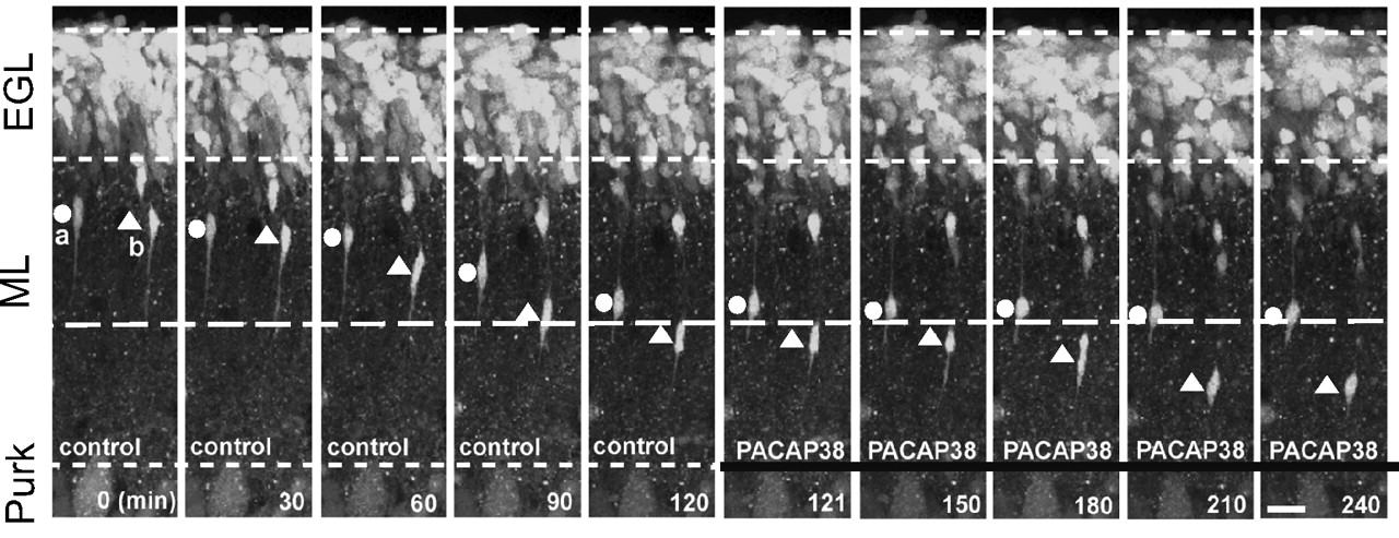
Time-lapse images showing that PACAP38 (1 μM) induces a rapid inhibition of cerebellar granule cell migration in the molecular layer on P10 mouse cerebellar slices. Elapsed time (in minutes) is indicated on the bottom of each microphotograph. [Reprinted from Cameron DB, Galas L, Jiang Y, Raoult E, Vaudry D, and Komuro H (2007) Cerebellar cortical-layer-specific control of neuronal migration by pituitary adenylate cyclase-activating polypeptide. Neuroscience 146:697–712. Copyright © 2007 Elsevier Science. Used with permission.]
In cortical neuron precursors, PACAP decreases the proportion of mitotic cells and promotes neuroblast differentiation (Lu and DiCicco-Bloom, 1997, 1998; Lu et al., 1998; Suh et al., 2001). PACAP has also been shown to contribute to the formation of the neuronal laminas in the developing cerebral cortex (Ohtsuka et al., 2008), and overexpression of PAC1-R leads to a dose-dependent hydrocephalus (Lang et al., 2006). The antimitogenic activity of PACAP on cortical neurons is mediated through the PAC1-R short variant expressed in these cells, which strongly inhibits p57Kip2-dependent CDK2 activity (Nicot and Dicicco-Bloom, 2001; Carey et al., 2002). Cortical neuroblasts, cultured in the presence of serum, turn into mature neurons that express glutamate and its receptors. It should be recalled that micromolar concentrations of glutamate exert a modest protective action on cortical neurons in primary culture, whereas millimolar concentrations of glutamate induce apoptotic cell death (Choi et al., 1987; Koh et al., 1991; Sagara and Schubert, 1998). In this model, PACAP potentiates the effect of otherwise marginally effective concentrations of glutamate (≈ 1 μM) on c-fos expression (Martin et al., 1995), arachidonic acid release (Stella and Magistretti, 1996; Magistretti et al., 1998), and brain-derived neurotrophic factor (BDNF) production (Pellegri et al., 1998; Zink et al., 2004). Moreover, PACAP protects cultured cortical neurons from the cytotoxic effect of high (≈1 mM) concentrations of glutamate (Morio et al., 1996). Excitotoxic doses of glutamate also substantially increase PACAP mRNA expression, and the PACAP receptor antagonist PACAP(6⇓⇓⇓⇓⇓⇓⇓⇓⇓⇓⇓⇓⇓⇓⇓⇓⇓⇓⇓⇓⇓⇓⇓⇓⇓⇓⇓⇓⇓⇓⇓–38) exacerbates the deleterious effect of glutamate (Shintani et al., 2005). Attenuation by PACAP of glutamate-induced neurotoxicity has also been reported in cultured retinal neurons (Shoge et al., 1999) and in neonatal brain lesions (Rangon et al., 2005; Favrais et al., 2007). Most of the actions of PACAP on cortical neurons are mediated through the cAMP pathway (Martin et al., 1995; Morio et al., 1996), although it has been reported that PACAP can directly modulate NMDA receptors independently of intracellular second messengers (Liu and Madsen, 1997, 1998). On these neurons, PACAP prevents the neurotoxic effect of lipopolysaccharide (Kong et al., 1999) and thrombin administration (Sanchez et al., 2009). The neuroprotective effect of PACAP is mediated at least in part indirectly through the release of BDNF (Frechilla et al., 2001; Shintani et al., 2005). Indeed, PACAP has been reported to induce the release of Rack1 from the NMDA receptor complex, which induces its translocation to the nucleus, where it activates BDNF expression (Yaka et al., 2003). In mesencephalic dopaminergic neurons, PACAP attenuates the neurotoxic effect of 6-hydroxydopamine (Takei et al., 1998; Reglödi et al., 2006) and 1-methyl-4-phenyl-1,2,3,6-tetrahydropyridine (Deguil et al., 2009). Ischemic death of hippocampal neurons can be prevented by intravenous infusion of PACAP (Uchida et al., 1996). PACAP38 protects hippocampal neurons from apoptosis by inhibiting the c-Jun N-terminal kinase 1 (JNK)/stress-activated protein and p38 signaling pathways (Dohi et al., 2002). It is noteworthy that PACAP38 is still effective in preventing cell death when administered several hours after ischemia (Reglodi et al., 2000), and it preserves the neurovascular reactivity after cerebral ischemia (Lenti et al., 2009), which suggests that the peptide may have therapeutic potency for the treatment of cerebral injuries. Pretreatment with PACAP38 also reduces the infarct size induced by stroke (Reglodi et al., 2002), and endogenous PACAP has been shown to contribute to neuron protection in case of stroke (Fig. 13) (Chen et al., 2006; Ohtaki et al., 2006). The antiapoptotic effect of PACAP38 after ischemia is indirect and involves IL-6 release (Ohtaki et al., 2006). Some of the neuroprotective effects of PACAP38 may also result from an inhibition of microglial activation (Delgado, 2002; Delgado et al., 2002a; Lee and Suk, 2004; Suk et al., 2004; Yang et al., 2006). After focal cerebral ischemia, the tumor suppressor protein p53 and the zinc finger protein Zac-1 (two genes controlling growth arrest and apoptosis) are up-regulated (Gillardon et al., 1998; Ciani et al., 1999). The p53 and Zac proteins have been demonstrated to regulate the PAC1-R gene, which, in the presence of PACAP38, can attenuate the damages of ischemia. Consistent with this finding, PACAP and PAC1-R mRNA expressions are transiently increased in the cortex and the hippocampus after traumatic brain injury (Skoglösa et al., 1999a; Stumm et al., 2007). Besides its neuroprotective activity, PACAP reduces the number of damaged axons after traumatic injury (Farkas et al., 2004; Tamás et al., 2006), favors dendrite outgrowth through the Rho GTPase and PI3-K pathways in response to neuronal activity (Henle et al., 2006) and enhances NMDA receptor activity (Macdonald et al., 2005), which probably contribute to functional recovery. PACAP also exerts a neuroprotective effect against retinal degeneration induced by carotid occlusion, kainic acid, and monosodium glutamate (Babai et al., 2005, Babai et al., 2006; Seki et al., 2006b; Atlasz et al., 2007, Atlasz et al., 2008). Considering the potential of PACAP for development into neuroprotective agent, stable analogs that can cross the blood-brain barrier are currently designed (Bourgault et al., 2008a; Dejda et al., 2008), and viral vectors for targeted delivery into the brain are being developed (Sanchez et al., 2008). It has been shown that the passage of PACAP38 across the blood brain barrier is transiently increased after ischemia (Somogyvári-Vigh et al., 2000), and antisense mRNA directed against the PACAP transporter PTS-6 have been successfully used to inhibit PACAP27 efflux (Nonaka et al., 2005; Dogrukol-Ak et al., 2009). Nevertheless, to foresee potential therapeutical applications, PACAP should be administered at doses as low as possible to avoid adverse effects notably on arterial blood pressure and heart rate (Ohtaki et al., 2004; Birk et al., 2007). To avoid such side effects, some lipophilic derivatives for intranasal administration have been developed (Gozes et al., 1999).

Brain tissue sections 24 h after permanent middle cerebral artery occlusion in PACAP(+/+), PACAP(+/−), and PACAP(−/−) mice, demonstrating the crucial role of endogenous PACAP in reducing neuronal damages caused by ischemia. [Reprinted from Ohtaki H, Nakamachi T, Dohi K, Aizawa Y, Takaki A, Hodoyama K, Yofu S, Hashimoto H, Shintani N, Baba A, Kopf M, Iwakura Y, Matsuda K, Arimura A, and Shioda S (2006) Pituitary adenylate cyclase-activating polypeptide (PACAP) decreases ischemic neuronal cell death in association with IL-6. Proc Natl Acad Sci U S A 103:7488–7493. Copyright © 2006 National Academy of Sciences of the United States of America.]
In dorsal root ganglia of embryos and newborn rats, the PACAP gene is expressed in sensory neurons (Lioudyno et al., 1998), and PACAP mRNA levels are up-regulated by axotomy (Zhang et al., 1996, 1998). Treatment of cultured ganglion neurons with PACAP increases cell survival and promotes neurite outgrowth (Lioudyno et al., 1998). In the same way, PACAP increases neuronal survival after spinal cord compression (Chen and Tzeng, 2005), suggesting that PACAP could have beneficial effects in tissue restoration after nerve injury. Consistent with this hypothesis, PACAP and PAC1-R mRNAs are up-regulated for as long as 30 days after facial motor neuron axotomy (Zhou et al., 1999b) and nerve regeneration is impaired in PACAP(−/−) animals (Armstrong et al., 2008).
PACAP contributes to synaptic transmission by enhancing NMDA receptor (Macdonald et al., 2005; MacDonald et al., 2007; Yang et al., 2009), increasing electrical activity (Di Mauro et al., 2003), and modulating AMPA receptor (Costa et al., 2009) in the hippocampus. PACAP has been shown to promote differentiation of embryonic stem cells into neurons and differentiation of neural stem cells into astrocytes (Vallejo and Vallejo, 2002; Cazillis et al., 2004; Ohno et al., 2005; Chafai et al., 2006; Hirose et al., 2006; Watanabe et al., 2006; Nishimoto et al., 2007). In cells that differentiate into neurons, expression of PAC1-R is increased, whereas the level of expression decreases in cells with a glial phenotype (Hirose et al., 2005). PACAP-evoked differentiation of precursor cells into astrocytes is mediated by cAMP, PKCβ, and calcium, involves coactivation of Ras and Rap1, and recruits the transcriptional repressor DREAM, an activator of GFAP gene expression (Cebolla et al., 2008; Lastres-Becker et al., 2008).
To summarize, during development, PACAP exerts neurotrophic activities to modulate cell proliferation, promote cell survival, inhibit cell migration, and stimulate cell differentiation. The effects of PACAP can be modulated during development according to the splice variants expressed. In adults, PACAP can rescue injured neurons from apoptosis, which suggests that it could be a useful molecule for the treatment of stroke or of neurodegenerative diseases.
5. Actions on Glial Cells.
Consistent with the occurrence of the PAC1-R-short and -hop splice variants in astroglial cells (Hashimoto et al., 2003), PACAP has been shown to stimulate cAMP production (Hashimoto et al., 2003; Masmoudi et al., 2003; Jozwiak-Bebenista et al., 2007; Nowak et al., 2007), to promote polyphosphoinositide turnover (Masmoudi et al., 2003; Dejda et al., 2006), to mobilize intracellular calcium stores (Tatsuno and Arimura, 1994), and to activate a quinine-sensitive potassium outward current (Ichinose et al., 1998) in rat astrocytes. The effect of PACAP on cAMP production involves neurofibromin, a protein controlling astrocyte proliferation (Dasgupta et al., 2003). In brain slices from newborn rat, PACAP enhances the number of glial precursor cells that express the proenkephalin-A gene in the neocortical subventricular zone (Just et al., 1998) and on cultured cells, PACAP promotes glutamate transport and metabolism (Figiel and Engele, 2000; Goursaud et al., 2008). In the presence of PACAP, glial cells also release interleukins (Seki et al., 2006a) and gliotransmitters (Masmoudi et al., 2003; Masmoudi-Kouki et al., 2006). Intraperitoneal administration of a VIP antagonist induces a marked reduction of the density of astrocytes in the cortex of E17 mouse embryos, and this effect is reversed by cotreatment with PACAP or the VPAC2-R agonist RO 25-1553 (Zupan et al., 1998), indicating that PACAP and/or VIP are involved in neocortical astrocytogenesis. In vitro, at picomolar concentrations, PACAP stimulates proliferation of astrocytes in a cAMP/ERK-dependent, PKA-independent manner (Moroo et al., 1998; Hashimoto et al., 2003; Li et al., 2005). In contrast, at nanomolar concentrations, PACAP has no more effect on ERK phosphorylation and reduces astrocyte proliferation by inhibiting the RhoA GTPase activity (Hashimoto et al., 2003; Meyer et al., 2005). RhoA and PI3-K inactivation are also involved in PACAP-induced astrocyte stellation (Ikeda et al., 2003; Perez et al., 2005).
In astrocytes, PACAP increases the production of various neurotrophic factors that can promote neuronal proliferation and/or differentiation (Ashur-Fabian et al., 1997). In particular, PACAP activates the expression and release of ciliary neurotrophic factor, activity-dependent neuroprotective protein, IL-6, glial cell line-derived neurotrophic factor, MIP, and regulated on activation normal T cell expressed and secreted (Gottschall et al., 1994; Nagao et al., 1995; Brenneman et al., 2002, 2003; Delgado et al., 2002a; Kimura et al., 2003; Zusev and Gozes, 2004; Dejda et al., 2005; Nakamachi et al., 2006; Nakatani et al., 2006). PACAP may also affect the expression of other neurotrophic factors, such as the protease nexin-1 or neurotrophin-3, known to be regulated by VIP. However, some of the neuroprotective effects of VIP that involve astrocytes are not mimicked by PACAP (Gressens et al., 1997, 1998a,b, 1999; Grimaldi and Cavallaro, 1999). These VIP-specific effects could be mediated through the PAC1-R splice variant hop2 (Pilzer and Gozes, 2006). Besides, the effect of PACAP on IL-6 release by astrocytes seems to be implicated in the neuroprotective action of the peptide in case of stroke (Ohtaki et al., 2006). The regulatory effect of PACAP on glycogen metabolism in astrocytes may also contribute to the neuroprotective effect of astrocytes (Masmoudi-Kouki et al., 2007). Finally, on these cells, PACAP attenuates histamine release, which may contribute to the anti-inflammatory activity of the peptide (Hansson et al., 2009).
Taken together, these data provide clear evidence that, in glial cells, PACAP plays a key role in the control of cell proliferation, plasticity, glycogen metabolism, and release of neurotrophic factors. It is noteworthy that PACAP acts at very low concentrations on astrocytes, that suggests that these cells may mediate many of the activities of PACAP in the brain.
B. Effects of Pituitary Adenylate Cyclase-Activating Polypeptide on the Pituitary Gland
The ability of PACAP to stimulate cAMP formation in pituitary cells provided the first evidence that the peptide may act as a hypophysiotropic neurohormone (Christophe, 1993; Arimura and Shioda, 1995; Nussdorfer and Malendowicz, 1998). The action of PACAP on the adenohypophysis has been reviewed in detail by Rawlings and Hezareh (1996). Among the different hypophysiotropic neuropeptides identified so far, the situation of PACAP is rather unique in that PACAP receptors are expressed by all endocrine cell types as well as by folliculostellate cells of the adenohypophysis (Vigh et al., 1993). Cytofluorimetric studies, conducted on dispersed rat pituitary cells, have shown that PACAP, acting through three different mechanisms, induces calcium elevation in all categories of endocrine cells (Canny et al., 1992; Gracia-Navarro et al., 1992; Rawlings et al., 1993, Rawlings et al., 1994; Rawlings and Hezareh, 1996; Alarcón and García-Sancho, 2000). Consistent with this observation, PACAP stimulates the release of GH, adrenocorticotropin, LH, follicle-stimulating hormone (FSH), PRL (Goth et al., 1992; Hart et al., 1992; Coleman and Bancroft, 1993; Koch and Lutz-Bucher, 1993; Perrin et al., 1993; Arbogast and Voogt, 1994; Hashizume et al., 1994; Velkeniers et al., 1994; Coleman et al., 1996; Martínez-Fuentes et al., 1998c; Ortmann et al., 1999) and somatolactin (Azuma et al., 2009). The effects of PACAP on the different pituitary cell types are summarized in Table 9.
Localization and relative abundance of PACAP receptor mRNA in rat peripheral tissues
Effects of PACAP on pituitary cells
1. Gonadotrope Cells.
Gonadotropin secretion is predominantly regulated by GnRH (Conn et al., 1981; Charlton, 2008). There is now evidence that PACAP acts either alone or synergistically with GnRH to stimulate LH and FSH mRNA expression (Tsujii and Winters, 1995; Tsujii et al., 1995; McArdle and Counis, 1996; Winters et al., 1997) and gonadotropin secretion (Culler and Paschall, 1991; Schomerus et al., 1994; Tsujii et al., 1994, 1995; Petersen et al., 1996; Ortmann et al., 1999; Counis et al., 2007). In the male rat, intra-atrial injection of PACAP, but not VIP, increases plasma LH level (Osuga et al., 1992). Perifusion of rat anterior pituitary cells with PACAP induces a transient stimulation of gonadotropin release and a concomitant increase in cytosolic calcium concentration (Canny et al., 1992; Rawlings et al., 1994; Tsujii et al., 1994). The effect of PACAP on gonadotropin, nitric-oxide synthase I, and estrogen receptor expression involves the cAMP/PKA pathway (Ishizaka et al., 1993; Winters et al., 1997; Schreihofer et al., 2001; Garrel et al., 2002), whereas its acute action on FSH/LH release is under the control of calcium elevation (Canny et al., 1992; Masumoto et al., 1995). Besides its direct action on gonadotropin release, PACAP has also been shown to increase rat GnRH receptor gene promoter activity through the cAMP/PKA pathway (Cheng and Leung, 2001; Ngan et al., 2001; Pincas et al., 2001; Sadie et al., 2003). Conversely GnRH stimulates PACAP gene expression (Grafer et al., 2009). Furthermore, GnRH agonists can inhibit PACAP-induced cAMP production by phosphorylation of PAC1-R through the PKC pathway (Larivière et al., 2006, 2008), illustrating the complex interplay between GnRH and PACAP in the regulation of gonadotrope cell functions.
2. Somatotrope Cells.
Secretion of GH is stimulated by GHRH and ghrelin, and inhibited by somatostatin (Castaño et al., 2005; Broglio et al., 2006). Administration of PACAP to cultured pituitary cells causes a significant increase in both GH mRNA expression and GH release (Velkeniers et al., 1994; Rousseau et al., 1999; Wong et al., 2005). In contrast, PACAP does not modify GH secretion from superfused cells (Velkeniers et al., 1994), suggesting that the stimulatory effect of PACAP on pituitary cells in static incubation involves the paracrine mediation of other hormones. PACAP exerts an additive effect on GHRH-stimulated GH output (Hashizume et al., 1994), and the stimulatory activity of PACAP on GH release is inhibited by addition of somatostatin (Goth et al., 1992; Hashizume et al., 1994). In fish, both PACAP and PRP stimulate GH secretion (Montero et al., 2000). In particular, PACAP provokes a robust increase of GH release from goldfish (Wong et al., 1998, 2000; Mitchell et al., 2008) and eel pituitary cells (Montero et al., 1998). PACAP increases intracellular calcium concentration in carp, frog, chicken, and rat somatotrope cells (Canny et al., 1992; Gracia-Navarro et al., 1992; Yada et al., 1993; Scanes et al., 2007), and the PACAP-evoked calcium response is blocked by the PKA antagonist 3′,5′-cyclic monophosphorothioate, Rp-isomer, indicating that the effect of PACAP is mediated through activation of the cAMP/PKA pathway and subsequent activation of the Ca2+/calmodulin-dependent protein kinase cascade (Rawlings et al., 1993, Rawlings et al., 1995; Alarcón and García-Sancho, 2000; Wong et al., 2005). The elevation of cytosolic calcium plays a pivotal role in PACAP-induced GH secretion (Martínez-Fuentes et al., 1998a,b,c). The maximal effect of PACAP on GH release is observed after 15 min of treatment, whereas prolonged incubation or pretreatment with PACAP causes desensitization of the secretory response (Goth et al., 1992; Wei et al., 1993).
3. Lactotrope Cells.
The secretion of PRL is predominantly under the tonic inhibitory control exerted by dopamine (Martinez de la Escalera and Weiner, 1992). The secretory activity of lactotrope cells is also regulated by various hypothalamic neuropeptides (Ruberg et al., 1981; Hinuma et al., 1998; Galas et al., 2009). In particular, VIP and to a lesser extent PHI and secretin stimulate PRL secretion (Vijayan et al., 1979; Enjalbert et al., 1980; Kimura et al., 1987; Muratori et al., 1994; Judd, 1995; Youngren et al., 1998). In fact, PACAP is more potent than other members of the family in stimulating PRL secretion because intravenous injection of PACAP to anesthetized rats induces a 4-fold increase of plasma PRL concentration (Yamauchi et al., 1995). Consistent with this notion, PRL levels are significantly reduced in PACAP knockout animals (Isaac and Sherwood, 2008). The effect of systemic administration of PACAP can be accounted for, at least in part, by a direct action at the pituitary level, because the peptide can also enhance plasma PRL level in hypothalamus-lesioned animals (Jarry et al., 1992). In vitro studies have confirmed that PACAP exerts a direct stimulatory effect on cytosolic calcium concentrations in fish, frog, and rat lactotrope cells (Canny et al., 1992; Gracia-Navarro et al., 1992; Matsuda et al., 2008). It should be noted, however, that PACAP increases the intracellular calcium level in 45% of PRL cells in frog (Gracia-Navarro et al., 1992) but only in 9% of PRL cells in rat (Canny et al., 1992). Studies aimed at investigating the effect of PACAP on PRL secretion by pituitary cells have led to controversial results. It has been initially reported that PACAP is devoid of PRL-releasing activity in cultured rat adenohypophysial cells (Miyata et al., 1989; Hart et al., 1992). PACAP was also found to have no effect on PRL release from cultured ovine (Sawangjaroen et al., 1997) and bovine (Hashizume et al., 1994) pituitary cells. In contrast, other studies have shown that PACAP can either slightly increase (Arbogast and Voogt, 1994) or inhibit (Jarry et al., 1992) PRL release from rat pituitary cells. To elucidate the origin of these apparent discrepancies, the effects of PACAP on PRL secretion have been compared in cultures of dispersed or aggregated cells and in pituitary fragments (Benter et al., 1995). In monolayer cultures, PRL release was inhibited by PACAP, whereas in cultures of aggregated cells and in pituitary fragments, PRL output was stimulated (Benter et al., 1995). These data suggest that cell-to-cell communication plays a crucial role in determining the type of action of PACAP on PRL secretion. Whereas intravenous injection of PACAP produces a significant increase in plasma PRL concentration in rat (Jarry et al., 1992; Yamauchi et al., 1995), systemic administration of PACAP has no effect on PRL level in sheep (Sawangjaroen and Curlewis, 1994), suggesting also the existence of marked species differences.
Besides its hypophysiotropic action at the pituitary level, PACAP may also regulate PRL secretion through modulation of various hypothalamic factors. In particular, in lactating rat, intracerebroventricular administration of the antagonist PACAP(6⇓⇓⇓⇓⇓⇓⇓⇓⇓⇓⇓⇓⇓⇓⇓⇓⇓⇓⇓⇓⇓⇓⇓⇓⇓⇓⇓⇓⇓⇓⇓–38) inhibits the PRL surge induced by suckling (Tohei et al., 2001). In sheep, injection of PACAP into the medial basal hypothalamus stimulates dopamine release from tuberoinfundibular neurons, leading to an inhibition of PRL secretion (Anderson and Curlewis, 1998). PACAP has also been found to decrease the activity of pyroglutamyl peptidase II (Vargas et al., 1998), a TRH-specific ectoenzyme that cleaves the pyroglutamyl-histidyl peptide bond of TRH (Charli et al., 1998). Because TRH is known to activate PRL secretion (Galas et al., 2009), the inhibition of pyroglutamyl peptidase II induced by PACAP may cause indirect stimulation of PRL release through reduction of TRH degradation. Taken together, these data indicate that PACAP may affect PRL secretion either via presynaptic action on hypothalamic neurons or via postsynaptic regulation of the activity of hypophysiotropic neurohormones.
4. Corticotrope Cells.
The secretion of adrenocorticotropin is primarily regulated by CRH (Rivier et al., 1982), and PACAP has been shown to activate CRH gene expression in the rat PVN (Grinevich et al., 1997). Intravenous administration of PACAP provokes a dose-related increase in plasma adrenocorticotropin level in human (Chiodera et al., 1996). The effect of PACAP on circulating adrenocorticotropin in human is not mimicked by VIP, indicating that the peptide acts through PAC1-R. In vitro, PACAP stimulates adrenocorticotropin secretion from superfused (Miyata et al., 1989) or cultured rat pituitary cells (Hart et al., 1992). However, in rat, the effect of PACAP on adrenocorticotropin secretion by cultured cells does not reach significance until 24 h, suggesting that PACAP does not exert a direct stimulatory action on corticotrope cells (Hart et al., 1992). Other in vitro studies have shown that PACAP does not stimulate adrenocorticotropin secretion from rat pituitary cells within 3 h of incubation (Culler and Paschall, 1991; Koch and Lutz-Bucher, 1993). In the frog R. ridibunda, PACAP causes an increase in cytosolic calcium concentration in 25% of corticotrope cells (Gracia-Navarro et al., 1992) and stimulates adrenocorticotropin secretion within 4 h (Martinez-Fuentes et al., 1994), indicating that, in amphibians, PACAP directly activates corticotrope cells.
5. Thyrotrope Cells.
In vivo administration of PACAP does not affect plasma thyroid-stimulating hormone (TSH) concentrations in rat (Hart et al., 1992) and human (Chiodera et al., 1996). Consistent with this observation, only a few thyrotrope cells express PACAP binding sites (Vigh et al., 1993), and PACAP does not modify TSH secretion from cultured rat anterior pituitary cells (Culler and Paschall, 1991). In frog, by contrast, PACAP increases free cytosolic calcium concentration in thyrotrope cells (Gracia-Navarro et al., 1992) and stimulates TSH release from dispersed pituitary cells (Okada et al., 2007, 2009).
6. Folliculostellate Cells.
Besides endocrine cells, the anterior pituitary encompasses a population of glial-like cells named folliculostellate cells. Incubation of cultured rat folliculostellate cells with PACAP causes stimulation of cAMP formation, IL-6 production, and follistatin secretion (Tatsuno et al., 1991c; Bilezikjian et al., 2003). Likewise, PACAP increases cAMP level and stimulates secretion of vascular endothelial growth factor, novel neurotrophin-1/B-cell-stimulating factor-3, and IL-6 in the mouse folliculostellate-like cell line TtT/GF (Matsumoto et al., 1993; Gloddek et al., 1999; Lohrer et al., 2001; Vlotides et al., 2004). Because IL-6 is involved in the differentiation of pituitary cells (Renner et al., 1998) and because it stimulates the release of various adenohypophysial hormones (Renner et al., 1998), several indirect effects of PACAP on endocrine pituitary cells may be mediated through activation of folliculostellate cells (Benter et al., 1995). In support of this notion, PACAP, added on cocultures of TtT/GF folliculostellate cells with rat anterior pituitary cells, significantly reduces the effect of activin-A on FSH secretion through follistatin release (Katayama et al., 2000).
7. Pituitary Fibroblasts.
The anterior pituitary gland also contains fibroblasts, a type of agranular connective cells (Gospodarowicz, 1979). PACAP has been shown to stimulate cAMP formation in cultured pituitary fibroblasts, suggesting that the peptide may modulate fibroblast proliferation (Koch and Lutz-Bucher, 1992b).
8. Melanotrope Cells.
The intermediate lobe of the pituitary is composed of a homogeneous population of cells, the melanotrope cells, which express the multifunctional precursor protein POMC (Crine et al., 1978). Post-translational processing of POMC in melanotrope cells gives rise to the formation of the melanotropic hormone α-MSH and the opioid peptide β-endorphin (Mains and Eipper, 1979). In rat, PACAP stimulates cAMP production and α-MSH release in cultured melanotrope cells (Koch and Lutz-Bucher, 1992a). PACAP has also been found to increase POMC mRNA levels in the rat pars intermedia (René et al., 1996). The stimulatory effect of PACAP on POMC gene expression and α-MSH secretion is associated with calcium influx through L-type calcium channels (Tanaka et al., 1997b). In the frog X. laevis, PACAP stimulates POMC gene expression in tissue culture of neurointermediate lobes and triggers the secretory activity of perifused isolated melanotrope cells (Kidane et al., 2007, 2008). Moreover, PACAP-LI in the neural lobe of X. laevis is higher when the animals are placed on an illuminated white environment, indicating that the peptide plays a physiological role in the neuroendocrine control of melanotrope cells during background color adaptation (Kidane et al., 2007, 2008). Indeed, the occurrence of PACAP mRNA in the neurointermediate lobe of rat (Tanaka et al., 1997b), frog (Alexandre et al., 2000b), and PAC1-R mRNA in the rat pars intermedia (Shioda et al., 1997a) strongly suggests that PACAP can act as a paracrine regulator of melanotrope cell activity. In agreement with this hypothesis, it has been shown that PACAP-LI in the neural lobe increases when frogs are placed on a white background and that PACAP, acting through VPAC1-R, induces the release of BDNF, which in turn stimulates POMC biosynthesis and MSH secretion (Kidane et al., 2007, 2008).
In conclusion, PACAP was initially discovered on the basis of its ability to activate the production of cAMP in rat anterior pituitary cells. Since then, PACAP has been shown to differentially regulate the activity of all cell types in the distal and intermediate lobes of the pituitary, including the nonendocrine folliculostellate cells, throughout the vertebrate phylum.
C. Effects of Pituitary Adenylate Cyclase-Activating Polypeptide on the Thyroid Gland
In the human and porcine thyroid, PACAP has been shown to stimulate cAMP production and to increase thyroxine secretion (Chen et al., 1993; Kouki et al., 1997; Bik et al., 2006).
D. Effects of Pituitary Adenylate Cyclase-Activating Polypeptide on the Gonads
The presence of PACAP and its receptors in the testis and ovary provides evidence that the peptide may operate as a local regulator of gonadal activity. In the rat testis, the concentration of PACAP is significantly reduced after hypophysectomy and is restored by FSH administration, indicating that the expression of PACAP is under the control of pituitary gonadotropins (Shuto et al., 1995). In vitro, PACAP induces a concentration-dependent stimulation of testosterone secretion from isolated rat Leydig cells (Romanelli et al., 1997; Rossato et al., 1997; El-Gehani et al., 1998c) and activates or inhibits protein synthesis in spermatocytes or spermatids, respectively (West et al., 1995). In Leydig cells, PACAP activates both AC and PLC through an interaction with PAC1-R (Romanelli et al., 1997). The effect of PACAP on Leydig cells may also be mediated via a novel receptor subtype coupled to a sodium channel through a pertussis toxin-sensitive G protein (Rossato et al., 1997). The effects of PACAP on protein synthesis in spermatocytes and spermatids are both mimicked by dibutyryl cAMP (West et al., 1995). In cultured Sertoli cells, PACAP increases cAMP concentration and stimulates estradiol and inhibin secretion (Heindel et al., 1992). In the epididymal epithelium, PACAP stimulates chloride secretion, which is important for sperm activation and storage (Zhou et al., 1997). The occurrence of PACAP-immunoreactive material in epididymal tubules indicates that PACAP is locally synthesized and thus may act as a paracrine regulator of sperm maturation (Zhou et al., 1997). The epithelium-derived PACAP may also stimulate epididymal spermatozoa that have lost the ability to produce PACAP (Shioda et al., 1994) but still possess PACAP binding sites (Shivers et al., 1991). In PACAP knock-out mice, testicular aging is delayed, probably because the expression of steroidogenic factors is impaired, which lowers the production of reactive oxygen species that are responsible for apoptosis (Lacombe et al., 2006). In the human cavernous tissue, PACAP dose dependently relaxes noradrenaline- and electrically contracted preparations, suggesting that the peptide may be involved in the induction and maintenance of penile erection (Hedlund et al., 1994, 1995). In line with this finding, a stearic acid VIP conjugate has been shown to increase the copulatory activity and penile reflex in castrated, testosterone-treated rats (Gozes and Fridkin, 1992). These data suggest that PACAP and/or VIP derivates could be developed for the treatment of impotence.
The PACAP-ergic system also seems to be involved in the reproductive function in female mice (Jamen et al., 2000b; Shintani et al., 2002; Sherwood et al., 2007). In the rat ovary, most granulosa and cumulus cells from large preovulatory follicles contain both PACAP mRNA and PACAP-LI (Gräs et al., 1996). Human chorionic gonadotropin (hCG) induces a transient increase of both PACAP and progesterone receptor mRNA expression (Ko et al., 1999; Barberi et al., 2007). The peak of expression of progesterone receptor mRNA occurs 3 h after hCG treatment and the peak of PACAP mRNA only after 6 to 12 h, suggesting that progesterone receptor activation is required for PACAP gene expression (Ko et al., 1999; Sayasith et al., 2007). In support of this hypothesis, it has been shown that blockage of progesterone receptors with the selective antagonist ZK98299 abrogates the effect of hCG on PACAP gene expression (Ko et al., 1999). The hCG-evoked stimulation of PACAP gene transcription is mediated through the PKA pathway and requires de novo protein synthesis (Ko et al., 1999; Sayasith et al., 2007). Exposure of cultured granulosa cells to PACAP causes a concentration-dependent increase in progesterone production (Zhong and Kasson, 1994; Apa et al., 1997a, Apa et al., 1997b; Gräs et al., 1999; Usuki and Kotani, 2001). Likewise, immunoneutralization of endogenous PACAP reduces progesterone formation and impairs subsequent luteinization, suggesting that PACAP plays an important role in LH-induced progesterone production during the periovulatory period (Gräs et al., 1999). Incubation of immature rat preovulatory follicles with PACAP or VIP induces a dose-dependent inhibition of follicle apoptosis (Flaws et al., 1995; Lee et al., 1999b), reduces cell proliferation promoted by FSH (Cecconi et al., 2004), and could be involved in the cyclic recruitment of immature follicles (Gräs et al., 2005). In luteinized granulosa cells, PACAP stimulates cAMP accumulation more potently than LH (Richards et al., 1995; Heindel et al., 1996), promotes survival (Barberi et al., 2007; Morelli et al., 2008), and increases plasminogen activator expression (Apa et al., 2002). In addition, neonatal administration of PACAP delays the first ovulation (Szabó et al., 2002). In the human female genital tract, PACAP is located in fibers innervating blood vessels and smooth muscle cells of the internal cervical os (Graf et al., 1995; Steenstrup et al., 1995). High concentrations of PACAP are also found throughout the human uteroplacental unit (Steenstrup et al., 1996). In vitro, PACAP induces relaxation of nonvascular smooth muscle strips from the fallopian tube and myometrium (Steenstrup et al., 1994, 1995) as well as stem villous and intramyometrial arteries (Steenstrup et al., 1996), suggesting that PACAP regulates the vascular tone in the human female reproductive tract. In placental cells, PACAP enhances cAMP formation, hCG and IL-6 production (Desai and Burrin, 1994).
Altogether, these data demonstrate the crucial role of PACAP in the regulation of the reproductive function. In particular, in the male PACAP facilitates sperm maturation, may contribute to penile erection and accelerates testicular aging, while in the female, PACAP stimulates progesterone production, prevents follicular apoptosis and improves fertility.
E. Effects of Pituitary Adenylate Cyclase-Activating Polypeptide on the Adrenal Gland
In adrenal chromaffin cells, PACAP exerts a stimulatory action on catecholamine secretion (Watanabe et al., 1992, Watanabe et al., 1995; Isobe et al., 1993; Chowdhury et al., 1994; Guo and Wakade, 1994; Houchi et al., 1994; Perrin et al., 1995; Neri et al., 1996; Jorgensen et al., 2000; Fukushima et al., 2002; Douglas et al., 2008; Valiante et al., 2008. PACAP also stimulates the release of brain natriuretic peptide and enkephalins, two regulatory peptides that are cosequestered with catecholamines in chromaffin granules (Babinski et al., 1996; Hahm et al., 1998) as well as a 15-fold increase in VIP mRNA expression (Lee et al., 1999a). The effect of PACAP on VIP biosynthesis and catecholamine secretion requires the coincident elevation of calcium and cAMP (Fukushima et al., 2001b; Hamelink et al., 2002a; Morita et al., 2002). It is noteworthy that the induction of VIP in septic shock is blocked in PACAP deficient mice (Ait-Ali et al., 2009). PACAP dose-dependently stimulates the release of the secretogranin II-derived peptides secretoneurin (Turquier et al., 2001) and EM66 (Guillemot et al., 2006). In vivo studies have shown that PACAP and VIP stimulate catecholamine release in anesthetized dogs through activation of PAC1-R coupled to dihydropyridine-sensitive L-type calcium channels (Gaspo et al., 1997; Geng et al., 1997; Lamouche et al., 1999; Lamouche and Yamaguchi, 2001). PACAP-induced catecholamine secretion is significantly greater after the induction of hypoglycemia (Yamaguchi and Lamouche, 1999) and PACAP(−/−) mouse exhibit a delayed normalization of plasma glucose levels in response to insulin injection (Hamelink et al., 2002b), suggesting that PACAP may play a beneficial role in glucose counter-regulatory mechanisms in the adrenal medulla during hypoglycemia. The effect of PACAP on catecholamine secretion is associated with activation of AC and elevation of cytosolic calcium concentrations (Isobe et al., 1993; Houchi et al., 1994; Isobe et al., 1994; Perrin et al., 1995; Chamoux et al., 1998). Incubation of adrenomedullary cells in calcium-free medium or blockage of voltage-operated calcium channels suppresses the PACAP-evoked stimulation of catecholamine secretion (Isobe et al., 1993; Houchi et al., 1995; Przywara et al., 1996; O'Farrell and Marley, 1997), indicating that the effect of PACAP on chromaffin cells is mediated through calcium influx. Concurrently, in bovine and human, PACAP increases calcium levels from ryanodine/caffeine-sensitive calcium stores (Houchi et al., 1995; Tanaka et al., 1996, 1998; Shibuya et al., 1999; Payet et al., 2003). The effect of PACAP on catecholamine release is associated with an increase in the expression of tyrosine hydroxylase, dopamine β-hydroxylase, and phenylethanolamine N-methyltransferase (Houchi et al., 1994; Rius et al., 1994; Isobe et al., 1996; Marley et al., 1996; Tönshoff et al., 1997; Hong et al., 1998; Choi et al., 1999; Park et al., 1999). It has been shown that the stimulatory effect of PACAP on tyrosine hydroxylase activity is mediated through activation of the AC/PKA transduction pathway (Marley et al., 1996) and can be accounted for by phosphorylation of TH at Ser40 (Bobrovskaya et al., 2007). The involvement of PACAP in adrenochromaffin cell development is not yet ascertained: on the one hand, PACAP has been reported to stimulate proliferation of rat chromaffin cells in primary culture (Tischler et al., 1995); on the other hand, PACAP inhibits the mitogenic effect of nerve growth factor on chromaffin cells (Frödin et al., 1995; Tischler et al., 1995). Finally, the adrenal medulla of PACAP(−/−) mouse exhibits normal catecholamine levels (Gray et al., 2002; Hamelink et al., 2002b).
Intravenous administration of PACAP causes elevation of plasma cortisol levels in dog and calf (Edwards and Jones, 1994; Kawai et al., 1994). PACAP stimulates corticosterone and aldosterone secretion from human, rat, and chicken adrenal slices, but does not affect the release of corticosteroids from dispersed fasciculata and glomerulosa cells (Andreis et al., 1995; Neri et al., 1996; Mazzocchi et al., 1997), suggesting that the response of adrenocortical cells to PACAP involves the contribution of another adrenal cell type. Exposure of human adrenal slices to the β-adrenergic receptor blocker l-alprenolol totally suppresses the steroidogenic effect of PACAP (Neri et al., 1996). Likewise, the action of PACAP on dehydroepiandrosterone and cortisol secretion by the fetal human adrenal gland is suppressed by the β-adrenoreceptor antagonist propranolol (Breault et al., 2000). Altogether, these observations indicate that, in several mammalian species, the effect of PACAP on corticosteroid secretion can be ascribed to the stimulatory action of the peptide on catecholamine secretion. In contrast, PACAP was found to stimulate corticosteroid release from dispersed bovine and frog adrenocortical cells (Yon et al., 1993b, 1994; Bodart et al., 1997). The fact that PACAP stimulates cAMP and inositol phosphate formation in bovine glomerulosa cells (Bodart et al., 1997) and calcium mobilization in individual frog adrenocortical cells (Yon et al., 1994) provides additional evidence for a direct stimulatory effect of the peptide on steroidogenesis in these two species.
In summary, PACAP stimulates the release of catecholamines and regulatory peptides from adrenochromaffin cells, and triggers steroid hormone secretion from adrenocortical cells. It has been suggested that PACAP may function as an emergency response factor in the case of prolonged metabolic stress. PACAP may also contribute to the development and differentiation of the adrenal gland.
F. Effects of Pituitary Adenylate Cyclase-Activating Polypeptide on the Gastrointestinal Tract
The presence of PACAP in various exocrine glands of the alimentary canal and in neuronal structures (ganglia, fibers) innervating smooth muscle layers suggests that the peptide plays an important role in the function of the gastrointestinal tract. In agreement with this notion, intravenous injection of PACAP to anesthetized rat stimulates secretion of saliva from the submandibular and parotid glands (Mirfendereski et al., 1997). In the ferret submandibular gland, PACAP and VIP exert a vasodilatory effect that contributes to trigger the secretory activity of the salivary glands (Tobin et al., 1995). In the hamster submandibular ganglion neurons, PACAP inhibits L-, N-, and P/Q-type Ca2+ channels via the Gs-protein γ subunit coupled to PAC1-R (Hayashi et al., 2002; Kamaishi et al., 2004) and induces potentiation of acetylcholine-evoked nicotinic currents through a PTX-sensitive G protein (Liu et al., 2000).
In the rat stomach, PACAP inhibits histamine- and pentagastrin-evoked gastric acid secretion but has no effect on carbachol-induced secretion (Mungan et al., 1992b, 1995; Li et al., 2000c). In contrast, on isolated rabbit parietal cells, PACAP potentiates the response to histamine and to carbachol (Healey et al., 1998). In the gastric mucosa, PACAP stimulates histamine synthesis, storage and release from enterochromaffin-like (ECL) cells (Lindström et al., 1997; Håkanson et al., 1998; Zeng et al., 1998, 1999a; Prinz et al., 2003) through activation of L-type calcium channels (Zeng et al., 1999b; Lindström et al., 2001) and triggers histidine decarboxylase activity (Bernsand et al., 2007). Consistent with these observations, the presence of PAC1-R has been detected on gastric ECL cells (Zeng et al., 1999a). Because histamine is a potent stimulator of chloride secretion (Helander and Keeling, 1993), these data suggest that the effect of PACAP on gastric acid production can be accounted for, at least in part, by an indirect stimulation of ECL cells (Sandvik et al., 2001). In support of this hypothesis, PACAP elevates [Ca2+]i in ECL cells and adjacent parietal cells in rabbit gastric glands, whereas histamine receptor antagonists abolish the Ca2+ response in adjacent parietal cells (Athmann et al., 2000). PACAP also stimulates proliferation of gastric ECL cells through activation of the PKA, protein tyrosine kinase, and MAPK pathways (Läuffer et al., 1999; Oh et al., 2005). Intracerebroventricular injection of PACAP stimulates gastric acid secretion (Ozawa et al., 1997), suggesting that PACAP may act centrally to regulate gastric acid release. The central effect of PACAP may involve peptide tyrosine tyrosine (PYY) as an indirect mediator (Guo et al., 1987), because intravenous injection of PACAP has been shown to increase plasma PYY concentrations (Zhang et al., 1993). In the antrum of mammals, PACAP regulates locally the secretion of a number of peptides, including atrial natriuretic peptide (Gower et al., 2003), gastrin, and somatostatin (Tornøe et al., 2001). In the guinea pig stomach, PACAP increases exocytosis of zymogen granules from isolated chief cells that release pepsinogen (Felley et al., 1992). Intravenous injection of PACAP also enhances bicarbonate secretion in the duodenum (Takeuchi et al., 1997; Konturek et al., 2004). In the distal colon, PACAP acts through cholinergic and noncholinergic neurons to evoke chloride secretions (Kuwahara et al., 1993). In human colonic T84 cells, PACAP also regulates chloride secretion in a Ca2+-dependent manner (Leung et al., 2001). One conspicuous feature is the superior potency of PACAP, compared with other gut neuropeptides, in stimulating gastrointestinal exocrine secretions (Läuff et al., 1999). In a model of experimental colitis induced by dextran sulfate sodium, PACAP inhibits the production of pro-inflammatory cytokines in the proximal and distal colon (Azuma et al., 2008). Using the same approach, it has been observed that PACAP(−/−) mice exhibit higher colonic inflammation on pathological examination than wild-type animals (Nemetz et al., 2008).
Besides its effects on the secretory activity of exocrine and endocrine cells, PACAP induces a concentration-dependent relaxation of gastric smooth muscles (Katsoulis et al., 1996; Robberecht et al., 1998; Mukai et al., 2006; Toyoshima et al., 2006), causing a decrease of gastric motility and a delay in stomach emptying (Ozawa et al., 1999). PACAP also exerts a relaxant effect on intestinal smooth muscles from rat (Mungan et al., 1992a; Schwörer et al., 1992; Katsoulis et al., 1993b; Grider et al., 1994; Ekblad and Sundler, 1997; Olsson and Holmgren, 2000), guinea pig (Mungan et al., 1992a; Schwörer et al., 1992; Katsoulis et al., 1993b; Grider et al., 1994; Ekblad and Sundler, 1997; Olsson and Holmgren, 2000) and Atlantic cod, Gadus morhua (Mungan et al., 1992a; Schwörer et al., 1992; Katsoulis et al., 1993b; Grider et al., 1994; Ekblad and Sundler, 1997; Olsson and Holmgren, 2000), thus reducing the motility of the bowel (Läuff et al., 1999). The mechanism by which PACAP induces muscle relaxation along the gastrointestinal tract has been extensively studied. In the mouse fundus, PACAP release, regulated by M2 muscarinic receptors (Takeuchi et al., 2006), induces a sustained relaxation that is suppressed by iberiotoxin, an inhibitor of big conductance calcium-activated K+ channels (Hagi et al., 2008). In circular smooth muscle cells of the pig jejunum, PACAP(6⇓⇓⇓⇓⇓⇓⇓⇓⇓⇓⇓⇓⇓⇓⇓⇓⇓⇓⇓⇓⇓⇓⇓⇓⇓⇓⇓⇓⇓⇓⇓–38) attenuates inhibitory junction potentials evoked by electrical field, suggesting that inhibitory neurotransmission is mediated at least in part by PACAP (Matsuda et al., 2004). In longitudinal muscle cells of the mouse ileum, PACAP inhibits spontaneous contractile activity through activation of PLC and Ca2+ release from intracellular stores, causing opening of apamin-sensitive Ca2+-dependent K+ channels (Zizzo et al., 2005). In the same tissue, PACAP also enhances nitric oxide (NO) production, which in turn may stimulate the release of PACAP from inhibitory neurons (Zizzo et al., 2004).
In the colon, the effect of PACAP on longitudinal muscle relaxation is mediated through PAC1-R (Mukai et al., 2002). On murine colonic smooth muscle cells, PACAP increases the frequency of Ca2+ transients, as well as the frequency and amplitude of spontaneous outward currents through activation of the AC pathway (Hagen et al., 2006). In colon-inferior mesenteric ganglion neurons, PACAP causes prolonged depolarization and intense generation of fast excitatory postsynaptic potentials and action potentials through PAC1-R (Ermilov et al., 2004). It is noteworthy that, in the rat distal colon, exogenous PACAP induces strong relaxation of the longitudinal muscle in 2-week-old rats but has no effect on tissues from 8-week-old rats, indicating that the effect of PACAP fades during postnatal maturation (Takeuchi et al., 2004).
At odds with the effects in rat colon is that in the guinea pig small intestine, PACAP stimulates normal peristalsis and counteracts drug-induced peristaltic arrest (Heinemann and Holzer, 1999). The contractile effect of PACAP on the guinea pig ileum is mediated through presynaptic stimulation of acetylcholine and substance P release (Katsoulis et al., 1993a). In the opossum internal anal sphincter, PACAP exerts a biphasic effect (i.e., an initial contraction followed by relaxation) (Rattan and Chakder, 1997; Chakder and Rattan, 1998). The contractile but not the relaxant effect of PACAP on the anal sphincter is abrogated by a substance P antagonist, confirming that the PACAP-evoked contraction is mediated through presynaptic activation of substance P afferents (Rattan and Chakder, 1997). In fish and amphibians, PACAP exerts an inhibitory control of peristalsis (Olsson and Holmgren, 2001). In the stargazer, PACAP inhibits rectum contractions stimulated by acetylcholine or potassium chloride (Matsudaa et al., 2000).
To summarize, in the gastrointestinal tract, PACAP stimulates the secretion of saliva, gastric acid and bicarbonate as well the release of other regulatory peptides including gastrin, somatostatin, atrial natriuretic factor and PYY. In addition, PACAP exerts a number of effects on the gastrointestinal tract motility through its action (mainly myorelaxation) on smooth muscles from the stomach, intestine, colon, rectum, and anal sphincter.
G. Effects of Pituitary Adenylate Cyclase-Activating Polypeptide on the Liver
It has long been known that VIP is a potent stimulator of AC activity in liver cells (Waelbroeck et al., 1981). Likewise, in cultured hepatocytes, PACAP causes a dose-dependent accumulation of cAMP but does not affect inositol phosphate turnover (el Fahime et al., 1996). The fact that VIP exerts a mitogenic action on rat hepatocytes (Kar et al., 1996) strongly suggests that PACAP could also be involved in the control of liver cell proliferation. Injection of PACAP to anesthetized dogs induces a 2-fold increase of plasma glucose concentration (Sekiguchi et al., 1994). Actually, PACAP is more potent than VIP in stimulating glucose output from the perfused rat liver (Yokota et al., 1995). The hyperglycemic action of PACAP observed in vivo can be ascribed to both a direct action on hepatocytes and an indirect effect via glucagon and/or adrenaline release (Sekiguchi et al., 1994). Finally, PACAP acting on VPAC2-R exerts a lipolytic effect on rat adipocytes (Akesson et al., 2003, Akesson et al., 2005) and PACAP knock-out animals exhibit microvesicular fat accumulation, indicating that PACAP acts as an important hormonal regulator of lipid and carbohydrate metabolism (Gray et al., 2001; Tomimoto et al., 2008). These observations support the view that PACAP agonists could be of therapeutic value for the treatment of obesity.
H. Effects of Pituitary Adenylate Cyclase-Activating Polypeptide on the Pancreas
In the pancreas, PACAP-immunoreactive fibers innervate both the exocrine acini and the islets of Langerhans, as well as the small arteries running within the connective tissue (Table 2) (Köves et al., 1993; Tornøe et al., 1997). Electrical stimulation of the vagus nerve causes the release of PACAP from the isolated perfused pig pancreas, suggesting that the peptide may control exocrine and/or endocrine pancreatic secretions (Tornøe et al., 1997). Nevertheless, overexpression of PACAP may be deleterious as it aggravates cerulean-induced pancreatitis in mice (Hamagami et al., 2009).
Intravenous injection of PACAP triggers amylase (Mungan et al., 1991; Alonso et al., 1994), pancreatic fluid, bicarbonate, and protein secretions (Naruse et al., 1992; Alonso et al., 1994; Zabielski et al., 1994; Rodríguez-López et al., 1995; Onaga et al., 1996; Wheeler et al., 1997; Lee et al., 1998; Glad et al., 2003). PACAP also induces vasodilation and increases pancreatic blood flow, notably in the exocrine part of the gland (Carlsson et al., 1996; Ito et al., 1998). The stimulatory effect of PACAP on juice flow is inhibited by the antagonist PACAP(6⇓⇓⇓⇓⇓⇓⇓⇓⇓⇓⇓⇓⇓⇓⇓⇓⇓⇓⇓⇓⇓⇓⇓⇓⇓⇓⇓⇓⇓⇓⇓–38) (Tornøe et al., 1997). Experiments conducted on isolated rat pancreatic acini have shown that PACAP exerts a direct increase on amylase and lipase secretions (Kashimura et al., 1991; Raufman et al., 1991; Schmidt et al., 1993). Coadministration of PACAP with cholecystokinin, carbachol or bombesin to dispersed guinea pig acinar cells causes additive stimulation of amylase secretion (Kimball and Mulholland, 1996). The effect of PACAP is probably mediated via the AC pathway and does not involve PLC activation or calcium mobilization (Kashimura et al., 1991; Kitagawa et al., 1995; Kimball and Mulholland, 1996). Besides its direct action on acinar cells, PACAP may also exert an indirect effect on pancreatic exocrine secretions through modulation of afferent nerve activity. In particular, PACAP has been shown to stimulate pancreatic enzyme secretion in sheep via activation of vagal cholinergic neurons (Onaga et al., 1997). PACAP also enhances electrically evoked stimulation of noradrenaline release in the canine pancreas (Yamaguchi and Fukushima, 1998), suggesting that the peptide may control juice flow through presynaptic modulation of the parasympathetic vagus nerve. Altogether, these data suggest that PACAP has to be added to the still growing list of secretagogues of the exocrine pancreas.
In the endocrine pancreas, PACAP seems to be much more potent than VIP or other regulatory peptides in stimulating pancreatic hormone secretion (Winzell and Ahrén, 2007; Ahrén, 2008). In vivo administration of PACAP causes a significant increase in plasma insulin level in mice (Fridolf et al., 1992; Filipsson et al., 1998a; Persson-Sjögren et al., 2006), calf (Edwards et al., 1997), dog (Kawai et al., 1992), and human (Filipsson et al., 1997). In support of a role of PACAP on islet hormone secretion (Yada et al., 1997a), PACAP knock-out mice or animals treated with the antagonist PACAP(6⇓⇓⇓⇓⇓⇓⇓⇓⇓⇓⇓⇓⇓⇓⇓⇓⇓⇓⇓⇓⇓⇓⇓⇓⇓⇓⇓⇓⇓⇓⇓–38) exhibit reduced insulin secretion after intraperitoneal glucose challenge (Shintani et al., 2003; Green et al., 2006; Tomimoto et al., 2008). PACAP acts at very low concentrations on cultured islets cells (Yada et al., 1994, 1997b,c; Filipsson et al., 1998b; Davalli et al., 1999; Filipsson et al., 1999), and its stimulatory effect on insulin secretion is mediated through activation of PAC1-R and VPAC2-R coupled to the AC pathway (Jamen et al., 2000a, 2002b; Asnicar et al., 2002; Persson and Ahrén, 2002). Furthermore, pancreatic β-cells express cell-surface ectopeptidases capable of degrading PACAP (Hupe-Sodmann et al., 1997), indicating that the action of PACAP on insulin secretion is finely regulated. The amplitude and kinetics of the PACAP-evoked stimulation of insulin release depends on glucose concentration in the incubation medium (Yokota et al., 1993; Bertrand et al., 1996; Edwards et al., 1997). PACAP induces a biphasic effect on insulin secretion [i.e., a rapid and transient stimulation (acute phase) followed by a rebound of the secretory response (plateau phase)]. The plateau phase could be ascribed to the ability of PACAP to regulate insulin gene expression (Borboni et al., 1999). The phosphatidylinositol 3-kinase inhibitor wortmannin inhibits the plateau phase but not the acute phase of the PACAP-evoked insulin release (Straub and Sharp, 1996). Exposure of pancreatic β-cells to PACAP causes calcium influx through L-type calcium channels (Yada et al., 1997c) and the stimulatory effect of PACAP on insulin secretion is abolished by nitrendipine (Komatsu et al., 1996), indicating that activation of voltage-sensitive L-type calcium channels is involved in the insulinotropic effect of PACAP. Strangely enough, the combination of glucose, PACAP, and carbachol stimulates insulin release but is unable to elevate intracellular calcium (Komatsu et al., 1996). Incubation of isolated rat islets with specific PACAP antisera inhibits the ability of glucose to stimulate insulin release (Yada et al., 1997b; Filipsson et al., 1999), suggesting that endogenous PACAP acts as a physiological regulator of pancreatic β-cell activity. PACAP is also a potent stimulator of glucagon secretion. Intravenous injection of PACAP increases plasma glucagon concentration in mice (Fridolf et al., 1992) and human (Filipsson et al., 1997). Likewise, in the perfused rat pancreas, PACAP enhances glucagon secretion (Yokota et al., 1993). The stimulatory effect of PACAP on insulin and glucagon release is completely abolished by somatostatin (Yokota et al., 1993). Besides its effect on hormone regulation, PACAP induces an antiapoptotic effect on rat insulinoma β-cells (Onoue et al., 2008), suggesting that impaired PACAP signaling during aging might contribute to the occurrence of type 2 diabetes.
To summarize, both endogenous and exogenous PACAP seem to be potent activators of pancreatic endocrine secretions. On the endocrine gland, PACAP stimulates insulin and glucagon secretion, which suggests that it could play an important role in prandial insulin secretion and contribute to the glucagon response to hypoglycemia. This ability of PACAP to stimulate insulin production may lead to the development of novel therapies for the treatment of type 1 diabetes (Kamiya et al., 2000; Herrera et al., 2006; Sakuma et al., 2009). Furthermore, the antiapoptotic effect of PACAP on insulinoma β-cells suggests that it could be of therapeutic interest for the treatment of type 2 diabetes.
I. Effects of Pituitary Adenylate Cyclase-Activating Polypeptide on the Respiratory System
The occurrence of PACAP and PACAP receptors has been reported at different levels of the airways (Tables 2 and 8). In rodents, PACAP causes relaxation of tracheal smooth muscles (Araki and Takagi, 1992; Conroy et al., 1995; Foda et al., 1995; Hiramatsu et al., 1995; Yoshihara et al., 1997; Berisha et al., 2002; Lindén et al., 2003), promotes bronchodilation (Lindén et al., 1995, Lindén et al., 1997, Lindén et al., 1999) and increases nasal airway resistance (Kinhult et al., 2003). The relaxant effect of PACAP on the trachea is mediated through activation of the cAMP/PKA-dependent (Araki and Takagi, 1992; Kanemura et al., 1993; Foda et al., 1995), NO/cGMP–dependent (Saotome et al., 1998), and carbon monoxide-dependent transduction pathways (Kinhult et al., 2001a). PACAP also plays a crucial role in the maintenance of normal pulmonary vascular tone during early postnatal life. Thus, deficiency of PACAP signaling leads to pulmonary hypertension (Otto et al., 2004), breathing defects (Wilson and Cumming, 2008), and sudden neonatal death (Cummings et al., 2004). PACAP may control breathing by acting either directly on carotid bodies (Xu et al., 2007, 2008) or indirectly via the respiratory centers in the CNS (Wilson and Cumming, 2008). PACAP is also a potent stimulator of airway mucus (Wagner et al., 1998; Liu et al., 1999) and chloride secretions (Dérand et al., 2004), suggesting a role in airway defense. PACAP exerts an antiapoptotic effect on the respiratory system and attenuates the cytotoxicity of cigarette smoke extracts on alveolar cells (Onoue et al., 2004). Owing to the broncho-relaxant and protective properties of PACAP and VIP, synthetic analogs have been developed for potential application in the treatment of asthma (Bolin et al., 1995; Meyer et al., 1996; Sergejeva et al., 2004; Yoshihara et al., 2004; Szema et al., 2006), and a VIP aerosol formulation, aviptadil, is currently under evaluation for the treatment of pulmonary hypertension (Leuchte et al., 2008). Thus, a therapy using the ventilatory effects of VIP could offer potential benefits for the treatment of obstructive and inflammatory diseases, and long-acting VIP-based compounds may represent a novel target for drug development.
J. Effects of Pituitary Adenylate Cyclase-Activating Polypeptide on the Cardiovascular System
The walls of blood vessels are richly innervated by PACAP-containing fibers (Table 2) (Köves et al., 1990; Cardell et al., 1991; Baeres et al., 2004) and a high density of PACAP binding sites is present in arteries (Table 6) (Amenta et al., 1991; Nandha et al., 1991; Huang et al., 1993). Although PACAP is able to exert an indirect hypertensive action mediated through the release of catecholamines (Ishizuka et al., 1992; Minkes et al., 1992a), this peptide, in very much the same way as VIP, is mainly considered as a highly potent vasorelaxant factor (Hirata et al., 1985; Ross-Ascuitto et al., 1993; Tong et al., 1993; Ascuitto et al., 1996). This vasodilatory activity, which can be ascribed at least in part to its activity on arterial smooth muscle cells (Huang et al., 1993; Naruse et al., 1993; Steenstrup et al., 1996; Bruch et al., 1997), is well documented in various organs, including the brain (Tong et al., 1993; Anzai et al., 1995), the eye (Nilsson, 1994; Elsås and White, 1997; Dorner et al., 1998), the pulmonary vascular bed (Minkes et al., 1992b; Cheng et al., 1993; Foda et al., 1995), the mesentery (Wilson and Warren, 1993), the pancreas (Bertrand et al., 1996; Ito et al., 1998), the testis (Lissbrant et al., 1999), the ovary (Steenstrup et al., 1994; Yao et al., 1996), the vagina (Steenstrup et al., 1994; Giraldi et al., 2002; Aughton et al., 2008), the kidney (Gardiner et al., 1994), the gastrointestinal tract (Portbury et al., 1995; Badawy and Reinecke, 2003), and the skin (Wallengren, 1997).
The intracellular mechanism of action of PACAP on blood vessel contractility is not fully understood. PACAP is known to increase cAMP formation in the isolated rabbit ovarian artery (Yao et al., 1996), the rat tail vein (Absood et al., 1992), and cerebral microvessels (Kobayashi et al., 1994; Wilderman and Armstead, 1997). Because cAMP has an inhibitory effect on smooth muscle cell contraction, the stimulatory effect of PACAP on cAMP production is likely to account for its vasorelaxation activity (Steer, 1976; Korenman and Krall, 1977; Farah, 1983). The action of PACAP on arterial smooth muscle cell relaxation requires the activation of ATP- and calcium-dependent potassium channels (Bruch et al., 1997). PACAP can also increase the amplitude of L-type Ca2+ channel currents in vascular smooth muscle cells through the activation of both PKA, PKC, and PLC (Chik et al., 1996; Markhotina et al., 2007) with a greater efficiency in spontaneously hypertensive rats than in normotensive animals (Li et al., 2001). The involvement of the endothelium in the vasodilatory activity of PACAP is still a matter of debate: two reports indicate that the vasorelaxant effect of PACAP on the aorta and coronary arteries is endothelium-independent (Warren et al., 1991; Kästner et al., 1995), whereas another study reveals that removal of the vascular endothelium abolishes the dilatory response induced by PACAP in pulmonary arteries (Cardell et al., 1997). Finally, some of the effects of PACAP on the vascular bed appears to be mediated through the release of vasculotropic factors. For example, in cerebral arteries, PACAP can activate a cyclooxygenase-independent mechanism (Lenti et al., 2007) but also a cyclooxygenase-dependent pathway leading to the release of the prostaglandin PGF2α (Anzai et al., 1995; Lenti et al., 2007). PACAP has also been found to stimulate the production of vascular endothelial growth factor, which plays an important role in angiogenesis and vascular permeability (Gloddek et al., 1999). The vasodilatory effect of PACAP is associated with a dose-dependent increase in blood flow in various organs (Nandha et al., 1991; Ishizuka et al., 1992; Minkes et al., 1992a; Warren et al., 1992a,b; Naruse et al., 1993; Santiago and Kadowitz, 1993; Mirfendereski et al., 1997; Whalen et al., 1999a,b,c), including brain (Uddman et al., 1993; Jansen-Olesen et al., 1994; Seki et al., 1995a; Reglodi et al., 2002; Ohtaki et al., 2004), and a decrease in mean arterial blood pressure (Ishizuka et al., 1992; Carlsson et al., 1996; Mirfendereski et al., 1997; Ohtaki et al., 2004). Altogether, these observations highlight the major vasorelaxant effect of PACAP and indicate that this neuropeptide may have a potential therapeutic value for the treatment of hypertension.
In the heart, PACAP produces positive inotropic, chronotropic, and dromotropic effects, making it a cardiotonic candidate for treatment of heart failure. For instance, intravenous injection of PACAP in cat, sheep, and human provokes an increase in heart rate and enhances the contractile ventricular force (Minkes et al., 1992a; Sawangjaroen et al., 1992; Sawangjaroen and Curlewis, 1994; Birk et al., 2007). PACAP also caused bradycardia in isolated perfused guinea pig heart through both PAC1-R and VPAC-R (Chang et al., 2005; Hoover et al., 2009). In the anesthetized dog and on the isolated canine heart, PACAP evokes a transient positive followed by negative chronotropic and inotropic responses (Hirose et al., 1997b; Hirose et al., 1998). The negative response can be ascribed to stimulation of cardiac parasympathetic neurons and acetylcholine release from cholinergic parasympathetic postganglionic nerves (Hirose et al., 1997c). In vitro studies on the isolated guinea pig heart have confirmed that the negative chronotropic effect of PACAP can be accounted for by an increase in acetylcholine release from parasympathetic neurons (Seebeck et al., 1996). Moreover, the response of the guinea pig heart was blocked by atropine, indicating that the negative chronotropic effect is mediated by cholinergic neurons (Chang et al., 2005). On the contrary, the positive effects of PACAP are attributable to direct stimulation of cardiac myocytes (Suzuki et al., 1993; Runcie et al., 1995; Hirose et al., 1997a; Chang et al., 2005). In isolated rat and guinea pig heart preparations, which lack sympathetic tone, PACAP causes tachycardia independently of adrenergic mechanisms (Chang et al., 2005). However, in the anesthetized rat, the PACAP-induced tachycardia is abolished by the β-adrenoreceptor antagonist propranolol but is not affected by the ganglion blocker chlorisondamine, indicating that PACAP can stimulate norepinephrine release from cardiac sympathetic nerve terminals (Whalen et al., 1999a).
In guinea pig, PACAP modulates the excitability of intracardiac neurons by enhancing a hyperpolarization-activated nonselective cationic conductance (Merriam et al., 2004). This effect is mediated by PAC1-R and cAMP production (Parsons et al., 2000; Tompkins and Parsons, 2008) and requires Ca2+ influx (Tompkins et al., 2006). In contrast, the increased excitability of rat neonatal cardiac neurons induced by PACAP requires coactivation of PAC1-R and VPAC-R and release of Ca2+ from intracellular stores (DeHaven and Cuevas, 2004). PACAP is localized to preganglionic parasympathetic nerves in rat and guinea pig hearts (Calupca et al., 2000; Richardson et al., 2003), and PACAP released at this site acts at postsynaptic PAC1-R to increase excitability of cardiac cholinergic neurons (Tompkins et al., 2007).
PACAP induces spontaneous atrial fibrillation in autonomically decentralized, anesthetized dogs and also causes arrhythmias in isolated guinea pig hearts (Hirose et al., 1997a; Chang et al., 2005). Muscarinic blockade with atropine prevents arrhythmias in both models, suggesting that this response is mediated by cholinergic neurons. In isolated dog atrium preparations, PACAP reduces action potential duration (Hirose and Chiba, 2003) and causes ectopic activity (Hirose and Laurita, 2007).
Taken together, these data indicate that PACAP exerts major stimulatory effects on the cardiovascular system. PACAP also exhibits protective properties against oxidative stress-induced apoptosis in cardiomyocytes (Gasz et al., 2006; Rácz et al., 2008; Roth et al., 2009) and endothelial cells (Rácz et al., 2007), pointing to the therapeutic interest of PACAP for the treatment of cardiovascular diseases.
K. Effects of Pituitary Adenylate Cyclase-Activating Polypeptide on Immune Cells
VIP and PACAP exert a broad spectrum of actions on cells of the systems of innate and acquired immunity. In cultured mast cells, PACAP stimulates histamine secretion (Schmidt-Choudhury et al., 1999a,b) and serotonin release (Seebeck et al., 1998). In mitogen-stimulated murine splenocytes, PACAP causes a dose-dependent inhibition of concanavalin A-induced cell proliferation (Tatsuno et al., 1991a). In murine spleen cells and thymocytes, PACAP inhibits IL-10 production via both cAMP-dependent and -independent pathways (Martinez et al., 1996; Wang et al., 1999). In CD4+CD8+ thymocytes, PACAP prevents glucocorticoid-induced apoptosis (Delgado et al., 1996b) through inhibition of Fas ligand expression (Delgado and Ganea, 2000a), suggesting involvement in intrathymic T-cell maturation.
In different animal models of chronic inflammatory diseases, treatment with PACAP attenuates the symptoms and modifies the cytokine profiles. For instance, in a murine model of experimental autoimmune encephalomyelitis, PACAP-deficient mice exhibit more severe clinical and pathological manifestations compared with wild-type animals. The increased sensitivity of PACAP-deficient mice is associated with enhanced production of proinflammatory cytokines, chemokines, and chemokine receptors, and reduced production of anti-inflammatory cytokines (Tan et al., 2009).
In unstimulated macrophages, PACAP and its agonist maxadilan inhibit the release of tumor necrosis factor-α and increase IL-6 production through activation of the PKA and PKC pathways (Delgado et al., 1998, 1999c,g; Martínez-Fuentes et al., 1998a; Soares et al., 1998). In contrast, PACAP inhibits the release of IL-6, IL-12, and tumor necrosis factor-α from lipopolysaccharide-stimulated macrophages (Martínez et al., 1998a,b; Delgado et al., 1999a,c,f). PACAP also inhibits NO production in a concentration- and time-dependent manner (Delgado et al., 1999e). The anti-inflammatory effects of PACAP can be ascribed to inhibition of nuclear factor-κB, interferon regulatory factor-1, and Ets, and blockage of the MEKK1/MEK4/JNK signaling pathways (Delgado et al., 1998, 1999e; Delgado and Ganea, 2000b, 2001b). Besides its inhibitory effect on the production of proinflammatory cytokines, PACAP also stimulates the synthesis and release of anti-inflammatory cytokines such as IL-10 (Bozza et al., 1998; Delgado et al., 1999d).
PACAP modulates the profile of chemokines produced by activated macrophages and the pattern of adhesion molecules expressed by granulocytes, thereby affecting the recruitment of polymorphonuclear cells, macrophages, and lymphocytes (Ganea and Delgado, 2002; El Zein et al., 2008). PACAP decreases chemotaxis of thymocytes and splenic lymphocytes through activation of the PKA pathway (Delgado et al., 1995; Garrido et al., 1996). In a model of acute peritonitis, PACAP inhibits the expression of MIP-2, IL-8, MIP-1α, MIP-1β, monocyte chemoattractant protein-1, and regulated on activation normal T-cell-expressed and secreted (RANTES), resulting in a decreased infiltration of polymorphonuclear cells, macrophages, and lymphocytes in the peritoneal cavity (Delgado and Ganea, 2001a). Both PACAP and VIP inhibit neutrophil chemotaxis in vitro and in vivo (Kinhult et al., 2001b, 2002; Martínez et al., 2005). In a model of septic shock, PACAP reduces leukocyte infiltration in target organs and induces a decrease of the mRNA encoding the adhesion molecules intercellular adhesion molecule-1 and vascular cell adhesion molecule-1 (Martínez et al., 2002, 2005, 2006). In spleen cells, PACAP inhibits the expression of IP-10 (CXCL10) but stimulates the expression MDC (CCL22), two chemokines attracting Th1 and Th2 cells, respectively, leading thereby to the preferential recruitment of the anti-inflammatory Th2 cell population (Delgado et al., 2002b; Jiang et al., 2002).
CD4+ T helper cells differentiate upon antigen recognition into four main cell subsets named Th1, Th2, Th17, and regulatory T cell. The differentiation of CD4+ T cells into these different subsets controls the immune response fate and thereby the pathogen clearance (Zhu and Paul, 2008). PACAP mediates, in vivo and in vitro, a skewing of Th responses toward an anti-inflammatory Th2 cell-mediated immune response (Delgado et al., 1999b). In vitro, PACAP-treated macrophages polarize antigen-primed T cells toward a Th2 phenotype characterized by IL-4 and IL-5 production while inhibiting Th1-type cytokine production (Delgado et al., 1999b). This effect of PACAP on Th cell differentiation relies not only on the alteration of cytokine production derived from macrophages and dendritic cells but also on the up-regulation of B7.2 but not B7.1 gene expression (Delgado et al., 1999b,h). In vivo administration of VIP or PACAP decreases the proinflammatory Th1 response and favors a Th2 response in antigen-immunized mice (Delgado et al., 1999b). VPAC-1 is expressed constitutively in T cells, whereas VPAC-2 is induced by T-cell receptor stimulation. Two distinct studies using opposite strategies (i.e., VPAC2-R-overexpressing transgenic mice and VPAC2-R-deficient mice), show a skewing of Th cells toward Th2 and Th1 phenotypes, respectively, illustrating the involvement of PACAP in the control of CD4+ T cell responses (Goetzl et al., 2001; Voice et al., 2001). In a model of experimental autoimmune encephalomyelitis, PACAP-deficient mice exhibit increased proliferation of Th1/Th17 cells associated with elevated production of pro-inflammatory cytokines and a decrease of TGF-β production and regulatory T cell proliferation after antigenic challenge (Tan et al., 2009). These observations suggest that PACAP may regulate in vivo the proliferation of these subsets of CD4+ T cells.
Through its action on cytokines, chemokines, cell adhesion molecules, and costimulatory molecules produced or expressed by activated antigen-presenting cells, and through its direct and/or indirect effects on Th cell responses, PACAP appears as an important endogenous immunomodulatory molecule that exerts protective anti-inflammatory actions in many different models of autoimmune diseases (Abad et al., 2001; Gomariz et al., 2006). Based on these observations, VIP and PACAP are currently raising interest as candidates for development into new therapeutically valuable anti inflammatory agents (Delgado et al., 1999b, 2000; Abad et al., 2006; Gonzalez-Rey et al., 2007; Tan et al., 2009).
L. Effects of Pituitary Adenylate Cyclase-Activating Polypeptide on Bones
PACAP has been detected in cartilage canals from newborn pigs (Strange-Vognsen et al., 1997), and functional receptors are expressed in human and mouse osteoblasts as well as in rat and mouse osteoclasts (Togari et al., 1997; Lundberg et al., 2000; Ransjö et al., 2000). Consistent with these observations, PACAP increases cAMP formation in mouse calvarial bones (Lerner et al., 1994) and synergizes with the proinflammatory bone-resorbing cytokine IL-1β in osteoblasts to promote the production of IL-6, another well known stimulator of bone resorption (Persson and Lerner, 2005). This effect of PACAP is mediated through VPAC2-R and involves the cAMP/PKA-dependent pathway (Persson et al., 2005; Nagata et al., 2009). PACAP also exerts an inhibitory effect on thyroid hormone-stimulated osteocalcin synthesis via blockade of the p38 MAPK in osteoblast-like MC3T3-E1 cells (Kanno et al., 2005). Besides its effect on osteoblasts, PACAP inhibits osteoclastogenesis and thus reduces bone resorption (Winding et al., 1997; Mukohyama et al., 2000). This effect can be ascribed, at least in part, to a decreased expression of the receptor activator of nuclear factor-κB ligand and its receptor, which play a role in osteoclast formation and activation, as well as an increased expression of the receptor activator of nuclear factor-κB ligand decoy receptor osteoprotegerin (Mukohyama et al., 2000).
M. Effects of Pituitary Adenylate Cyclase-Activating Polypeptide on Tumor Cells
As mentioned in sections II.G and III.G, PACAP and its receptors are actively expressed in a number of tumor cell lines. In agreement with this observation, PACAP has been found to exert either stimulatory or inhibitory effects on tumor cells (Lelièvre et al., 2003). In the small-cell lung cancer cell line NCI-H345, PACAP stimulates cell proliferation through activation of type II binding sites (Moody et al., 1993, 1997). In rat pancreatic carcinoma AR4–2J and human pancreatic carcinoid BON cells, PACAP induces gene expression (Schäfer et al., 1996; Hofsli et al., 2005) and increases cell proliferation (Buscail et al., 1992; Douziech et al., 1998; Hofsli et al., 2005). The effect of PACAP on AR4–2J cells is mediated through activation of tyrosine kinase and phospholipase D (Morisset et al., 1995). PACAP also promotes c-fos expression in lung cancer cells (Draoui et al., 1996). PACAP(6⇓⇓⇓⇓⇓⇓⇓⇓⇓⇓⇓⇓⇓⇓⇓⇓⇓⇓⇓⇓⇓⇓⇓⇓⇓⇓⇓⇓⇓⇓⇓–38) reduces tumor growth in nude mice transplanted with lung tumor cell (Zia et al., 1995) and breast cancer cell (Leyton et al., 1999) xenografts, which indicates that PACAP exerts a tonic stimulatory effect on cell proliferation. Likewise, PACAP transiently increases c-fos gene expression in prostate cancer cells in vitro, and PACAP(6⇓⇓⇓⇓⇓⇓⇓⇓⇓⇓⇓⇓⇓⇓⇓⇓⇓⇓⇓⇓⇓⇓⇓⇓⇓⇓⇓⇓⇓⇓⇓–38) markedly inhibits tumor growth in mice bearing PC-3 xenografts (Leyton et al., 1998). In contrast, PACAP slackens proliferation of colonic adenocarcinoma cells (Vertongen et al., 1996; Lelièvre et al., 1998a). Although PACAP was initially reported to inhibit T98G glioblastoma cell division (Vertongen et al., 1996), it can also stimulate proliferation of U87, U118, U373, and C6 cell lines (Sharma et al., 2001; Dufes et al., 2003). On neuroblastoma cell lines, PACAP exerts a biphasic, concentration-dependent effect, with stimulation of proliferation occurring at subnanomolar doses and differentiation at higher concentrations (Deutsch et al., 1993; Hoshino et al., 1993; Lilling et al., 1994; Lelièvre et al., 1996, 1998b; Monaghan et al., 2008b). PACAP also protects neuroblastoma cells from apoptosis (Deguil et al., 2007). The effect of PACAP on neuroblastoma differentiation involves a cAMP/ERK-dependent, PKA-independent pathway (Monaghan et al., 2008a). In these cells, PACAP promotes alpha secretase activity, which might contribute to its neuroprotective properties (Kojro et al., 2006). In some tumor cells, the antiproliferative effect of PACAP would come from its ability to antagonize hedgehog overexpression (Waschek et al., 2000, 2006). On LNCaP prostate tumor cells, short-term exposure to PACAP stimulates proliferation, whereas long-term treatment leads to cell differentiation toward a neuroendocrine phenotype (Juarranz et al., 2001; Farini et al., 2003). In the PC-3 cell line, PACAP acts as an autocrine factor to protect cancer cells from apoptosis (Gutiérrez-Cañas et al., 2003). PACAP also prevents renal proximal tubule cell injury and inhibits myeloma cell growth both in vitro and in vivo (Arimura et al., 2006; Li et al., 2007, Li et al., 2008).
In PC12 cells, PACAP promotes cell survival, inhibits proliferation, and induces neurite outgrowth (Deutsch and Sun, 1992; Hernandez et al., 1995; Lazarovici et al., 1998; Vaudry et al., 2002a). The effect of PACAP on PC12 cell neuritogenesis involves translocation of PAC1-R into caveolae, where both AC and the regulating G-proteins reside (Zhang et al., 2007b). Although PAC1-R is known to be coupled to the PKA- and PKC-signaling cascades (Watanabe et al., 1990; Deutsch and Sun, 1992; Cavallaro et al., 1995; Kozawa et al., 1995), the action of PACAP on neuritogenesis seems to be mediated through a noncanonical cAMP-Rap1-dependent, PKA-independent pathway (Bouschet et al., 2003; Stessin et al., 2006; Gerdin and Eiden, 2007; Ravni et al., 2008). Via mechanisms that are probably connected, PACAP induces a transient activation of Rac1 at filamentous actin-rich protrusions, which is likely to contribute to neurite formation (Sakai et al., 2004). It has also been shown that the extracellular ERK MAPK cascade is required for initiating the effect of PACAP on PC12 cell differentiation into sympathetic-like neurons (Vaudry et al., 2002e; Traverse et al., 1992; Frödin et al., 1994; Barrie et al., 1997; Tanaka et al., 1997a). In addition, PACAP prevents apoptosis of PC12 cells provoked by serum depletion, glutamate, prion protein fragment 106–126, amyloid, or rotenone, through stimulation of the PKA pathway and subsequent activation of the MAPK cascade (Tanaka et al., 1997a; Onoue et al., 2002a,b,c; Wang et al., 2005). PACAP also prevents ceramide-induced apoptosis of PC12 cells by affecting signaling events downstream of the JNK (Hartfield et al., 1998). Finally, it has recently been reported that the neuroprotective effect of nicotine on differentiated PC12 cells could involve PACAP expression (Tominaga et al., 2008). Besides its effects on PC12 cell differentiation and survival, PACAP has been shown to stimulate catecholamine secretion, to induce cell excitability, and to enhance the biosynthesis of other neuropeptides in PC12 cells (Corbitt et al., 1998; Grumolato et al., 2003b; Ravni et al., 2006b). The PACAP-evoked increase in TH expression is regulated through both the PKA and PKC pathways (Corbitt et al., 2002). PACAP also enhances chromogranin A gene expression (Taupenot et al., 1998); activates the transcription of transfected neuropeptide Y, NPY-Y1 receptor gene, and proenkephalin A (Colbert et al., 1994; Monnier and Loeffler, 1998); and regulates genes bearing a CRE or 12-O-tetradecanoylphorbol 13-acetate response element motif via the cAMP/PKA and PLC/inositol 1,4,5-trisphosphate pathways (Schadlow et al., 1992; Monnier and Loeffler, 1998; Bournat and Allen, 2001). Microarray studies have provided a comprehensive view of the genes activated by PACAP in PC12 cells (Vaudry et al., 2002b; Grumolato et al., 2003b; Ishido and Masuo, 2004; Eiden et al., 2008; Ravni et al., 2008). Many of the known genes and proteins regulated by PACAP are associated with neuritogenesis [i.e., DISC1-binding zinc-finger protein or early growth response 1 (Hattori et al., 2007; Ravni et al., 2008)], hormone secretion [i.e., selenoprotein T (Grumolato et al., 2003a, 2008)], cell growth [i.e., growth arrest specific 1 or cyclin B2 (Vaudry et al., 2002b)], and cell survival [i.e., caspase3 or serum/glucocorticoid regulated kinase (Lebon et al., 2006; Ravni et al., 2006a, 2008; Samal et al., 2007)]. It should be pointed out, however, that during differentiation, PACAP probably synergizes with other growth factors to induce the full functional phenotype of neuroendocrine cells (Lazarovici and Fink, 1999; Hashimoto et al., 2000a; Sakai et al., 2001; Vaudry and Taupenot, 2002; Beaujean et al., 2003; Greene and Angelastro, 2005).
In tumor pituitary cells, PACAP modulates hormone secretion and/or cell proliferation. For instance, PACAP stimulates the catalytic and regulatory subunits of PKA in the mouse gonadotrope αT3–1 cell line (Garrel et al., 1997) and inhibits TGF-β-induced apoptosis in the human pituitary adenoma cell line HP75 (Oka et al., 1999). In the lactotrope 235–1 cell line, PACAP stimulates prolactin release through activation of the PLC pathway and exerts mitogenic effects (Hammond et al., 1996). In the lactotrope/somatotrope GH3 cell lines, nanomolar concentrations of PACAP stimulate GH and PRL release through activation of type II receptors and recruitment of voltage-gated sodium channels (Propato-Mussafiri et al., 1992; Murakami et al., 1995). In GH3 cells, PACAP also stimulates the expression of the pituitary-specific variant of estrogen receptor α (TERP-1) (Bryant et al., 2006). The increase of PRL mRNA level induced by PACAP is mediated through a cAMP/PKA/ERK-dependent pathway that is distinct from the mechanisms involved for PRL and GH secretion (Coleman and Bancroft, 1993; Murakami et al., 1995; Koshimura et al., 1997; Yonehara et al., 2001). Similar mechanisms have been reported with the somatolactotrope GH4C1 cell line, in which PACAP activates PRL gene expression through VPAC2-R in a cAMP/PKA/ERK/Rap1-dependent manner (Le Péchon-Vallée et al., 2000; Romano et al., 2003). In the corticotrope AtT20 cell line, PACAP mimics the effect of CRH; i.e., it stimulates AC activity and triggers both POMC gene transcription and adrenocorticotropin release (Koch and Lutz-Bucher, 1992a, Koch and Lutz-Bucher, 1995; Boutillier et al., 1994; Braas et al., 1994; Aoki et al., 1997). In the folliculostellate cell line TtT/GF, PACAP increases IL-6 secretion (Matsumoto et al., 1993). PACAP has also been found to activate human pituitary adenomas: in actively secreting adenoma, PACAP exhibits a modest stimulatory effect on adrenocorticotropin, GH, or gonadotropin release (Desai et al., 1994), whereas in nonfunctional pituitary tumors, PACAP stimulates cAMP formation and induces calcium influx through L-type calcium channels (Lania et al., 1995).
Taken together, these observations indicate that PACAP either stimulates or inhibits the proliferation of many tumor cell types. In some tumor cells, PACAP has also been shown to promote survival and to enhance hormone secretion. Thus, selective PACAP agonists or antagonists are now raising interest for development into therapeutically valuable antitumoral agents (Jiang et al., 1997; Frühwald et al., 1999).
Acknowledgments
This work was supported by grants from the Institut National de la Santé et de la Recherche Médicale (INSERM; U413), the Institut pour la Recherche sur la Moelle épinière et l'Encéphale, l'Agence Nationale de la Recherche, the Institut de Recherches Scientifiques sur les Boissons, the European Regional Development Fund (Interreg IVA, project AdMiN), an INSERM-Fonds de la Recherche en Santé du Québec (FRSQ) exchange program (to A.F. and D.V.), a PROCORE exchange program (to B.K.C.C. and H.V.), a SAKURA exchange program (to H.H. and D.V.), and the Région de Haute-Normandie. H.V. is Affiliated Professor at the Institut National de la Recherche Scientifique-Institut Armand Frappier. We thank Dr. Jan Fahrenkrug (Bispebjerg University Hospital, Copenhagen, Denmark), Dr. Donald Hoover (East Tennessee State University, Johnson City, TN), Dr. Rodney L. Parsons (University of Vermont College of Medicine, Burlington, VT), and Dr. Emma Persson (Section for Histology and Cell Biology, Umeå University, Umeå, Sweden) for valuable discussion and comments on the manuscript and Donovan Liot for skillful secretarial assistance. We dedicate this review to Akira Arimura, the “father” of PACAP, who passed away in 2007.
Footnotes
-
This article is available online at http://pharmrev.aspetjournals.org.
doi:10.1124/pr.109.001370.
-
↵1 Abbreviations:
-
↵2 In this review the abbreviations PACAP38 and PACAP27 have been used to refer to specific properties of each molecular form. The abbreviation PACAP has been used to refer to properties that should be common to both peptides.
- © 2009 by The American Society for Pharmacology and Experimental Therapeutics
References
- ↵
- ↵
- ↵
- ↵
- ↵
- ↵
- ↵
- ↵
- ↵
- ↵
- ↵
- ↵
- ↵
- ↵
- ↵
- ↵
- ↵
- ↵
- ↵
- ↵
- ↵
- ↵
- ↵
- ↵
- ↵
- ↵
- ↵
- ↵
- ↵
- ↵
- ↵
- ↵
- ↵
- ↵
- ↵
- ↵
- ↵
- ↵
- ↵
- ↵
- ↵
- ↵
- ↵
- ↵
- ↵
- ↵
- ↵
- ↵
- ↵
- ↵
- ↵
- ↵
- ↵
- ↵
- ↵
- ↵
- ↵
- ↵
- ↵
- ↵
- ↵
- ↵
- ↵
- ↵
- ↵
- ↵
- ↵
- ↵
- ↵
- ↵
- ↵
- ↵
- ↵
- ↵
- ↵
- ↵
- ↵
- ↵
- ↵
- ↵
- ↵
- ↵
- ↵
- ↵
- ↵
- ↵
- ↵
- ↵
- ↵
- ↵
- ↵
- ↵
- ↵
- ↵
- ↵
- ↵
- ↵
- ↵
- ↵
- ↵
- ↵
- ↵
- ↵
- ↵
- ↵
- ↵
- ↵
- ↵
- ↵
- ↵
- ↵
- ↵
- ↵
- ↵
- ↵
- ↵
- ↵
- ↵
- ↵
- ↵
- ↵
- ↵
- ↵
- ↵
- ↵
- ↵
- ↵
- ↵
- ↵
- ↵
- ↵
- ↵
- ↵
- ↵
- ↵
- ↵
- ↵
- ↵
- ↵
- ↵
- ↵
- ↵
- ↵
- ↵
- ↵
- ↵
- ↵
- ↵
- ↵
- ↵
- ↵
- ↵
- ↵
- ↵
- ↵
- ↵
- ↵
- ↵
- ↵
- ↵
- ↵
- ↵
- ↵
- ↵
- ↵
- ↵
- ↵
- ↵
- ↵
- ↵
- ↵
- ↵
- ↵
- ↵
- ↵
- ↵
- ↵
- ↵
- ↵
- ↵
- ↵
- ↵
- ↵
- ↵
- ↵
- ↵
- ↵
- ↵
- ↵
- ↵
- ↵
- ↵
- ↵
- ↵
- ↵
- ↵
- ↵
- ↵
- ↵
- ↵
- ↵
- ↵
- ↵
- ↵
- ↵
- ↵
- ↵
- ↵
- ↵
- ↵
- ↵
- ↵
- ↵
- ↵
- ↵
- ↵
- ↵
- ↵
- ↵
- ↵
- ↵
- ↵
- ↵
- ↵
- ↵
- ↵
- ↵
- ↵
- ↵
- ↵
- ↵
- ↵
- ↵
- ↵
- ↵
- ↵
- ↵
- ↵
- ↵
- ↵
- ↵
- ↵
- ↵
- ↵
- ↵
- ↵
- ↵
- ↵
- ↵
- ↵
- ↵
- ↵
- ↵
- ↵
- ↵
- ↵
- ↵
- ↵
- ↵
- ↵
- ↵
- ↵
- ↵
- ↵
- ↵
- ↵
- ↵
- ↵
- ↵
- ↵
- ↵
- ↵
- ↵
- ↵
- ↵
- ↵
- ↵
-
- ↵
- ↵
- ↵
- ↵
- ↵
- ↵
- ↵
- ↵
- ↵
- ↵
- ↵
- ↵
- ↵
- ↵
- ↵
- ↵
- ↵
- ↵
- ↵
- ↵
- ↵
- ↵
- ↵
- ↵
- ↵
- ↵
- ↵
- ↵
- ↵
- ↵
- ↵
- ↵
- ↵
- ↵
- ↵
- ↵
- ↵
- ↵
- ↵
- ↵
- ↵
- ↵
- ↵
- ↵
- ↵
- ↵
- ↵
- ↵
- ↵
- ↵
- ↵
- ↵
- ↵
- ↵
- ↵
- ↵
- ↵
-
- ↵
- ↵
- ↵
- ↵
- ↵
- ↵
- ↵
- ↵
- ↵
- ↵
- ↵
- ↵
- ↵
- ↵
- ↵
- ↵
- ↵
- ↵
- ↵
- ↵
- ↵
- ↵
- ↵
- ↵
- ↵
- ↵
- ↵
- ↵
- ↵
- ↵
- ↵
- ↵
- ↵
- ↵
- ↵
- ↵
- ↵
- ↵
- ↵
- ↵
- ↵
- ↵
- ↵
- ↵
-
- ↵
- ↵
- ↵
- ↵
- ↵
- ↵
- ↵
- ↵
- ↵
- ↵
- ↵
- ↵
- ↵
- ↵
- ↵
- ↵
- ↵
- ↵
- ↵
- ↵
- ↵
- ↵
- ↵
- ↵
- ↵
- ↵
- ↵
- ↵
- ↵
- ↵
- ↵
- ↵
- ↵
- ↵
- ↵
- ↵
- ↵
- ↵
- ↵
- ↵
- ↵
- ↵
- ↵
- ↵
- ↵
- ↵
- ↵
- ↵
- ↵
- ↵
- ↵
- ↵
- ↵
- ↵
- ↵
- ↵
- ↵
- ↵
- ↵
- ↵
- ↵
- ↵
- ↵
- ↵
- ↵
- ↵
- ↵
- ↵
- ↵
- ↵
- ↵
- ↵
- ↵
- ↵
- ↵
- ↵
- ↵
- ↵
- ↵
- ↵
- ↵
- ↵
- ↵
- ↵
- ↵
- ↵
- ↵
- ↵
- ↵
- ↵
- ↵
- ↵
- ↵
- ↵
-
- ↵
- ↵
- ↵
- ↵
- ↵
- ↵
- ↵
- ↵
- ↵
- ↵
- ↵
- ↵
- ↵
- ↵
- ↵
- ↵
- ↵
- ↵
-
-
- ↵
- ↵
- ↵
- ↵
- ↵
- ↵
- ↵
- ↵
- ↵
- ↵
- ↵
- ↵
- ↵
- ↵
-
- ↵
- ↵
- ↵
- ↵
- ↵
- ↵
- ↵
- ↵
- ↵
- ↵
- ↵
- ↵
- ↵
- ↵
- ↵
- ↵
- ↵
- ↵
- ↵
- ↵
- ↵
- ↵
- ↵
- ↵
- ↵
- ↵
- ↵
- ↵
- ↵
- ↵
- ↵
- ↵
- ↵
- ↵
- ↵
- ↵
- ↵
-
-
- ↵
- ↵
- ↵
- ↵
- ↵
- ↵
- ↵
- ↵
- ↵
- ↵
- ↵
- ↵
- ↵
- ↵
- ↵
- ↵
- ↵
- ↵
- ↵
- ↵
- ↵
- ↵
- ↵
- ↵
- ↵
- ↵
- ↵
- ↵
- ↵
- ↵
- ↵
- ↵
- ↵
- ↵
- ↵
- ↵
- ↵
- ↵
- ↵
- ↵
- ↵
-
- ↵
- ↵
- ↵
- ↵
- ↵
- ↵
- ↵
- ↵
- ↵
- ↵
- ↵
-
-
- ↵
- ↵
- ↵
- ↵
- ↵
- ↵
- ↵
- ↵
- ↵
- ↵
- ↵
- ↵
- ↵
- ↵
- ↵
- ↵
- ↵
-
- ↵
-
- ↵
- ↵
- ↵
- ↵
- ↵
- ↵
- ↵
- ↵
- ↵
- ↵
- ↵
- ↵
- ↵
- ↵
- ↵
- ↵
- ↵
- ↵
- ↵
- ↵
- ↵
- ↵
- ↵
- ↵
- ↵
- ↵
- ↵
- ↵
- ↵
- ↵
- ↵
- ↵
- ↵
- ↵
- ↵
- ↵
- ↵
- ↵
- ↵
- ↵
- ↵
- ↵
- ↵
- ↵
- ↵
- ↵
- ↵
- ↵
- ↵
- ↵
- ↵
- ↵
- ↵
- ↵
- ↵
- ↵
- ↵
- ↵
- ↵
- ↵
-
-
-
- ↵
- ↵
- ↵
- ↵
- ↵
- ↵
-
- ↵
- ↵
- ↵
- ↵
- ↵
- ↵
- ↵
- ↵
- ↵
- ↵
- ↵
- ↵
- ↵
- ↵
- ↵
- ↵
- ↵
- ↵
- ↵
- ↵
- ↵
- ↵
- ↵
- ↵
- ↵
- ↵
- ↵
-
-
- ↵
- ↵
- ↵
- ↵
- ↵
- ↵
- ↵
- ↵
- ↵
- ↵
- ↵
- ↵
- ↵
- ↵
- ↵
- ↵
- ↵
- ↵
- ↵
- ↵
- ↵
- ↵
-
- ↵
- ↵
- ↵
- ↵
- ↵
- ↵
-
- ↵
- ↵
- ↵
- ↵
- ↵
- ↵
- ↵
- ↵
- ↵
- ↵
- ↵
- ↵
- ↵
- ↵
- ↵
- ↵
- ↵
-
- ↵
- ↵
- ↵
- ↵
- ↵
- ↵
- ↵
- ↵
- ↵
- ↵
- ↵
- ↵
- ↵
- ↵
- ↵
- ↵
-
-
- ↵
- ↵
- ↵
- ↵
- ↵
- ↵
- ↵
- ↵
- ↵
- ↵
- ↵
- ↵
- ↵
- ↵
-
- ↵
- ↵
- ↵
- ↵
- ↵
- ↵
- ↵
- ↵
- ↵
- ↵
- ↵
- ↵
- ↵
- ↵
- ↵
- ↵
- ↵
- ↵
- ↵
- ↵
-
- ↵
- ↵
- ↵
- ↵
- ↵
- ↵
- ↵
- ↵
- ↵
- ↵
- ↵
- ↵
- ↵
- ↵
- ↵
- ↵
- ↵
- ↵
- ↵
- ↵
- ↵
- ↵
- ↵
- ↵
- ↵
- ↵
- ↵
- ↵
- ↵
- ↵
- ↵
- ↵
- ↵
- ↵
- ↵
- ↵
- ↵
- ↵
- ↵
- ↵
- ↵
- ↵
- ↵
- ↵
- ↵
- ↵
- ↵
-
- ↵
- ↵
- ↵
- ↵
- ↵
- ↵
- ↵
- ↵
- ↵
- ↵
- ↵
- ↵
-
- ↵
- ↵
- ↵
-
- ↵
-
- ↵
- ↵
- ↵
- ↵
- ↵
- ↵
- ↵
- ↵
- ↵
- ↵
- ↵
- ↵
- ↵
- ↵
- ↵
- ↵
- ↵
- ↵
- ↵
- ↵
- ↵
- ↵
- ↵
- ↵
- ↵
- ↵
- ↵
- ↵
- ↵
- ↵
- ↵
- ↵
-
-
- ↵
- ↵
- ↵
- ↵
- ↵
-
- ↵
- ↵
-
-
-
- ↵
- ↵
- ↵
- ↵
- ↵
- ↵
- ↵
- ↵
-
- ↵
- ↵
- ↵
- ↵
- ↵
- ↵
- ↵
- ↵
- ↵
-
- ↵
- ↵
-
- ↵
- ↵
- ↵
- ↵
-
- ↵
- ↵
- ↵
- ↵
- ↵
- ↵
- ↵
- ↵
- ↵
-
-
-
- ↵
- ↵
- ↵
- ↵
- ↵
- ↵
- ↵
- ↵
- ↵
- ↵
- ↵
- ↵
- ↵
-
-
- ↵
- ↵
- ↵
- ↵
- ↵
- ↵
- ↵
- ↵
- ↵
- ↵
- ↵
- ↵
- ↵
- ↵
- ↵
- ↵
- ↵
- ↵
- ↵
- ↵
- ↵
-
-
- ↵
- ↵
- ↵
- ↵
- ↵
- ↵
- ↵
- ↵
-
- ↵
- ↵
- ↵
- ↵
- ↵
- ↵
- ↵


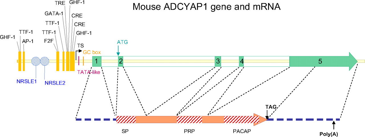
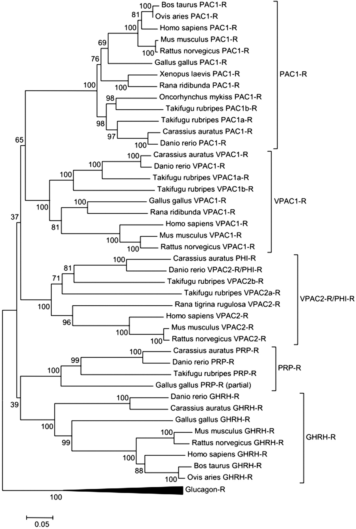
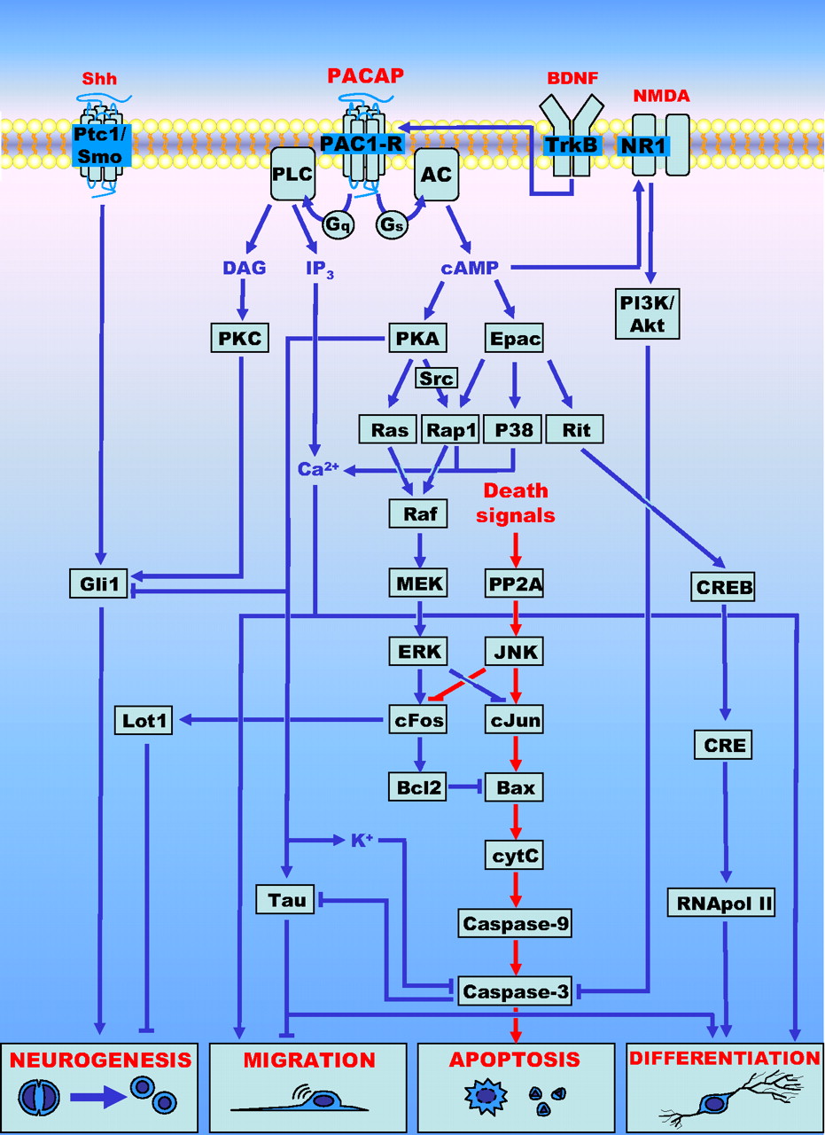
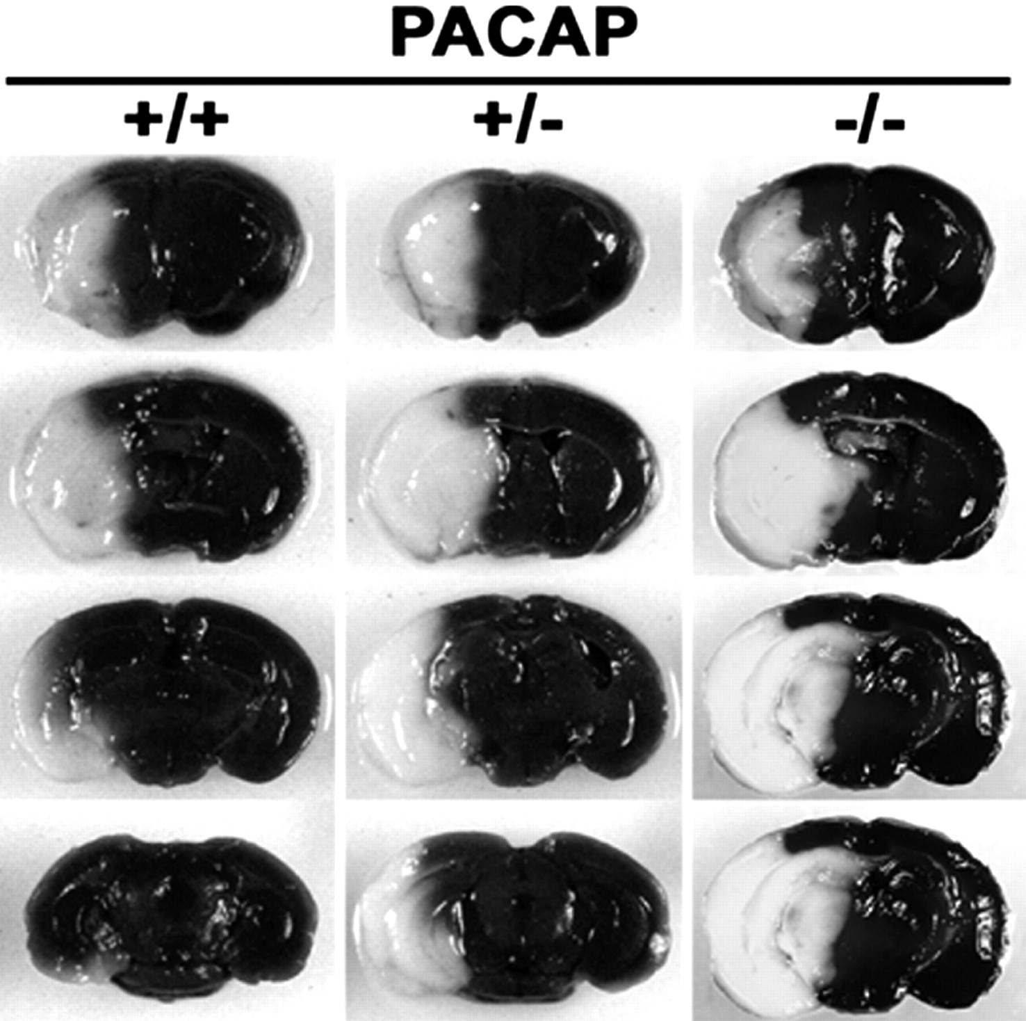